


Báo cáo hóa học: "Comparison of anti-CD3 and anti-CD28-coated beads with soluble anti-CD3 for expanding human T cells: Differing impact on CD8 T cell phenotype and responsiveness to restimulation"
lượt xem 5
download
 Download
Vui lòng tải xuống để xem tài liệu đầy đủ
Download
Vui lòng tải xuống để xem tài liệu đầy đủ
Tuyển tập báo cáo các nghiên cứu khoa học quốc tế ngành hóa học dành cho các bạn yêu hóa học tham khảo đề tài: Comparison of anti-CD3 and anti-CD28-coated beads with soluble anti-CD3 for expanding human T cells: Differing impact on CD8 T cell phenotype and responsiveness to restimulation
Bình luận(0) Đăng nhập để gửi bình luận!
Nội dung Text: Báo cáo hóa học: "Comparison of anti-CD3 and anti-CD28-coated beads with soluble anti-CD3 for expanding human T cells: Differing impact on CD8 T cell phenotype and responsiveness to restimulation"
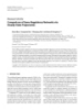
- Li and Kurlander Journal of Translational Medicine 2010, 8:104 http://www.translational-medicine.com/content/8/1/104 RESEARCH Open Access Comparison of anti-CD3 and anti-CD28-coated beads with soluble anti-CD3 for expanding human T cells: Differing impact on CD8 T cell phenotype and responsiveness to restimulation Yixin Li, Roger J Kurlander* Abstract Background: The ability to expand virus- or tumor-specific T cells without damaging their functional capabilities is critical for success adoptive transfer immunotherapy of patients with opportunistic infection or tumor. Careful comparisons can help identify expansion methods better suited for particular clinical settings and identify recurrent deficiencies requiring new innovation. Methods: We compared the efficacy of magnetic beads coated with anti-CD3 and anti-CD28 (anti-CD3/CD28 beads), and soluble anti-CD3 plus mixed mononuclear cells (designated a rapid expansion protocol or REP) in expanding normal human T cells. Results: Both anti-CD3/CD28 beads and soluble anti-CD3 promoted extensive expansion. Beads stimulated greater CD4 cell growth (geometric mean of 56- versus 27-fold (p < 0.01) at day 21) but both stimulated similar CD8 expansion (189- versus 186-fold). Phenotypically, bead-treated CD4 and CD8 T cells and anti-CD3-treated CD4 cells typically assumed an effector/effector memory phenotype by day 14. By comparison, a subset of anti-CD3-treated CD8 cells, derived from naïve cells, retained much greater expression of CD45RA, CD27 and CCR7, than matched bead-treated cells despite comparable expansion. These cells were clearly distinguishable from CD45RA+ terminally differentiated effector cells by the presence of CD27, the absence of CD57 and their inability to produce cytokines after stimulation. When used to expand previously stimulated cells, anti-CD3 plus autologous MNCs produced much less antigen-induced cell death of CD8 cells and significantly more CD8 expansion than beads. Conclusions: Anti-CD3/CD28 beads are highly effective for expanding CD4 cells, but soluble anti-CD3 has significant potential advantages for expanding CD8 T cells, particularly where preservation of phenotypically “young” CD8 cells would be desirable, or where the T cells of interest have been antigen-stimulated in vitro or in vivo in the recent past. Background for transfer can only be retrieved from blood or tissue With advances in the methods for selecting and manip- sites in relatively small numbers, consequently they ulating T cells there is increasing interest in the adop- usually are expanded specifically or nonspecifically prior tive transfer of bioactive T cells as a treatment for to transfer. Such ex vivo manipulations, however, poten- infections and cancer. This approach has been used suc- tially can damage T cell homing, proliferation, and sur- cessfully to transfer antiviral immunity after stem cell vival after infusion [3,4]. Given this risk, the choice of transplantation [1], and is under active investigation in methods may have important implications for clinical treating malignancy [2]. Antigen-specific T cells suitable efficacy. Antibodies against CD3 are a central element in many T cell proliferation protocols. Immobilized on a surface, * Correspondence: rkurlander@mail.cc.nih.gov anti-CD3 delivers a strong proliferative signal through Department of Laboratory Medicine, NIH Clinical Center, National Institutes of Health, Bethesda, Maryland, USA © 2010 Li and Kurlander; licensee BioMed Central Ltd. This is an Open Access article distributed under the terms of the Creative Commons Attribution License (http://creativecommons.org/licenses/by/2.0), which permits unrestricted use, distribution, and reproduction in any medium, provided the original work is properly cited.
- Li and Kurlander Journal of Translational Medicine 2010, 8:104 Page 2 of 15 http://www.translational-medicine.com/content/8/1/104 the T cell receptor complex (signal 1) but in the absence situations where preservation of the CD8 T cell response of additional costimulatory signals (signal 2), the result- in important. ing proliferation is often followed by premature T cell Methods apoptosis or anergy [5]. By immobilizing anti-CD3 and anti-CD28 to simultaneously deliver signal 1 and a costi- Antibodies, beads, and chemicals mulatory signal 2, proliferation can be increased without CD45RA/FITC, CD45RA/PE, CD57/FITC, CD28/PE, provoking early cell death [6]. The expanding cells also CD4/PerCP, CD8/PerCP, CD27/APC, brefeldin A, anti- IFNg/FITC, anti-TNFa/PE, 7-Amino-Actinomycin D (7- demonstrate enhanced ability to release cytokines and lyse targets cells in an MHC unrestricted manner [7]. AAD), and appropriate isotype controls were purchased Consequently, magnetic beads coated with anti-CD3 from BD Biosciences. Anti-human CCR7-phycoerythrin and anti-CD28 (anti-CD3/CD28 beads) have proved a was obtained from R&D Systems. Biotinylated anti-CD3 convenient reagent for expansion which has been used and anti-CD28 antibodies were purchased from experimentally to boost T cell immunity in immunosup- eBioscience. Anti-CD3 (Orthoclone OKT3) was pro- pressed cancer patients [8-10] and enhance the anti- vided by Stephen Migueles (NIAID, Bethesda, MD). tumor effect of donor lymphocyte infusions after Flow-Check Fluorospheres were purchased from Beck- allotransplantation [11]. These studies have established man Coulter. Streptavidin-labeled Dynabeads (M280) that beads can be used to expand functional T cells, and and CD3/CD28 T cell expander beads were obtained that some of these cells can persist in vivo postinfusion. from Invitrogen. Carboxyfluorescein succinimidyl ester While these results are encouraging, the bead expan- (CFSE) was purchased from Molecular Probes (Eugene, sion technique has limitations. Ex vivo expansion stimu- OR) and recombinant human IL-2 was purchased from lates the generation of effector T cells with increased PeproTech (Rocky Hill NJ). lytic and cytokine producing capability [7], but the capa- city of these cells for additional homing and prolifera- Preparation of anti-CD3/CD28 beads tion after infusion is uncertain [3]. While CD4 cells To prepare antibody-coated beads of varying composi- respond very well to anti-CD3/CD28 stimulation, CD8 tion, streptavidin-labeled beads were coated with varying cells proliferate less extensively with an increased rate of mixtures of biotinylated anti-CD3 and anti-CD28 anti- apoptosis [12]. Given the importance of CD8 T cells in bodies. To this end, streptavidin-M280 beads were the anti-tumor response, this is a significant concern. washed once with sterile PBS/BSA and resuspended at One commonly used alternative approach for stimu- 10-50 millions beads/ml. Preliminary dose response stu- lating proliferation is the incubation of T cells with dies, using FITC-labeled anti-mouse IgG and flow cyto- soluble anti-CD3 antibody in the presence of Fc recep- metry to monitor biotinylated antibody binding to tor bearing accessory cells [13-15], an approach desig- beads, established that beads were saturated by 100 ng nated the “Rapid Expansion Protocol” (REP). Antibody of biotinylated antibody/million beads. Consequently “presented” to T cells in this manner clearly generates a this total immunoglobulin/bead ratio was routinely used more effective proliferative signal than soluble anti-CD3 for bead coating. To vary the ratio of antibody coating alone or anti-CD3 immobilized on a plastic surface [16]. on beads equimolar solutions of anti-CD3 and anti- This presumably reflects the dual benefit of more exten- CD28 were mixed at 1:0, 1:5, 1:10, 1:20, 1:40, 1:80, 2 sive anti-CD3-T cell receptor crosslinking on a surface, 1:160, and 0:1 ratios. Control beads were coated with and the costimulation provided by cell-cell interaction biotinylated IgG1 isotype. Coating was performed on a between T cells and Fc receptor positive accessory cells rotator stand at room temperature for 2-3 hours. Beads such as monocytes which constitutively express CD80 were then washed two times with filtered PBS/BSA, [17], CD86 [17], and CD137 [18]. These complex inter- once with complete medium, and then resuspended in actions in some respects mimic events during physiolo- RPMI 1640 complete medium. Antibody coating was gic antigen presentation. Given its efficacy, this performed as needed, but preliminary studies established approach has been used extensively for expansion of that beads could be stored 4°C for at least one week T cell clones and lines for in vitro and clinical adoptive without any change in potency. In selected studies, transfer studies [13-15,19]. T cells were also stimulated using commercially To gain further insight into the similarities and differ- prepared anti-CD3 and anti-CD28 coated beads (CD3/ ences between the T cell responses produced by beads CD28 T cell Expander, Invitrogen). and REP, the current studies critically compare their impact on T cell survival, proliferation, and phenotype. Flow cytometry While both beads and anti-CD3 are effective in expanding Flow cytometry was performed using a 4-color Facscali- T cells, our studies demonstrate substantial differences in bur (BD biosciences). The standard phenotypic analysis their impact on CD8 cells that merit consideration in was performed at different time point using antibody
- Li and Kurlander Journal of Translational Medicine 2010, 8:104 Page 3 of 15 http://www.translational-medicine.com/content/8/1/104 p anels as described in the results. The flow data was cell composition and number were performed in analyzed using Flow-Jo software. quadruplicate. The absolute number of cells per well at each time point was calculated based on the number of calibrator Human leukocyte acquisition and purification Normal healthy donors gave informed consent to blood beads and the number of viable cells detected per well donation or leukapheresis procedures performed as spe- by flow cytometry using the formula: cified in clinical protocols approved by the Institutional The proportion of cells undergoing 0-6 divisions could Review Board of the Clinical Center of the National then be quantitated based on the pattern of CFSE fluor- Institutes of Health. Mixed mononuclear cells (MNCs) escence using Flo-Jo software, and the total number of obtained by leukepheresis or prepared from buffy coats viable cells per well. using Ficoll-Paque density gradient centrifugation, were cryopreserved, and thawed as previously described [20]. Bulk stimulation of T cells in vitro To prepare T cell subsets for selected experiments, To monitor cell phenotype and cell expansion over a 3 MNCs from freshly collected buffy coat cells were puri- week period, fresh MNC or cells expanded previously fied by negative selection using CD8+ Memory T Cell using anti-CD3/CD28 beads or anti-CD3 were cultured in Isolation and CD8 + Naive T Cell Isolation Kits pur- 12 well plates (5 × 10⁶ cells in 2 ml/well) with anti-CD3/ chased from Miltenyi Biotech. CD28 beads (three beads/cell), anti-CD3 (30 ng/ml), or medium alone. Previously expanded cells restimulated with anti-CD3 routinely also received irradiated autolo- Monitoring T cell division and early expansion using CFSE gous MNC (2 cells/responder) as a source of Fc receptor labeled cells To monitor cells division and expansion during the early positive accessory cells. Medium containing recombinant days after stimulation, cells were CFSE-labeled and moni- human IL2 (50 units/ml) was added to freshly cultured tored using methods described by Hawkins, et al. [21]. In cells at day 2 and to restimulated cells throughout the pro- brief, to label cells, 2-5 × 10⁷ mixed mononuclear cells or cess. Beads were removed using a magnet on day 7 post cultured T cells maintained in RPMI 1640 containing stimulation. Cell counts of freshly stimulated cells were 10% fetal calf serum plus 100 unit/ml penicillin, 100 ug/ monitored at least twice weekly and cultures were fed ml streptomycin, and 2 mM glutamine (RPMI/FCS) were every other day with fresh RPMI/FCS and IL2, and trans- incubated with 2 μ M CFSE at 37°C for 10 min. Cells ferred to flasks or frozen as needed to maintain cell num- bers between 0.75 and 2 × 10 6 /ml. Because of the were then washed three times to remove unbound CFSE, resuspended in fresh RPMI/FCS, and incubated over- presence of irradiated autologous feeder cells in REP trea- night. Labeled cells were then distributed (50,000/well) in ted cells, viable cell counts were not used to monitor cell a 96 well round bottom plate in wells also containing growth in restimulated cultures until after day 7 by which anti-CD3/CD28 coated beads (three beads/cell), anti- time no more viable irradiated cells were present. CD3 (30 ng/ml), or no additional stimulator. When using anti-CD3 to re-treat previously stimulated cells, 100,000 Measurement of Intracytoplasmic cytokine Production irradiated MNCs (accessory cell:responder ratio of 2:1) T cells harvested 14 days after stimulation with anti- were also added as a source of Fc receptor positive acces- CD3/CD28 beads or soluble anti-CD3 were treated for 4 sory cells suitable for “ presentation ” of anti-CD3 to T hours with phorbol myristate acetate (PMA, 35 nM) and the calcium ionophore A23187 (0.5 μM) or with medium cells. Fresh cells received IL2 (50 U/ml) on day 2. Resti- mulated cells were maintained with 50 U/ml of IL2 from alone in the presence of brefeldin (Golgiplug, BD day 1. Wells were fed with additional medium containing Bioscience). Cells were then incubated with anti-CD8 IL2 at day 4 or 5 and every 2-3 days thereafter. With con- PerCP and CD27 APC for 30 minutes, fixed and permea- tinued growth, the contents of wells were diluted 4 fold bilized using Cytofix/cytoperm solution (BD Bioscience) into new wells with fresh medium and IL2 as needed to as recommended by the manufacturer, and stained intra- cellularly using anti-IFNg FITC and antiTNFa PE. Dupli- prevent overcrowding. To monitor cell growth, at selected time points after cate samples were stained with an appropriate isotype stimulation, 10,000 calibrator beads/well (Flow-Check control. Cytokine expression in treated and control cells Fluorospheres, Beckman Coulter, CA) were added to was then assessed using flow cytometry. wells along with PE labeled anti-CD4 or anti-CD8 anti- bodies. The contents of the well were mixed, incubated Statistics for one half hour, and then washed twice with PBS/ Paired t-tests and nonparametric 2-tail Wilcoxon BSA (1%) before addition of 7-AAD to exclude dead matched pairs tests were performed using Graphpad cells in flow cytometry analysis. All measurements of Prism Software.
- Li and Kurlander Journal of Translational Medicine 2010, 8:104 Page 4 of 15 http://www.translational-medicine.com/content/8/1/104 expanded more rapidly in response to beads than anti- Results CD3 (Figure 2A) and this difference was statistically sig- Time course for T cell response to stimulation nificant at days 7, 14, and 21. There was a trend to The CFSE-labeled CD4 and CD8 T cells in MNCs began more rapid expansion of CD8 cells in response to anti- dividing 40-50 hours after stimulation with anti-CD3/ CD3, particularly at days 7 and 14, but these differences CD28 beads or soluble anti-CD3. CD8 cells divided did not achieve statistical significance (Figure 2B). Con- slightly more rapidly than CD4 cells, and there were no sistent with these reciprocal trends in CD4 and CD8 consistent differences in early response to the two sti- expansion, cultures stimulated with anti-CD3 accumu- muli (Figure 1A and 1C). The number of viable cells at lated a significantly higher proportion of CD8 cells at all hour 40 (just before proliferation began) was similar in three time points than matched bead-treated cultures unstimulated, bead-stimulated, and anti-CD3-stimulated (Table 1). wells indicating neither stimulus caused extensive early These studies were performed with beads coated with activation-induced cell death (AICD) (Figure 1B and anti-CD3 and anti-CD28 at a ratio of 1:20 but similar 1D). Consistent with the timing of cell division, expan- results were obtained using beads coated at ratios of 1:5, sion in cell number in response to either stimulus was and 1:80 and with commercially available T cell expan- delayed until 50-60 hours after stimulation. der beads (data not shown). Comparisons of expansion To compare the expansion produced by anti-CD3/ produced by anti-CD3 at 30 and 300 ng/ml also yielded CD28 beads and anti-CD3, we monitored changes in essentially identical results (data not shown). cell number in bulk cultures over a 21-day period in 11 separate studies (Figure 2) and noted several persistent Phenotypic changes in T cells during in vitro expansion trends. Consistent with the more rapid rate of early cell Peripheral blood T cells are usually subclassified as division noted in Figures 1A and 1C, CD8 cells naïve (CD45RA+, CCR7+), central memory (CD45RA-, expanded more rapidly than CD4 cells in response to CCR7+), effector memory (CD45RA-, CCR7-), or either stimulus. Focusing on CD4 cells, this subset Figure 1 Early time course of T cell division and expansion in response to anti-CD3/CD28 beads and soluble anti-CD3. CFSE-labeled CD4 (A) and CD8 (C) cells began dividing 40-60 hours after exposure to beads (solid lines) or anti-CD3 (dashed lines), but divided minimally in the absence of stimulation (2× solid lines). The number of stimulated and control CD4 (B) and CD8 (D) T cells remained comparable until about 60 hours after stimulation when measureable cell expansion began.
- Li and Kurlander Journal of Translational Medicine 2010, 8:104 Page 5 of 15 http://www.translational-medicine.com/content/8/1/104 The phenotype of CD4 T cells was similarly affected by exposure to beads or anti-CD3 (Figure 3A) with sub- stantial reductions in the expression of CD45RA, CCR7, and CD27. CD8 T cells, on the other hand, were affected differently by beads and anti-CD3. By day 14, CD45RA expression on bead-treated cells was markedly reduced, but a substantial fraction of anti-CD3-treated cells retained CD45RA expression (Figure 3B). This sub- population was strongly CD27 positive, and (to a lesser extent) CCR7 positive. The same pattern of CD45RA and CD27 expression was seen at day 21, but CCR7 expression often diminished substantially by this time point. On the other hand, bead-treated cells usually retained a higher proportion of CD28 positive cells both at day 14 and 21. The size of the CD45RA+, CD27+ subset of CD8 cells in anti-CD3-treated cultures varied somewhat from donor to donor, but the underlying pat- tern was qualitatively consistent. To clarify the origin of the persistent CD45RA +/CD27+ subset, we stimulated purified naïve and mem- ory CD8 populations with anti-CD3/CD28 beads or anti-CD3 plus irradiated MNCs as a source of FcR+ accessory cells. A substantial proportion of anti-CD3- treated naïve CD8 cells maintained the CD45RA+, CCR7+, CD27+ phenotype noted above, but much fewer bead-treated cells demonstrated this phenotype (Figure Figure 2 Comparison of CD4 (A) and CD8 (B) expansion after 4A and 4C). Memory cells did not consistently differ in stimulation with anti-CD3/CD28 beads (squares) or anti-CD3 their pattern of CD45RA and CD27 retention in (triangles). Beads stimulated significantly greater CD4 expansion response to either stimulus (Figure 4B and 4C). Thus than anti-CD3 (p values for statistical significance at each time point is indicated at the top of each box). CD8 expansion was slightly the phenotypic differences noted in mixed populations greater in response to anti-CD3 at 7 and 14 days, but this trend was can be largely attributed to variations in the response of not statistically significant. The p values were calculated using the naïve CD8 T cells to soluble anti-CD3 plus accessory Wilcoxon matched pairs test. cells versus beads. CD45RA expression on cultured T cells is a typical effector cells (CD45RA+, CCR7-)[22]. Naïve cells also characteristic of terminally differentiated effector T cells, express CD27 and CD28, which are both progressively but unlike conventional effectors [22], the CD45RA+ lost with post-thymic proliferation and “differentiation” CD8 T cells noted after anti-CD3 treatment were con- towards an effector phenotype [23-26]. To compare the sistently CD27+ (Figure 3) and CD57- (data not shown). impact of beads and anti-CD3, the expression of these Effector T cells typically produce intracellular cytokines markers on CD4 and CD8 cells was assessed before and within 4 hours after stimulation in vitro [22], but the after 2-3 weeks of in vitro stimulation. The results from CD27+ anti-CD3-expanded CD8 cells (in contrast with one representative experiment are illustrated in Figure 3. the CD27- cells in the same preparation) produced little intracellular IFNg or TNFa in response to PMA/A23187 stimulation (Figure 5). Table 1 % CD8 T cells in primary cultures at 7-21 days T cell response to restimulation after stimulation with anti-CD3/CD28 beads and OKT3 On occasion, the number of cells generated by one cycle Day after Stimulation of T cell expansion may be insufficient for the desired Cells stimulated with: 7 14 21 purpose, and further expansion would be desirable. To Anti-CD3/CD28 beads 34.0 ± 6.8 (11)* 47.6 ± 6.3 (11) 48.9 ± 5.4 (9) compare impact of restimulation, cells previously OKT3-treated 53.7 ± 7.0 (11) 68.0 ± 5.4 (11) 63.3 ± 4.4 (9) expanded using anti-CD3/CD28 beads were CFSE- P < 0.005 P < 0.005 P < 0.05 labeled 6 to 63 days later, and restimulated with fresh *The results represent the mean ± SE for matched pairs of cultures with the anti-CD3/CD28 beads, anti-CD3 plus irradiated autolo- number of experiments marked in parentheses. Paired t-test was used to gous MNCs, or, as a control, maintained in medium assess significance.
- Li and Kurlander Journal of Translational Medicine 2010, 8:104 Page 6 of 15 http://www.translational-medicine.com/content/8/1/104 Figure 3 Comparison of T cell phenotype 14 and 21 days after primary stimulation of MNCs with CD3/CD28 beads and soluble anti- CD3. CD4 T cells (A) showed a similar pattern of changes in response to either stimulus, but CD8 cells (B) demonstrated significant differences in phenotype. At comparable levels of expansion, anti-CD3 treated CD8 cells retained higher levels of CD45RA, CD27, and CCR7 expression than anti-CD3/CD28 bead treated cells and bead-treated CD8 cells expressed higher levels of CD28. p lus IL2 alone. Like fresh cells, restimulated cells relative to control cells over the first 40 hours of culture showed an increased rate of division 40-50 hours post reflecting early AICD (Figure 7B and 7D). stimulation (Figure 6A and 6C). Unlike fresh cells (Fig- The findings from 11 experiments comparing T cell ure 1B and 1D), however, restimulated cells (particularly expansion 3-14 days after restimulation are summarized those retreated with beads) often decreased in number in Figure 7. Despite the initial AICD in many cases,
- Li and Kurlander Journal of Translational Medicine 2010, 8:104 Page 7 of 15 http://www.translational-medicine.com/content/8/1/104 Figure 4 Comparison of the impact of anti-CD3/CD28 beads and soluble anti-CD3 plus irradiated MNCs on expansion by purified naive (A) and memory (B) CD8 cells. Significant differences in the phenotype of expanded naïve T cells, but not in memory cells, were noted. Panel C collates the results of 3 experiments quantitating changes in CD45RA, CCR7, CD27, and CD28 surface antigen (expressed as geometric mean channel fluorescence) when naïve and memoryCD8 cells were expanded using beads (squares) or anti-CD3/MNCs(circles) for 14 days. CD4 cells incubated with either stimulus expanded 10- showed significantly less expansion at all 3 time points 100 fold by day 7 (figure 7A). CD8 T cells incubated (Figure 7B). with soluble anti-CD3 plus irradiated MNCs demon- Although the gross expansion of restimulated cells in strated a similar pattern, but bead-treated CD8 cells some experiments (Figure 7) was comparable in
- Li and Kurlander Journal of Translational Medicine 2010, 8:104 Page 8 of 15 http://www.translational-medicine.com/content/8/1/104 after primary stimulation) was associated with greater early cell loss at day 3, and poor expansion at day 7 (Figure 8B and 8D). This was most striking for bead- treated cells (particularly CD8 cells), but soluble anti- CD3 stimulated cells showed a similar, albeit less marked, trend. In these studies, only cells rested > 30 days between stimulations demonstrated substantial expansion relative to control cells. In sum, these studies emphasize that even when gross expansion is observed, restimulation (particularly with beads) may actually impede cell expansion. While figures 5, 6, 7 focused on the responses of cells initially expanded using anti-CD3/CD28 beads, analo- gous studies performed using cells initially expanded using soluble anti-CD3 gave qualitatively identical results. Comparison of T cell size after stimulation with anti-CD3/ CD28 beads or anti-CD3 To gain additional insight into the relative impact of beads and anti-CD3 on cells, we serially monitored for- ward scatter (a relative measure of cell size) in CD4 and CD8 cells after stimulation and restimulation. CD4 and CD8 T cell size increased in a similar manner after pri- mary treatment with either stimulus (Figure 9A and 9B). Cells restimulated with anti-CD3/CD28 beads however increased in size more slowly and maintained a larger size for a longer period than matched anti-CD3 treated cells, even when they were expanding poorly. These dif- ferences in size could not be simply explained by differ- ences in IL2 feeding schedule or cell concentration within flasks. Figure 5 Intracytoplasmic cytokine production by day 14 anti- Impact of variations in bead coating with anti-CD3 and CD3 stimulated CD8 T cells stimulated for 4 hours with PMA/ A23187. By comparison to CD27- cells, the CD27+ subset produced CD28 on T cell responses to restimulation little intracytoplasmic (A) tumor necrosis factor (TNF-a) or (B) To assess whether bead-mediated expansion could be interferon- g (IFN-g). Similar results were obtained in each of 4 improved by modifying the ratio of anti-CD3 to anti- studies. CD28 coating, we restimulated cells with beads coated using a variety of antibody ratios (Figure 10). Restimu- lated CD4 cells expanded better (overlapping in efficacy m agnitude to that observed after primary expansion with anti-CD3 plus irradiated MNCs) in response to (Figure 2), matched control cells also often expanded as beads coated using lower anti-CD3: anti-CD28 ratios. well. To distinguish true restimulation-dependent CD8 cell expansion was also improved by reducing the growth from persistent expansion still attributable to anti-CD3:anti-CD28 ratio but even using the most primary stimulation, we calculated the ratio of expan- lightly coated beads (or beads coated with anti-CD 28 sion by stimulated cells/expansion by matched control alone), expansion remained substantially inferior to that cells and plotted this as a function of the time interval produced using anti-CD3/MNCs. The poor response of between first and second stimulation (Figure 8A-D). CD8 cells to beads was not appreciably improved by These plots make two important points. First, the adding irradiated MNCs and sufficient beads to main- expansion ratio for cells re-exposed to beads was almost tain a 3:1 bead to total cell ratio (data not shown). always less than 1 at day 3 poststimulation (Figure 8A and 8C) reflecting AICD, and this effect was particularly Discussion severe for CD8 cells. By comparison soluble anti-CD3- Anti-CD3/CD28 beads and soluble anti-CD3 both sti- treated cells seldom demonstrated this degree of early mulate extensive polyclonal expansion of human cell loss. Second, early restimulation (less than 20 days
- Li and Kurlander Journal of Translational Medicine 2010, 8:104 Page 9 of 15 http://www.translational-medicine.com/content/8/1/104 Figure 6 Early time course for expansion of bead-expanded T cells after restimulation 14 days later with anti-CD3/CD28 beads or anti-CD3 plus irradiated MMCs. Restimulated CD4 (A) and CD8 (C) both divided more extensively in response to anti-CD3 (dashed lines) than anti-CD3/CD 28 (solid lines), but this difference was more pronounced for CD8 cells. CD4 T cell expansion (C) was similar in response to either stimulus, but CD8 T cells (D) expanded substantially more rapidly than anti-CD3/CD28 bead-treated cells. peripheral blood T cells. Beads show a small but signifi- shown) respond in the same manner. This sensitivity cant advantage in expanding CD4 cells and anti-CD3 persists even in cells rested for more than 3 weeks demonstrates a trend towards more rapid early CD8 cell between stimulations. This was not an isolated observa- expansion. Proliferation of anti-CD3 treated cells slows tion using one particular bead formulation. Similar after day 14, while bead-mediated proliferation typically results were observed using commercially available beads and “home-brew” beads anti-CD3 and anti-CD28 continues for more than 21 days. Judged solely by their efficacy in promoting expansion, beads are more effec- coated at a variety of ratios. tive. Our studies, however, identify two qualitative dif- Soluble anti-CD3 used in conjunction with irradiated ferences that may merit consideration in tailoring an MNCs to restimulate cells produced significantly less expansion method for any particular clinical situation. AICD and more CD8 T cell growth. The difference was First, beads and anti-CD3 have quite different effects particularly striking when CD8 T cells were retreated before cells had “rested” sufficiently after primary stimu- in restimulating CD8 T cells. AICD, mediated at least in part through Fas/FasL interaction and activation of the lation. While anti-CD3 might fail to increase the growth proapoptotic molecule BIM, is a well-described compli- rate of still expanding cells, it did not produce the strik- cation of T cell stimulation [27,28]. Although CD28 ing AICD and extended growth retardation associated costimulation enhances expression of the anti-apoptotic with anti-CD3/CD28 beads. molecule BCL-XL, concurrent anti-CD3 and anti-CD28 The mechanism underlying this difference was not signaling can promote CD8 apoptosis [12]. The current addressed in these studies, but a variety of factors may studies demonstrate that previously stimulated CD8 contribute. Judging by the differences in time course for cells are particularly susceptible to AICD and growth changes in cell size after restimulation (Figure 9), solu- retardation after bead exposure. This effect is not lim- ble anti-CD3 generates a less pronounced and pro- ited to cells previously exposed to beads. Cells initially longed T cell perturbation in restimulated cells than stimulated using soluble anti-CD3 or PHA (data not anti-CD3/CD28 beads. Equally important, Fc receptor
- Li and Kurlander Journal of Translational Medicine 2010, 8:104 Page 10 of 15 http://www.translational-medicine.com/content/8/1/104 Figure 7 Comparison of CD4 (A) and CD8 (B) growth when bead-treated T cells were restimulation with anti-CD3/CD28 beads (squares) or anti-CD3/irradiated MMCs (triangles). T cell expansion was monitored using CFSE labeled cells and flow cytometry as described in the methods. CD4 expansion was similar in response to either stimulus. By contrast, bead-treated CD8 cells expanded significantly less well than anti-CD3-treated cells at all time points. The p values with obtain using the Wilcoxon matched pairs test are indicated at the top of each box. b earing monocytes “ presenting ” anti-CD3 to T cells, more complex costimulatory environment than an inert express not only the CD28 ligands CD80 and CD86, antibody-coated bead. Whatever the specific signaling but CD137L which can activate CD137 [18], another events, the adverse impact of beads on activated CD8 potent costimulatory molecule for CD8 T cell expansion cells can not be simply ameliorated by reducing the [29]. After interaction with stimulated T cells, mono- concentration of anti-CD3 on the bead surface or by cytes binding anti-CD3 may express additional costimu- the presence of irradiated autologous MNCs at the time latory molecules and cytokines as well, generating a of restimulation.
- Li and Kurlander Journal of Translational Medicine 2010, 8:104 Page 11 of 15 http://www.translational-medicine.com/content/8/1/104 Figure 8 Evaluation of the interrelationship between the type and timing of the second stimulus and expansion of restimulated CD4 (A and B) and CD8 (C and D) T cells. To normalize for the impact of persistent growth attributable to primary stimulation, the ratio of growth after restimulation to growth by matched control cells in the absence of restimulation was calculated. Regardless of the interval between stimulations, by day 3, CD4 (A) and CD8 (B) cells re-incubated with anti-CD3/CD28 beads usually had a stimulation ratio of less than 1 i.e. cells had diminished in number compared to control untreated cells. Even at day 7 (B and D), the stimulation ratio seldom exceeded 1 for cells which had been rested in vitro between stimulations for less than 20 days. By comparison, the stimulation ratio for anti-CD3/MMC treated cells seldom dropped below 1 at day 3, and increased further with time, even when the rest interval between stimulations was less than 14 days. antigen-specific cell subset may have been “preactivated” The propensity of anti-CD3/CD28 beads to damage previously activated CD8 T cells has important implica- in vivo. tions. A second round of stimulation with anti-CD3/ We also noted significant phenotypic differences CD28 beads is a risky strategy for enhancing the yield of between the CD8 T cells expanded in response to beads antigen-specific cells. Any increase in absolute cell num- and soluble anti-CD3. Consistent with most [22,30,31], ber must be balanced against damage to surviving cells but not all [32] prior studies of in vitro stimulated T and unwanted pruning of the repertoire from AICD cells bead and soluble anti-CD3 treated CD4 cells and before expansion. Equally important, if cells activated bead-treated CD8 cells assume predominantly an effec- physiologically behave like in vitro stimulated cells, T tor or effector memory phenotype, markedly downregu- cells responding to antigen in vivo (for example tumor- lating CD45RA and CCR7 by day14 post stimulation. By specific CD8 T cells in tumor infiltrating lymphocytes contrast, a subset of anti-CD3 treated CD8 cells retained preparations or antiviral CD8 T cells retrieved from the CD45RA, CD27, and, to lesser extent, CCR7 and CD28 blood of a patient with active viral infection) may be expression at day 14. By studying purified CD8 subset vulnerable to bead-induced AICD or growth retardation cells, we could establish that these phenotypically dis- during even an initial round of ex vivo stimulation. If tinctive cells were derived from the naïve CD8 T cell so, bead exposure may selectively damage or destroy an subset. The pattern of strong CD45RA expression in important subset of harvested T cells. For this reason, cultured CD8 T cells, is often associated with terminally stimulation using soluble anti-CD3 plus MNCs may differentiated effectors [22], but based on their deriva- deserve greater consideration as an alternative approach tion from naïve cells, the preservation of CD27, and for expanding of CD8 cells in situations where a desired CCR7 (at week 2 with gradual disappearance by week
- Li and Kurlander Journal of Translational Medicine 2010, 8:104 Page 12 of 15 http://www.translational-medicine.com/content/8/1/104 Figure 9 Comparison of changes in forward scatter measured by flow cytometry (a measure of cell size) in response to anti-CD3/ CD28 beads and anti-CD3. Primary CD4 (A) and CD8 cells (B) showed similar changes in forward scatter after exposure to either stimulus, but CD4 (C) and CD8 (D) cells restimulated on day 30 increased in size more slowly and remained large for longer after restimulation with beads. This difference in size was noted even when the enlarged cells were expanding poorly. 3), in the absence of CD57 expression, or rapid intracel- The functional properties of the CD45RA+ CD8 cells lular cytokine production in response to restimulation, produced during anti-CD3 mediated expansion merit we suggest these cells evolve towards a phenotype ana- further study. Derived from naïve cells, these relatively “young” cells retain expression of both CCR7, a crucial logous to the CD45RA+, CCR7-, CD27+ CD8 T cells noted previously in human blood [25]. This subset has receptor in lymphoid trafficking to central lymphoid tis- been shown to have a proliferative history and effector sues for several weeks, and CD27, an antigen positively function intermediate between naïve and effector mem- associated with T cell proliferation in vitro and engraft- ory cells. ment after adoptive transfer in vivo [34,36] for even The explanation for these differences in phenotypic longer despite substantial expansion. Cells with these evolution of bead and soluble anti-CD3 stimulated cells properties could have an advantage in trafficking into remains uncertain. Since CD45RA expression can be and proliferating within central lymphoid tissues in vivo reversibly downregulated in response to T cell receptor compared to bead treated cells expressing an effector stimulation [33], reduced expression of this antigen phenotype. On the other hand, although these cells do could, in part, be explained by the prolonged duration express more CD27 and CCR7, they also often express of bead-mediated stimulation noted in these and prior less CD28 than comparable bead-treated cells, and studies [6]. The more prolonged impact of bead stimula- CD28 expression in some settings has been a surrogate tion could also contribute to reduced CD27 expression marker for proliferative potential and telomerase expres- by enhancing and sustaining expression of CD70, a nat- sion [37,38]. Clearly, additional in vitro and ultimately ural ligand, which can downregulate CD27 expression in vivo function studies addressing the homing and pro- on stimulated T cells [34]. It is unlikely however, that liferative capacity of this subset would be needed to such reversible effects could fully explain CD45RA and accurately assess its properties. CD27 loss. Neither CD45RA nor CD27 were re- Anti-CD3/CD28 coated beads and soluble anti-CD3 expressed on bead-treated cells even after several weeks have each been used clinically to expand cells for adop- after bead removal suggesting more durable differences tive transfer with promising results in some settings in CD8 T cell expression. Such divergence would not be [8-11,13-15,19]. While beads are extremely effective in surprising since differences in the quality, magnitude, stimulating CD4 T cell expansion and in generating and duration of T cells signaling have been well docu- bioactive effector cells, the current studies raise the pos- mented to influence T cell differentiation [35]. sibility that soluble anti-CD3 may be a safer reagent for
- Li and Kurlander Journal of Translational Medicine 2010, 8:104 Page 13 of 15 http://www.translational-medicine.com/content/8/1/104 memory cells, and CMV-specfic responses are often dominated by effector CD8 T cells [40,41]. These dif- ferences in phenotype and biology may substantially shape how each antigen-specific population respond to any given expansion protocol. Future studies systemati- cally comparing the efficacy of anti-CD3/CD28 beads, soluble anti-CD3, and potentially other expansion pro- tocols in expanding defined populations of antigen- specific naïve, memory, and effector T cells could be helpful in beginning to develop more nuanced guide- lines for selecting the best cell expansion method for any particular clinical settings. Looking to the future, with growing interest in using adoptive transfer to treat human disease, many promis- ing new approaches are under active study for improv- ing T cell expansion. These include the use of other cytokines as well as or instead of IL2 as growth factors, tumor-derived cell lines engineered to express bioactive costimulatory molecules and cytokines as accessory cells [42], and retroviral or lentiviral vectors coding chimeric antigen receptors to confer defined antigenic specificity on T cells [42-44]. In judging the impact of these newer approaches and cell products, the current studies defin- ing the in vitro performance characteristics of “standard” expansion protocols hopefully may serve as a useful “measuring stick” with which to judge the value of new approaches for stimulating and restimulating human T Figure 10 Impact of variation in the anti-CD3:anti-CD28 cells. coating ratio on the efficacy of anti-CD3/CD28 beads in restimulating previously expanded CD4 (upper panel) and CD8 Conclusions (lower panel) T cells. CD4 cells expanded most extensively in Anti-CD3 in the presence of Fc receptor positive acces- response to soluble anti-CD3 plus irradiated MMCs or beads coated sory cells, is an effective method for expanding CD4 and at a very low anti-CD3:anti-CD28 ratio. Expansion was inhibited by higher levels of anti-CD3 coating. CD8 cells also responded better CD8 T cells which could have potential advantages over to beads coated at a low anti-CD3:anti-CD28 ratio, but even at the anti-CD3/CD28 coated beads in expanding CD8 T cells lowest ratio tested, beads were considerably less effective than anti- which may have been recently activated or antigen- CD3/MMCs in promoting cell growth. exposed in vitro or in vivo without provoking antigen induced cell death. This method also may be advanta- geous in maintaining CCR7 expression on expanding expanding CD8 cells which may have been recently anti- CD8 cells in settings where central lymphoid trafficking gen-stimulated. The findings also suggest this approach of adoptively transferred CD8 cells may be particularly may be more effective in preserving expression of func- desireable. tionally important surface antigens such as CCR7 and CD27 on naive T cells during expansion. While these studies have focused broadly on the List of Abbreviations expansion and phenotype of CD4 and CD8 T cell AICD: activation-induced cell death; Anti-CD3/CD28 beads-beads coated populations, in practical applications it is the ability of with anti-CD3 and anti-CD28; CFSE: Carboxyfluorescein succinimidyl ester; MNCs: mixed mononuclear cells; REP: Rapid Expansion Protocol. any given method to expand functionally active, anti- gen-specific T cells directed against a defined target Acknowledgements pathogen or tumor that is most critical. Evaluating effi- We wish to acknowledge Fran Hakim for her many helpful suggestions and for reviewing the manuscript. This research was supported by the Intramural cacy in achieving this goal is more complicated parti- Research Program of the NIH and the Clinical Center cularly given the potential phenotypic heterogeneity of Authors’ contributions antigen-specific populations. Whereas naturally occur- YL performed studies, participated in their design, and reviewed the ring melan A-responsive CD8 T cells are predomi- manuscript. RK participated in the design of the studies, performed the nantly naïve in phenotype [39], EBV-specific CD8 cell statistical analysis and wrote the manuscript. populations contain a high proportion of effector Both authors read and approved the final manuscript.
- Li and Kurlander Journal of Translational Medicine 2010, 8:104 Page 14 of 15 http://www.translational-medicine.com/content/8/1/104 17. Fleischer J, Soeth E, Reiling N, GrageGriebenow E, Flad HD, Ernst M: Competing interests Differential expression and function of CD8O (B7-1) and CD86 (B7-2) on The authors declare that they have no competing interests. human peripheral blood monocytes. Immunology 1996, 89:592-598. 18. Ju SW, Ju SG, Wang FM, Gu ZJ, Qiu YH, Yu GH, Ma HB, Zhang XG: A Received: 4 June 2010 Accepted: 26 October 2010 functional anti-human 4-1BB ligand monoclonal antibody that enhances Published: 26 October 2010 proliferation of monocytes by reverse signaling of 4-1BBL. Hybridoma and Hybridomics 2003, 22:333-338. References 19. Rosenberg SA, Dudley ME: Cancer regression in patients with metastatic 1. Heslop HE, Slobod KS, Pule MA, Hale GA, Rousseau A, Smith CA, Bollard CM, melanoma after the transfer of autologous antitumor lymphocytes. Proc Liu H, Wu MF, Rochester RJ, et al: Long-term outcome of EBV-specific T- Natl Acad Sci USA 2004, 101(2):14639-14645. cell infusions to prevent or treat EBV-related lymphoproliferative disease 20. Kurlander RJ, Tawab A, Fan Y, Carter CS, Read EJ: A functional comparison in transplant recipients. Blood 115:925-935. of mature human dendritic cells prepared in fluorinated ethylene- 2. Rosenberg SA, Dudley ME: Adoptive cell therapy for the treatment of propylene bags or polystyrene flasks. Transfusion 2006, 46:1494-1504. patients with metastatic melanoma. Curr Opin Immunol 2009, 21:233-240. 21. Hawkins ED, Hommel M, Turner ML, Battye FL, Markham JF, Hodgkin PD: 3. Gattinoni L, Klebanoff CA, Palmer DC, Wrzesinski C, Kerstann K, Yu Z, Measuring lymphocyte proliferation, survival and differentiation using Finkelstein SE, Theoret MR, Rosenberg SA, Restifo NP: Acquisition of full CFSE time-series data. Nature Protocols 2007, 2:2057-2067. effector function in vitro paradoxically impairs the in vivo antitumor 22. Sallusto F, Lenig D, Forster R, Lipp M, Lanzavecchia A: Two subsets of efficacy of adoptively transferred CD8+ T cells. J Clin Invest 2005, memory T lymphocytes with distinct homing potentials and effector 115:1616-1626. functions. Nature 1999, 401:708-712. 4. Casado JG, DelaRosa O, Pawelec G, Peralbo E, Duran E, Barahona F, 23. Appay V, Dunbar PR, Callan M, Klenerman P, Gillespie GM, Papagno L, Solana R, Tarazona R: Correlation of effector function with phenotype Ogg GS, King A, Lechner F, Spina CA, et al: Memory CD8+ T cells vary in and cell division after in vitro differentiation of naive MART-1-specific differentiation phenotype in different persistent virus infections. Nature CD8+ T cells. Int Immunol 2009, 21:53-62. medicine 2002, 8:379-385. 5. Schwartz RH: A cell-culture model for lymphocyte-T clonal anergy. 24. Hamann D, Kostense S, Wolthers KC, Otto SA, Baars PA, Miedema F, van Science 1990, 248:1349-1356. Lier RA: Evidence that human CD8+CD45RA+CD27- cells are induced by 6. Levine BL, Bernstein WB, Connors M, Craighead N, Lindsten T, antigen and evolve through extensive rounds of division. Int Immunol Thompson CB, June CH: Effects of CD28 costimulation on long-term 1999, 11:1027-1033. proliferation of CD4(+) T cells in the absence of exogenous feeder cells. 25. Rufer N, Zippelius A, Batard P, Pittet MJ, Kurth I, Corthesy P, Cerottini JC, Journal of Immunology 1997, 159:5921-5930. Leyvraz S, Roosnek E, Nabholz M, Romero P: Ex vivo characterization of 7. Garlie NK, LeFever AV, Siebenlist RE, Levine BL, June CH, Lum LG: T cells human CD8+ T subsets with distinct replicative history and partial coactivated with immobilized anti-CD3 and anti-CD28 as potential effector functions. Blood 2003, 102:1779-1787. immunotherapy for cancer. J Immunother 1999, 22:336-345. 26. Romero P, Zippelius A, Kurth I, Pittet MJ, Touvrey C, Iancu EM, Corthesy P, 8. Rapoport AP, Stadtmauer EA, Aqui N, Badros A, Cotte J, Chrisley L, Veloso E, Devevre E, Speiser DE, Rufer N: Four functionally distinct populations of Zheng ZH, Westphal S, Mair R, et al: Restoration of immunity in human effector-memory CD8+ T lymphocytes. J Immunol 2007, lymphopenic individuals with cancer by vaccination and adoptive T-cell 178:4112-4119. transfer. Nature medicine 2005, 11:1230-1237. 27. Green DR, Droin N, Pinkoski M: Activation-induced cell death in T cells. 9. Stadtmauer EA, Rapoport AP, Levine BL, Badros A, Porter DL, Luger SM, Immunol Rev 2003, 193:70-81. Mann D, Cross A, June CH: Co-stimulated autologous T-cell infusion after 28. Snow AL, Oliveira JB, Zheng L, Dale JK, Fleisher TA, Lenardo MJ: Critical role autologous stem cell transplantation (SCT) for myeloma accelerates for BIM in T cell receptor restimulation-induced death. Biol Direct 2008, lymphocyte recovery and augments response to pneumoccal vaccine: 3:34. Results of a randomized trial. Journal of Clinical Oncology 2005, 29. Zhu YW, Zhu GF, Luo LQ, Flies AS, Chen LP: CD137 stimulation delivers an 23:568S-568S. antigen-independent growth signal for T lymphocytes with memory 10. Laport GG, Levine BL, Stadtmauer EA, Schuster SJ, Luger SI, Grupp S, phenotype. Blood 2007, 109:4882-4889. Bunin N, Strobl FJ, Cotte J, Zheng ZH, et al: Adoptive transfer of 30. Geginat J, Lanzavecchia A, Sallusto F: Proliferation and differentiation costimulated T cells induces lymphocytosis in patients with relapsed/ potential of human CD8+ memory T-cell subsets in response to antigen refractory non-Hodgkin lymphoma following CD34(+)-selected or homeostatic cytokines. Blood 2003, 101:4260-4266. hernatopoietic cell transplantation. Blood 2003, 102:2004-2013. 31. Duarte RF, Chen FE, Lowdell MW, Potter MN, Lamana ML, Prentice HG, 11. Porter DL, Levine BL, Bunin N, Stadtmauer EA, Luger SM, Goldstein S, Madrigal JA: Functional impairment of human T-lymphocytes following Loren A, Phillips J, Nasta S, Perl A, et al: A phase 1 trial of donor PHA-induced expansion and retroviral transduction: implications for lymphocyte infusions expanded and activated ex vivo via CD3/CD28 gene therapy. Gene Therapy 2002, 9:1359-1368. costimulation. Blood 2006, 107:1325-1331. 32. Mercier-Letondal P, Montcuquet N, Sauce D, Certoux JM, Jeanningros S, 12. Laux I, Khoshnan A, Tindell C, Bae D, Zhu XM, June CH, Effros RB, Nel A: Ferrand C, Bonyhadi M, Tiberghien P, Robinet E: Alloreactivity of ex vivo- Response differences between human CD4+ and CD8+ T-cells during expanded T cells is ecorrelated with expansion and CD4/CD8 ratio. CD28 costimulation: Implications for immune cell-based therapies and Cytotherapy 2008, 10:275-288. studies related to the expansion of double-positive T-cells during aging. 33. Carrasco J, Godelaine D, Van Pel A, Boon T, van der Bruggen P: CD45RA on Clin Immunol 2000, 96:187-197. human CD8 T cells is sensitive to the time elapsed since the last 13. Riddell SR, Greenberg PD: The Use of Anti-Cd3 and Anti-Cd28 antigenic stimulation. Blood 2006, 108:2897-2905. Monoclonal-Antibodies to Clone and Expand Human Antigen-Specific T- 34. Huang J, Kerstann KW, Ahmadzadeh M, Li YF, El-Gamil M, Rosenberg SA, Cells. Journal of Immunological Methods 1990, 128:189-201. Robbins PF: Modulation by IL-2 of CD70 and CD27 expression on CD8+ 14. Dudley ME, Wunderlich JR, Shelton TE, Even J, Rosenberg SA: Generation T cells: importance for the therapeutic effectiveness of cell transfer of tumor-infiltrating lymphocyte cultures for use in adoptive transfer immunotherapy. J Immunol 2006, 176:7726-7735. therapy for melanoma patients. Journal of Immunotherapy 2003, 35. Sallusto F, Geginat J, Lanzavecchia A: Central memory and effector 26:332-342. memory T cell subsets: function, generation, and maintenance. Annual 15. Klapper JA, Thomasian AA, Smith DM, Gorgas GC, Wunderlich JR, Smith FO, review of immunology 2004, 22:745-763. Hampson BS, Rosenberg SA, Dudley ME: Single-pass, closed-system rapid 36. Ochsenbein AF, Riddell SR, Brown M, Corey L, Baerlocher GM, Lansdorp PM, expansion of lymphocyte cultures for adoptive cell therapy. Journal of Greenberg PD: CD27 expression promotes long-term survival of Immunological Methods 2009, 345:90-99. functional effector-memory CD8+ cytotoxic T lymphocytes in HIV- 16. Clement LT, Tilden AB, Dunlap NE: Analysis of the Monocyte-Fc Receptors infected patients. J Exp Med 2004, 200:1407-1417. and Antibody-Mediated Cellular Interactions Required for the Induction 37. Speiser DE, Migliaccio M, Pittet MJ, Valmori D, Lienard D, Lejeune F, of T-Cell Proliferation by Anti-T3 Antibodies. Journal of Immunology 1985, Reichenbach P, Guillaume P, Luscher I, Cerottini JC, Romero P: Human CD8 135:165-171. (+) T cells expressing HLA-DR and CD28 show telomerase activity and
- Li and Kurlander Journal of Translational Medicine 2010, 8:104 Page 15 of 15 http://www.translational-medicine.com/content/8/1/104 are distinct from cytolytic effector T cells. European Journal of Immunology 2001, 31:459-466. 38. Valenzuela HF, Effros RB: Divergent telomerase and CD28 expression patterns in human CD4 and CD8 T cells following repeated encounters with the same antigenic stimulus. Clin Immunol 2002, 105:117-125. 39. Pittet MJ, Valmori D, Dunbar PR, Speiser DE, Lienard D, Lejeune F, Fleischhauer K, Cerundolo V, Cerottini JC, Romero P: High frequencies of naive Melan-A/MART-1-specific CD8(+) T cells in a large proportion of human histocompatibility leukocyte antigen (HLA)-A2 individuals. J Exp Med 1999, 190:705-715. 40. Appay V, Dunbar PR, Callan M, Klenerman P, Gillespie GMA, Papagno L, Ogg GS, King A, Lechner F, Spina CA, et al: Memory CD8(+) T cells vary in differentiation phenotype in different persistent virus infections. Nat Med 2002, 8:379-385. 41. van Lier RAW, ten Berge IJM, Gamadia LE: Human CD8(+) T-cell differentiation in response to viruses. Nat Rev Immunol 2003, 3:931-938. 42. Suhoski MM, Golovina TN, Aqui NA, Tai VC, Varela-Rohena A, Milone MC, Carroll RG, Riley JL, June CH: Engineering artificial antigen-presenting cells to express a diverse array of co-stimulatory molecules. Molecular Therapy 2007, 15:981-988. 43. Dotti G, Savoldo B, Brenner M: Fifteen Years of Gene Therapy Based on Chimeric Antigen Receptors: “Are We Nearly There Yet?’’. Human Gene Therapy 2009, 20:1229-1239. 44. Heemskerk MH, Griffioen M, Falkenburg JH: T-cell receptor gene transfer for treatment of leukemia. Cytotherapy 2008, 10:108-115. doi:10.1186/1479-5876-8-104 Cite this article as: Li and Kurlander: Comparison of anti-CD3 and anti- CD28-coated beads with soluble anti-CD3 for expanding human T cells: Differing impact on CD8 T cell phenotype and responsiveness to restimulation. Journal of Translational Medicine 2010 8:104. Submit your next manuscript to BioMed Central and take full advantage of: • Convenient online submission • Thorough peer review • No space constraints or color figure charges • Immediate publication on acceptance • Inclusion in PubMed, CAS, Scopus and Google Scholar • Research which is freely available for redistribution Submit your manuscript at www.biomedcentral.com/submit
CÓ THỂ BẠN MUỐN DOWNLOAD

-
Báo cáo hóa học: " Research Article Comparison of Error Protection Methods for Audio-Video Broadcast over DVB-H"
 12 p |
  48
|
48
|  10
10
-

Báo cáo sinh học: " Comparisons of the M1 genome segments and encoded µ2 proteins of different reovirus isolates"
17 p | 68
| 8
-

Báo cáo hóa học: " Research Article Modeling Signal Transduction Leading to Synaptic Plasticity: Evaluation and Comparison of Five Models"
12 p | 52
| 8
-
Báo cáo hóa học: " Comparison of Antenna Array Systems Using OFDM for Software Radio via the SIBIC Model"
9 p | 45
| 6
-

Báo cáo hóa học: " Research Article Energy Efficiency Comparison of MIMO-Based and Multihop Sensor Networks"
13 p | 36
| 6
-

báo cáo hóa học:" A comparison of Leg Length and Femoral Offset discrepancies in Hip Resurfacing, Large Head Metal-on-Metal and Conventional Total Hip Replacement: a case series"
27 p | 45
| 6
-
Báo cáo hóa học: " Research Article A Comparison of Detection Performance for Several Track-before-Detect Algorithms"
10 p | 42
| 5
-
Báo cáo hóa học: " Research Article Comparison of Image Transform-Based Features for Visual Speech Recognition in Clean and Corrupted Videos"
9 p | 43
| 5
-
báo cáo hóa học:" Comparison of the effects of vitamin D products in a psoriasis plaque test and a murine psoriasis xenograft model"
9 p | 57
| 5
-
báo cáo hóa học:" A comparison of classification methods for predicting Chronic Fatigue Syndrome based on genetic data"
8 p | 50
| 5
-

báo cáo hóa học: " Managing variability in the summary and comparison of gait data"
20 p | 58
| 4
-

Báo cáo hóa học: " COMPARISON OF FASTNESS OF THE CONVERGENCE AMONG KRASNOSELSKIJ, MANN, AND ISHIKAWA ITERATIONS IN ARBITRARY REAL BANACH SPACES"
12 p | 46
| 4
-

Báo cáo hóa học: " Performance Analysis and Comparison of Time-Hopping and Direct-Sequence UWB-MIMO Systems"
18 p | 40
| 4
-

Báo cáo hóa học: " Research Article Comparison of Spectral-Only and Spectral/Spatial Face Recognition for Personal Identity Verification"
6 p | 100
| 3
-

Báo cáo hóa học: " Research Article Comparison of OQPSK and CPM for Communications at 60 GHz with a Nonideal Front End"
14 p | 41
| 3
-
Báo cáo hóa học: " Comparison of three rapamycin dosing schedules in A/J Tsc2+/- mice and improved survival with angiogenesis inhibitor or asparaginase treatment in mice with subcutaneous tuberous sclerosis related tumors"
18 p | 67
| 3
-
Báo cáo hóa học: " Research Article Comparison of Gene Regulatory Networks via Steady-State Trajectories"
11 p | 34
| 3


Chịu trách nhiệm nội dung:
Nguyễn Công Hà - Giám đốc Công ty TNHH TÀI LIỆU TRỰC TUYẾN VI NA
LIÊN HỆ
Địa chỉ: P402, 54A Nơ Trang Long, Phường 14, Q.Bình Thạnh, TP.HCM
Hotline: 093 303 0098
Email: support@tailieu.vn
Giấy phép Mạng Xã Hội số: 670/GP-BTTTT cấp ngày 30/11/2015 Copyright © 2022-2032 TaiLieu.VN. All rights reserved.

