


Báo cáo hóa học: " Detection of EGFR mutations with mutation-specific antibodies in stage IV non-small-cell lung cancer"
lượt xem 6
download
 Download
Vui lòng tải xuống để xem tài liệu đầy đủ
Download
Vui lòng tải xuống để xem tài liệu đầy đủ
Tuyển tập báo cáo các nghiên cứu khoa học quốc tế ngành hóa học dành cho các bạn yêu hóa học tham khảo đề tài: Detection of EGFR mutations with mutation-specific antibodies in stage IV non-small-cell lung cancer
Bình luận(0) Đăng nhập để gửi bình luận!
Nội dung Text: Báo cáo hóa học: " Detection of EGFR mutations with mutation-specific antibodies in stage IV non-small-cell lung cancer"
- Simonetti et al. Journal of Translational Medicine 2010, 8:135 http://www.translational-medicine.com/content/8/1/135 RESEARCH Open Access Detection of EGFR mutations with mutation-specific antibodies in stage IV non-small-cell lung cancer Sara Simonetti1, Miguel Angel Molina1, Cristina Queralt2, Itziar de Aguirre2, Clara Mayo1, Jordi Bertran-Alamillo1, José Javier Sanchez3, Jose Luis Gonzalez-Larriba4, Ulpiano Jimenez5, Dolores Isla6, Teresa Moran2, Santiago Viteri1, Carlos Camps7, Rosario Garcia-Campelo8, Bartomeu Massuti9, Susana Benlloch1, Santiago Ramon y Cajal1,10, Miquel Taron1,2*, Rafael Rosell1,2 Abstract Background: Immunohistochemistry (IHC) with mutation-specific antibodies may be an ancillary method of detecting EGFR mutations in lung cancer patients. Methods: EGFR mutation status was analyzed by DNA assays, and compared with IHC results in five non-small-cell lung cancer (NSCLC) cell lines and tumor samples from 78 stage IV NSCLC patients. Results: IHC correctly identified del 19 in the H1650 and PC9 cell lines, L858R in H1975, and wild-type EGFR in H460 and A549, as well as wild-type EGFR in tumor samples from 22 patients. IHC with the mAb against EGFR with del 19 was highly positive for the protein in all 17 patients with a 15-bp (ELREA) deletion in exon 19, whereas in patients with other deletions, IHC was weakly positive in 3 cases and negative in 9 cases. IHC with the mAb against the L858R mutation showed high positivity for the protein in 25/27 (93%) patients with exon 21 EGFR mutations (all with L858R) but did not identify the L861Q mutation in the remaining two patients. Conclusions: IHC with mutation-specific mAbs against EGFR is a promising method for detecting EGFR mutations in NSCLC patients. However these mAbs should be validated with additional studies to clarify their possible role in routine clinical practice for screening EGFR mutations in NSCLC patients. Background arginine in the exon 21. The remaining mutants are Non-small-cell lung cancer (NSCLC) is one of the most insertions in exon 20 (5%) and uncommon substitutions frequent human malignancies, constituting about 80% of spanning exons from 18 to 21, such as L861Q [3,4]. all lung tumors. NSCLC can be divided into genetic These specific mutations are related to a higher sensitivity subsets on the basis of the activating mutations that to the tyrosine kinase inhibitors (TKIs) erlotinib and gefiti- they harbor; each of these subsets may correspond to nib [4-7], whereas the EGFR T790 M mutation in exon 20 patient cohorts that are likely to benefit from treatment is observed in 50% of cases with acquired resistance to erlo- with specific inhibitors [1]. tinib and gefitinib [8] and has also been detected in 38% of Activating mutations in the epidermal growth factor patients with de novo drug resistance [9]. receptor (EGFR), affecting hotspots within exons that Molecular biology techniques, such as SARMS or code for the tyrosine kinase domain, can be found in direct automatic sequencing, are currently used to 10-40% of NSCLC patients, mostly in adenocarcinomas, detect EGFR mutations in formalin-fixed, paraffin- with the higher frequency observed in Asian patients embedded tissues (FFPET). In our experience, in-frame [1,2]. About 50% of mutated patients harbor in-frame deletions in exon 19 are detected by fragment analysis deletions in exon 19, (around codons 746 to 750) and of fluorescently labeled PCR products, and L858R muta- 35-45% show the substitution of leucine 858 by an tions in exon 21 by TaqMan assay. Mutations are then confirmed by direct sequencing [10,11]. However, the routine use of these methods in clinical laboratories is * Correspondence: taron.miquel@gmail.com still often limited by financial and technical constraints. 1 Pangaea Biotech, USP Dexeus University Institute, Barcelona, Spain Full list of author information is available at the end of the article © 2010 Simonetti et al; licensee BioMed Central Ltd. This is an Open Access article distributed under the terms of the Creative Commons Attribution License (http://creativecommons.org/licenses/by/2.0), which permits unrestricted use, distribution, and reproduction in any medium, provided the original work is properly cited.
- Simonetti et al. Journal of Translational Medicine 2010, 8:135 Page 2 of 8 http://www.translational-medicine.com/content/8/1/135 Moreover, their sensitivity depends on the quality and Table 1 Clinicopathological features of the patients analyzed for EGFR mutations by IHC assay the quantity of tumoral cells in FFPET. In a previous study, we developed a highly sensitive molecular method Patients (N = 78) for detecting EGFR mutations in NSCLC samples con- Characteristic No. % taining as few as eight tumor cells [10]. Age, years The development of antibodies that specifically detect Mean 64 mutant EGFR protein by IHC would be an easy pre- Range 36-85 screening test to complement the molecular assays cur- Sex rently used for the assessment of EGFR mutations in Male 28 36 NSCLC. Yu et al [12] have developed mutation-specific Female 50 64 rabbit monoclonal antibodies (mAb) against EGFR with Race the E746_A750 deletion in exon 19 or the L858R point Caucasian 78 100 mutation in exon 21 for IHC application (Cell Signaling Smoker Technology Inc., Danvers, MA, USA). In the present study, these two rabbit mAbs were used Ex-smoker 26 33 to assess EGFR mutations in five NSCLC cell lines and Current smoker 7 9 in tumor biopsies from 78 stage IV NSCLC patients. Never smoker 45 58 The results were then compared with those obtained by Histology other molecular analyses [10,11]. Adenocarcinoma 69 88.4 Large-cell carcinoma 5 7.1 Methods Squamous cell carcinoma 1 1.4 Sources of cell lines and culture Others 3 4.3 The PC-9 lung tumor cell line was kindly provided by Adenocarcinoma subtype Roche (Basel, Switzerland); the A549 and H460 cell lines Glandular 36 52.2 were purchased from the American Type Culture Col- Solid 20 29 lection. Tissue culture materials were obtained from Papillary 6 8.7 Biological Industries (Kibbutz Beit Haemek, Israel) and Micropapillary 1 1.4 Invitrogen (Paisley, Scotland, UK). H1650 and H1975 BAC 6 8.7 were kindly provided by Dr. Herbert Haack and Dr. Katherine Crosby (Cell Signaling Technology, Inc.). We received five slides of the H1975 cell line and five of follow: 36 adenocarcinomas with a glandular pattern, 20 the H1650 cell line with 4-μm sections for IHC analysis with a solid aspect, 6 with a partial papillary differentia- from the Cell Signaling Technology laboratory. tion, 1 with micropapillary aspects and 6 with a partial bronchioloalveolar pattern (Table 1). Study population and tumor pathology Twenty-six stage IV NSCLC patients had been seen at DNA extraction and mutation analyses the USP Dexeus University Institute, and 52 had been Tumor cells (8 to 150) were captured by laser microdis- previously screened for EGFR mutations and treated section (Carl Zeiss MicroImaging GmbH, München, Germany) into 10 μL of PCR buffer (Ecogen, Barcelona, with erlotinib as part of the Spanish Lung Adenocarci- noma Data Base (SLADB) [11]. All of these 52 patients Spain) plus proteinase K and incubated 4 hours to over- were known to have EGFR mutations, while the remain- night at 60°C. Proteinase was inactivated at 95°C for ing 26 patients had not been previously screened. All 10 min, and the cell extract submitted to PCR. DNA patients provided written informed consent. Approval from the cell line PC-9 was used as a mutated control was obtained from the institutional review board and for exon 19, and wt control for exons 20 and 21. DNA the ethics committee at each hospital. Table 1 shows from the H1975 cell line was used as a wt control for patient characteristics. exon 19, and mutated control for exons 21/20. Four- μ m sections of the FFPET specimens were EGFR gene mutations in exons 19 and 21 were ana- stained with H/E and histologically examined. All sam- lyzed by our sensitive methodology as previously ples were classified according to the 2004 WHO classifi- described [10]. Exons 19 and 21 of the EGFR gene were cation [13]: 5 undifferentiated large cell carcinomas and amplified by a nested PCR. Sequencing was performed 3 small cell neuroendocrine carcinomas, 1 squamous using forward and reverse nested primers with the ABI cell carcinoma and 69 adenocarcinomas, of which 55 Prism 3100 DNA Analyzer (Applied Biosystems, Foster showed a single pattern and 14 presented mixed aspects. City, CA, USA). In addition to sequencing, EGFR dele- We further evaluated the adenocarcinoma subtype as tions in exon 19 were determined by length analysis of
- Simonetti et al. Journal of Translational Medicine 2010, 8:135 Page 3 of 8 http://www.translational-medicine.com/content/8/1/135 fluorescently labeled PCR products. The collected data (37%) had a deletion in exon 19, and 27 (35%) had muta- were evaluated with the GeneScan Analysis Software tions in exon 21. Of the 29 patients with the exon 19 (Applera, Norwalk, CT, USA). Finally, EGFR mutation deletion, 17 (59%) had 15-bp deletions (16 with del E746- (L858R) in exon 21 was also determined by TaqMan® A750 [ELREA] and 1 with del E746-A750 [ELREA] + Assay (Applied Biosystems). The L861Q mutation was T751I), and 12 (41%) had rare deletions of 9-bp, 12-bp, detected by direct sequencing. 18-bp, 21-bp or 24-bp. Of the 27 patients with exon 21 mutations, 25 (93%) had the L858R mutation and 2 (7%) had the L861Q mutation (Additional File 1, Table S1). Immunohistochemical analysis The following antibodies were used for the IHC analysis (Cell Signaling Technology, Inc.): EGF Receptor IHC analysis of mutation-specific mAbs against EGFR in (D38B1), EGFR E746-A750 deletion specific (6B6) and human NSCLC cell lines EGFR L858R mutant-specific (43B2). The FFPET sam- We analyzed by IHC five human NSCLC cell lines with ples were cut serially at 4 μ m and the sections were known EGFR gene status. In the two cell lines with wt introduced in the stainer and automatically deparaffi- EGFR (H460 and A549), we found positive (score 3) nized (Leica Microsystems BondMAX Automated expression of EGFR (D38B1) protein (100%) and nega- Immunostainer, Wetzlar, Germany). The reactives were tive (score 0) expression of EGFR E746-A750 deletion added automatically, treating the samples with EDTA specific (6B6) and EGFR L858R mutant-specific (43B2). buffer (pH 9.0) (Bond Epitope Retrieval Solution 2, In the two cell lines with exon 19 deletion (15 bp) Leica Microsystems) as antigen retriever and processed (H1650 and PC9), expression of EGFR (D38B1) protein for 30 min. The slides were incubated with the antibo- and EGFR E746-A750 deletion specific (6B6) was posi- dies against EGF receptor and EGFR mutations at a tive (score 3) (100%) and expression of EGFR L858R dilution of 1:100 for 60 minutes. After the sections were mutant-specific (43B2) was negative (score 0). treated with the streptavidin-biotin-peroxidase complex The cell line H1975 with exon 21 mutation (L858R) method (Bond Polymer Refine Detection, Leica Micro- showed positivity (score 3) for EGFR (D38B1) and EGFR systems) with diaminobenzidine (DAB) as a chromogen L858R mutant-specific (43B2) (100%) and negativity and counterstained with hematoxylin. (score 0) for EGFR E746-A750 deletion specific (6B6). IHC expression of mAbs against EGFR was evaluated (Table 2, Figure 1). using the following scoring, as previously described [14]: 0 = negative or faint staining in 10% of cancer cells; 2 = moderate NSCLC patients staining; 3 = strong staining. A score of 0 was consid- In 22 tumor tissues with wt EGFR, we found high ered negative, a score of 1 was considered weakly expression of EGFR (D38B1) protein (score 2 or 3) in 8 positive, and a score of 2 or 3 was considered highly cases (36%) and weak positivity (score 1) in 4 cases positive (Additional File 1, Figure S1). (18%). All the cases were negative for EGFR E746-A750 deletion specific (6B6) and EGFR L858R mutant-specific (43B2) proteins. Statistical analyses The absolute and relative frequencies of qualitative vari- In the 29 patients with exon 19 deletions, high expres- ables were calculated in percentages. The sensitivity and sion of EGFR E746-A750 deletion-specific (6B6) protein specificity of the EGFR test by IHC was determined in (score 2 or 3) was observed in 17/17 cases (100%) with comparison with PCR-based results. All analyses were 15-bp deletion (16 with ELREA and 1 with ELREA + performed using SPSS v 16.0 software (SPSS Inc., T751I). Of the 12 cases showing uncommon deletions Chicago, IL). in exon 19, nine (75%) samples were completely nega- tive (score 0) and 3 (25%) were weakly positive (score Results 1). All cases were negative (score 0) for EGFR L858R mutant-specific (43B2) protein. EGFR mutation analysis in NSCLC patients We screened EGFR mutations in 78 FFPET samples from In the 27 patients with exon 21 mutations, IHC with NSCLC patients by a methodology described elsewhere the mAb against the L858R mutation showed high posi- [10], which involves fragment analysis (exon 19), Taqman tivity for the protein (score 2 or 3) in 25/27 (93%) and assay (exon 21) and sequencing. Twenty-six samples were in 100% of the 25 samples with the L858R substitution; analyzed in the Pangaea Biotech Oncology Laboratory however, it failed to identify the L816Q mutation (0/ and 52 from a previous study [11] were analyzed in the 2 cases). In addition, all 27 samples were negative for Catalan Institute of Oncology, Hospital Germans Trias i the EGFR E746-A750 deletion-specific (6B6) protein Pujol. Twenty-two samples (28%) were wt EGFR, 29 (Tables 2 and 3, Figures 2 and 3).
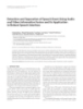
- Simonetti et al. Journal of Translational Medicine 2010, 8:135 Page 4 of 8 http://www.translational-medicine.com/content/8/1/135 Table 2 IHC expression of EGFR mutation antibodies in human NSCLC cell lines and in NSCLC tumor tissues EGFR mutation status EGFR (D38B1) EGFR E746-A750 deletion-specific EGFR L858R mutant-specific Ab (+) (6B6) Ab (+) (43B2) Ab (+) H460 and A549 (WT) 2/2 (100%) 0/2 (0%) 0/2 (0%) H1650 and PC9 (DEL 19) 2/2 (100%) 2/2 (100%) 0/2 (0%) H1975 (L858R + T790M) 1/1 (100%) 0/1 (0%) 1/1 (100%) Tumor Tissue (WT; N = 22) 8/22 (36%) 0/22 (0%) 0/22(0%) Tumor Tissue (DEL 19;N = 29) 28/29 (97%) 20/29 (69%) 0/29 (0%) Tumor Tissue (MUT 21;N = 27) 26/27 (96%) 0/27 (0%) 25/27 (93%) Abbreviations: WT: wild-type. DEL 19: Exon 19 deletion; MUT 21: Exon 21 mutation. and in recent years many efforts have been made to find a Discussion more specific and sensitive methodology to detect them EGFR is a member of the ErbB family of receptor tyrosine [10,16-18]. Nevertheless, these techniques are relatively kinases, which also includes HER2/neu, HER3, and HER4 expensive for routine use in clinical laboratories, and [15]. Activating mutations in the tyrosine kinase domain, depend on the quality of the samples. IHC is a standar- involving mainly exons 19 and 21, play an important role dized assay of simple methodology and high sensitivity in lung oncogenesis and tumor progression and are related and specificity, and the development of specific antibodies to the clinical efficacy of EGFR TKIs such as gefitinib or against EGFR mutation proteins might be useful for the erlotinib [5,9,11]. Analysis of these mutations has become an important tool for targeted therapy in lung cancer3, diagnosis and treatment of lung cancer. Figure 1 IHC analysis of EGFR mutations in five human NSCLC cell lines. A549 and H460 showed negativity for both mutation-specific antibodies. EGFR E746-A750 deletion specific (6B6) antibody stained H1650 and PC9 harboring the exon 19 deletion, and EGFR L858R mutant- specific (43B2) antibody stained the H1975 cell line with exon 21 mutation.
- Simonetti et al. Journal of Translational Medicine 2010, 8:135 Page 5 of 8 http://www.translational-medicine.com/content/8/1/135 Table 3 Correlation of IHC expression of mutation-specific antibodies and EGFR exon 19 deletion subtype analyzed by GeneScan, TaqMan and direct sequencing EGFR EXON 19 DELETION SUBTYPE 0 1+ 2+ 3+ 15 bp 0/17 (0%) 0/17 (0%) 2/17 (11%) 15/17 (89%) N = 17 9 bp 2/4 (50%) 2/4(50%) 0/4 (0%) 0/4 (0%) N=4 12 bp 1/1 (100%) 0/1 (0%) 0/1 (0%) 0/1 (0%) N=1 18 bp 4/5 (80%) 1/5 (20%) 0/5 (0%) 0/5 (0%) N=5 21 bp 1/1 (100%) 0/1 (0%) 0/1 (0%) 0/1 (0%) N=1 24 bp 1/1 (100%) 0/1 (0%) 0/1 (0%) 0/1 (0%) N=1 In 2009 Yu et al [12] first generated two mAbs against unknown EGFR mutations, comparing the IHC results E746-A750del and L858R point mutation from New with DNA sequencing. They found that IHC with these Zealand rabbits and evaluated them by Western blot- mutation-specific antibodies for EGFR mutations ting, immunofluorescence and IHC. They tested these showed a sensitivity of 92% and a specificity of 99%. antibodies in a series of cell lines and in tumor tissues Recently, five studies [14,19-22] examined the presence from patients with primary NSCLC, with known and of EGFR mutations in NSCLC by IHC using the same Figure 2 IHC staining of tumor samples from lung cancer patients. EGFR E746-A750 deletion specific (6B6) antibody detected 100% of cases with the 15-bp exon 19 deletion, and EGFR L858R mutant-specific (43B2) antibody detected 100% of cases harboring L858R mutation of exon 21.
- Simonetti et al. Journal of Translational Medicine 2010, 8:135 Page 6 of 8 http://www.translational-medicine.com/content/8/1/135 Figure 3 Expression of EGFR E746-A750 deletion specific (6B6) protein in the different types of exon 19 deletions. Among samples (12/ 29) showing negative or weak protein expression (score 0 or 1) 4 cases had a 9-bp deletion, 1 case had a 12-bp deletion, 5 cases had 18-bp deletion, 1 case had 21-bp deletion, and 1 case had a 24-bp deletion. two rabbit mAbs and reported sensitivity ranging from L858R and a sensitivity of 40% and a specificity of 99% 36% to 100% and specificity ranging from 94% to 99% for E746-A750. In the present study, we found a sensitiv- (Table 4). Kato et al [20] analyzed 70 gefitinib-treated ity of 100% and a specificity of 100% for the L858R exon NSCLC patients. Although a high sensitivity and specifi- 21 mutation antibody and a sensitivity of 63% and a spe- city for these mAbs were described, IHC staining was not cificity of 100% for the 15-bp deletion antibody. Table 4 significantly correlated with overall survival. A very summarizes the clinicopathological characteristics of exhaustive analysis of the role of EGFR in NSCLC was patients and the findings of seven studies examining recently reported by Ilie et al [19]. They assessed EGFR EGFR mutations by IHC, including the present study. status in a tissue microarray (TMA) of 61 lung adenocar- Although the most common EGFR mutations are the cinomas by IHC, fluorescent in situ hybridization (FISH) 15-bp ELREA deletion in exon 19 and the L858R substi- and direct sequencing and compared their results with tution in exon 21 [2,3], other less frequent deletions those of conventional methods performed on whole-tis- have been identified [4,6,8,23]. Using DNA sequencing, sue sections. The authors reported a specificity of 92% Yu et al [12] detected only two cases with uncommon for the mAb against the E746-750 deletion. Kawahara et deletions in exon 19; E746-T751 del stained positive and al [21] reported a sensitivity of 83% for the L858R muta- L747-A750 negative for IHC. In the present study, we tion antibody and 79% for the exon 19 deletion antibody. had 12 samples with uncommon deletions in exon 19 Brevet et al [14] reported a sensitivity of 84.6% and a spe- (9-bp, 12-bp, 18-bp, 21-bp and 24-bp) and 2 samples cificity of 98.9% for E746-A750 and a sensitivity of 95.2% with the uncommon exon 21 L816Q mutation. In these and a specificity of 98.8% for L858R. Kitamura et al [22] samples, IHC for both mutation-specific antibodies was reported a sensitivity of 36% and a specificity of 97% for not able to detect the alteration.
- Simonetti et al. Journal of Translational Medicine 2010, 8:135 Page 7 of 8 http://www.translational-medicine.com/content/8/1/135 Table 4 Patient characteristics and EGFR mutation status in seven studies examining EGFR mutations by IHC. (Blank cells indicate that information is not available) Simonetti et al Ilie et al [19] Kato et al [20] Kitamura et al [22] Brevet et al [14] Yu et al [12] Kawahara et al [21] Total cases 78 61 70 343 194 340 60 Age, years range 36-85 42-83 27-88 median 64 67 59.9 Sex male 28 31 36 female 50 30 34 Ethnicity Caucasian 78 61 0 0 0 Asian 0 0 70 343 60 Smoking history smokers 33 37 41 non-smokers 45 24 29 Histology adeno 69 61 57 217 60 SCC 1 0 7 112 0 LCC 5 0 4 11 0 others 3 0 2 0 0 EGFR exon 19 29 10 18 21 55 58 21 exon21 27 0 12 14 18 56 23 wild-type 22 51 29 296 145 167 16 IHC sensitivity overall 92% delE746-A750 Ab 63% 22.86% 81.1% 99% 84.6% 79% l858r Ab 100% 75% 97% 95.2% 83% IHC specificity overall 99% delE746-A750 Ab 100% 92% 100% 40% 98.8% L858R Ab 100% 96.6% 36% 98.8% 20 could lead to the universal application of IHC for Conclusions detecting EGFR mutations in NSCLC patients, as part of IHC with the mutation-specific rabbit mAbs against the routine IHC work-up of lung adenocarcinomas. EGFR is a simple and standardized assay which could prove useful as a first, quick screening of NSCLC patients. However, although these antibodies seem to be Additional material quite reliable for the detection of patients carrying the most common EGFR mutations [12], they were not able Additional file 1: Table S1. Table showing EGFR mutation status as detected by our sensitive methodology. Figure S1. Images showing to detect other EGFR gene mutations, such as 9-bp, 12- scoring of IHC staining of human NSCLC cell lines and lung cancer bp, 18-bp, 21-bp or 24-bp deletions or the L861Q sub- patient tumor tissues. A score of 0 was considered negative, a score of 1 stitution [14]. In consequence, if the antibodies are to was considered weakly positive, and a score of 2 or 3 was considered strongly positive be used in clinical practice, molecular biology techni- ques will be needed to further analyze the IHC-negative patients. However, the generation of a refined panel of antibodies able to detect both the frequent and the Acknowledgements The authors thank Herbert Haack and Katherine Crosby (Cell Signaling uncommon EGFR exon 19 deletions and exon 21 muta- Technology, Inc., Danvers, MA, USA) for providing the monoclonal antibodies tions as well as the resistance mutation T790 M in exon used in the study and Ignacio Wistuba (Departments of Thoracic/Head and
- Simonetti et al. Journal of Translational Medicine 2010, 8:135 Page 8 of 8 http://www.translational-medicine.com/content/8/1/135 Neck Medical Oncology and Pathology, The University of Texas M. D. EGFR mutations in non-small-cell lung cancer. Clin Cancer Res 2009, Anderson Cancer Center, Houston, TX, USA) for comments on an earlier 15:3023-3028. version of the manuscript. 13. Motoi N, Szoke J, Riely GJ, Seshan VE, Kris MG, Rusch VW, Gerald WL, Travis WD: Lung adenocarcinoma: modification of the 2004 WHO mixed Author details subtype to include the major histologic subtype suggests correlations 1 Pangaea Biotech, USP Dexeus University Institute, Barcelona, Spain. 2Catalan between papillary and micropapillary adenocarcinoma subtypes, EGFR Institute of Oncology, Hospital Germans Trias i Pujol, Badalona, Barcelona, mutations and gene expression analysis. Am J Surg Pathol 2008, Spain. 3Autonomous University of Madrid, Madrid, Spain. 4Hospital San 32:810-827. Carlos, Madrid, Spain. 5Hospital La Princesa, Madrid, Spain. 6Hospital Lozano 14. Brevet M, Arcila M, Ladanyi M: Assessment of EGFR mutation status in Blesa, Zaragoza, Spain. 7Hospital General de Valencia, Valencia, Spain. lung adenocarcinoma by immunohistochemistry using antibodies 8 Hospital Juan Canalejo, La Coruña, Spain. 9Hospital General de Alicante, specific to the two major forms of mutant EGFR. J Mol Diagn 2010, Alicante, Spain. 10Hospital Vall d’Hebron, Barcelona, Spain. 12:169-176. 15. Olayioye MA, Neve RM, Lane HA, Hynes NE: The ErbB signaling network: Authors’ contributions receptor heterodimerization in development and cancer. Embo J 2000, SS, MT, RR participated in the design of the study and its writing. MAM, CQ, 19:3159-3167. IDA, CM, JBA, SB carried out the molecular genetic studies. JLGL, UJ, DI, TM, 16. Ohnishi H, Ohtsuka K, Ooide A, Matsushima S, Goya T, Watanabe T: A SV, CC, RGC, BM have made substantial contributions to acquisition of data. simple and sensitive method for detecting major mutations within the JJS, MT, RR, SS have made substantial contributions to analysis and tyrosine kinase domain of the epidermal growth factor receptor gene in interpretation of data. SS, SRC carried out the immunoassays. JJS performed non-small-cell lung carcinoma. Diagn Mol Pathol 2006, 15:101-108. the statistical analysis. All authors read and approved the final manuscript. 17. Uhara M, Matsuda K, Taira C, Higuchi Y, Okumura N, Yamauchi K: Simple polymerase chain reaction for the detection of mutations and deletions Competing interests in the epidermal growth factor receptor gene: applications of this The authors declare that they have no competing interests. method for the diagnosis of non-small-cell lung cancer. Clin Chim Acta 2009, 401:68-72. Received: 16 September 2010 Accepted: 18 December 2010 18. Yatabe Y, Hida T, Horio Y, Kosaka T, Takahashi T, Mitsudomi T: A rapid, Published: 18 December 2010 sensitive assay to detect EGFR mutation in small biopsy specimens from lung cancer. J Mol Diagn 2006, 8:335-341. 19. Ilie MI, Hofman V, Bonnetaud C, Havet K, Lespinet-Fabre V, Coelle C, Gavric- References Tanga V, Venissac N, Mouroux J, Hofman P: Usefulness of tissue 1. Sharma SV, Haber DA, Settleman J: Cell line-based platforms to evaluate microarrays for assessment of protein expression, gene copy number the therapeutic efficacy of candidate anticancer agents. Nat Rev Cancer and mutational status of EGFR in lung adenocarcinoma. Virchows Arch 2010, 10:241-253. 2010, 457:483-495. 2. Sharma SV, Bell DW, Settleman J, Haber DA: Epidermal growth factor 20. Kato Y, Peled N, Wynes MW, Yoshida K, Pardo M, Mascaux C, Ohira T, receptor mutations in lung cancer. Nat Rev Cancer 2007, 7:169-181. Tsuboi M, Matsubayashi J, Nagao T, et al: Novel epidermal growth factor 3. Kosaka T, Yatabe Y, Endoh H, Kuwano H, Takahashi T, Mitsudomi T: receptor mutation-specific antibodies for non-small cell lung cancer: Mutations of the epidermal growth factor receptor gene in lung cancer: immunohistochemistry as a possible screening method for epidermal biological and clinical implications. Cancer Res 2004, 64:8919-8923. growth factor receptor mutations. J Thorac Oncol 2010, 5:1551-1558. 4. Lynch TJ, Bell DW, Sordella R, Gurubhagavatula S, Okimoto RA, 21. Kawahara A, Yamamoto C, Nakashima K, Azuma K, Hattori S, Kashihara M, Brannigan BW, Harris PL, Haserlat SM, Supko JG, Haluska FG, et al: Aizawa H, Basaki Y, Kuwano M, Kage M, et al: Molecular diagnosis of Activating mutations in the epidermal growth factor receptor activating EGFR mutations in non-small cell lung cancer using mutation- underlying responsiveness of non-small-cell lung cancer to gefitinib. N specific antibodies for immunohistochemical analysis. Clin Cancer Res Engl J Med 2004, 350:2129-2139. 2010, 16:3163-3170. 5. Paez JG, Janne PA, Lee JC, Tracy S, Greulich H, Gabriel S, Herman P, 22. Kitamura A, Hosoda W, Sasaki E, Mitsudomi T, Yatabe Y: Kaye FJ, Lindeman N, Boggon TJ, et al: EGFR mutations in lung cancer: Immunohistochemical detection of EGFR mutation using mutation- correlation with clinical response to gefitinib therapy. Science 2004, specific antibodies in lung cancer. Clin Cancer Res 2010, 16:3349-3355. 304:1497-1500. 23. de Gunst MM, Gallegos-Ruiz MI, Giaccone G, Rodriguez JA: Functional 6. Paz-Ares L, Soulieres D, Melezinek I, Moecks J, Keil L, Mok T, Rosell R, analysis of cancer-associated EGFR mutants using a cellular assay with Klughammer B: Clinical outcomes in non-small-cell lung cancer patients YFP-tagged EGFR intracellular domain. Mol Cancer 2007, 6:56. with EGFR mutations: pooled analysis. J Cell Mol Med 2009, 14(1-2):51-69. 7. Marchetti A, Martella C, Felicioni L, Barassi F, Salvatore S, Chella A, doi:10.1186/1479-5876-8-135 Camplese PP, Iarussi T, Mucilli F, Mezzetti A, et al: EGFR mutations in non- Cite this article as: Simonetti et al.: Detection of EGFR mutations with small-cell lung cancer: analysis of a large series of cases and mutation-specific antibodies in stage IV non-small-cell lung cancer. Journal development of a rapid and sensitive method for diagnostic screening of Translational Medicine 2010 8:135. with potential implications on pharmacologic treatment. J Clin Oncol 2005, 23:857-865. 8. Rosell R, Moran T, Carcereny E, Quiroga V, Molina MA, Costa C, Benlloch S, Taron M: Non-small-cell lung cancer harbouring mutations in the EGFR kinase domain. Clin Transl Oncol 2010, 12:75-80. 9. Gazdar AF: Activating and resistance mutations of EGFR in non-small-cell Submit your next manuscript to BioMed Central lung cancer: role in clinical response to EGFR tyrosine kinase inhibitors. and take full advantage of: Oncogene 2009, 28(Suppl 1):S24-31. 10. Molina-Vila MA, Bertran-Alamillo J, Reguart N, Taron M, Castella E, Llatjos M, Costa C, Mayo C, Pradas A, Queralt C, et al: A sensitive method for • Convenient online submission detecting EGFR mutations in non-small cell lung cancer samples with • Thorough peer review few tumor cells. J Thorac Oncol 2008, 3:1224-1235. 11. Rosell R, Moran T, Queralt C, Porta R, Cardenal F, Camps C, Majem M, • No space constraints or color figure charges Lopez-Vivanco G, Isla D, Provencio M, et al: Screening for epidermal • Immediate publication on acceptance growth factor receptor mutations in lung cancer. N Engl J Med 2009, • Inclusion in PubMed, CAS, Scopus and Google Scholar 361:958-967. 12. Yu J, Kane S, Wu J, Benedettini E, Li D, Reeves C, Innocenti G, Wetzel R, • Research which is freely available for redistribution Crosby K, Becker A, et al: Mutation-specific antibodies for the detection of Submit your manuscript at www.biomedcentral.com/submit
CÓ THỂ BẠN MUỐN DOWNLOAD

-

Báo cáo hóa học: "Detection of carcinoembryonic antigen messenger RNA in blood using quantitative real-time reverse transcriptase-polymerase chain reaction to predict recurrence of gastric adenocarcinoma"
 8 p |
8 p |  53
|
53
|  8
8
-

Báo cáo hóa học: "Radio Frequency Interference Suppression for Landmine Detection by Quadrupole Resonance"
14 p | 75
| 7
-
Báo cáo hóa học: "Research Article Detecting and Georegistering Moving Ground Targets in Airborne QuickSAR via Keystoning and Multiple-Phase Center Interferometry"
11 p | 116
| 7
-

Báo cáo hóa học: " Detection of epithelial apoptosis in pelvic ileal pouches for ulcerative colitis and familial adenomatous polyposis"
6 p | 73
| 7
-

báo cáo hóa học:" Validation of a HLA-A2 tetramer flow cytometric method, IFNgamma real time RT-PCR, and IFNgamma ELISPOT for detection of immunologic response to gp100 and MelanA/MART-1 in melanoma patients"
25 p | 52
| 6
-

báo cáo hóa học:" Clinical values of multiple Epstein-Barr virus (EBV) serological biomarkers detected by xMAP technology"
8 p | 49
| 5
-
Báo cáo hóa học: " Detection of Point Sources on Two-Dimensional Images Based on Peaks"
11 p | 35
| 5
-
Báo cáo hóa học: " Research Article Unsupervised Video Shot Detection Using Clustering Ensemble with a Color Global Scale-Invariant Feature Transform Descriptor"
10 p | 73
| 5
-
Báo cáo hóa học: " Research Article A Motion-Adaptive Deinterlacer via Hybrid Motion Detection and Edge-Pattern Recognition"
10 p | 93
| 5
-
Báo cáo hóa học: " Research Article A Comparison of Detection Performance for Several Track-before-Detect Algorithms"
10 p | 42
| 5
-

báo cáo hóa học:" Detection of postoperative granulation tissue with an ICG-enhanced integrated OI-/X-ray System"
10 p | 75
| 4
-

Báo cáo hóa học: " Research Article Simultaneous Eye Tracking and Blink Detection with Interactive Particle Filters"
17 p | 59
| 4
-

Báo cáo hóa học: " Research Article New Structured Illumination Technique for the Inspection of High-Reflective Surfaces: Application for the Detection of Structural Defects"
14 p | 70
| 4
-
Báo cáo hóa học: " Evaluating Edge Detection through Boundary Detection "
15 p | 43
| 4
-
Báo cáo hóa học: " Detecting Impulses in Mechanical Signals by Wavelets"
7 p | 48
| 4
-
báo cáo hóa học:" Of gastro and the gold standard: evaluation and policy implications of norovirus test performance for outbreak detection"
9 p | 65
| 3
-
Báo cáo hóa học: " Detection and Separation of Speech Event Using Audio and Video Information Fusion and Its Application to Robust Speech Interface"
12 p | 44
| 2


Chịu trách nhiệm nội dung:
Nguyễn Công Hà - Giám đốc Công ty TNHH TÀI LIỆU TRỰC TUYẾN VI NA
LIÊN HỆ
Địa chỉ: P402, 54A Nơ Trang Long, Phường 14, Q.Bình Thạnh, TP.HCM
Hotline: 093 303 0098
Email: support@tailieu.vn
Giấy phép Mạng Xã Hội số: 670/GP-BTTTT cấp ngày 30/11/2015 Copyright © 2022-2032 TaiLieu.VN. All rights reserved.

