


Báo cáo khoa học: "A case of matrix-producing carcinoma of the breast"
lượt xem 3
download
 Download
Vui lòng tải xuống để xem tài liệu đầy đủ
Download
Vui lòng tải xuống để xem tài liệu đầy đủ
Tuyển tập báo cáo các nghiên cứu khoa học quốc tế ngành y học dành cho các bạn tham khảo đề tài: A case of matrix-producing carcinoma of the breast
Bình luận(0) Đăng nhập để gửi bình luận!
Nội dung Text: Báo cáo khoa học: "A case of matrix-producing carcinoma of the breast"
- World Journal of Surgical Oncology BioMed Central Open Access Case report A case of matrix-producing carcinoma of the breast Toshiyuki Hirose1, Junko Honda2, Yoshimi Bando3, Mitsunori Sasa*4, Yukiko Hirose2, Taeko Nagao2 and Akira Tangoku2 Address: 1Department of Surgery, National Higashi Tokushima Hospital, 1-1, Ohmukai-kita, Ootera, Itano, Tokushima 779-0193, Japan, 2Department of Oncological and Regenerative Surgery, Institute of Health Biosciences, The University of Tokushima, 3-18-15, Kuramoto-Cho, Tokushima 770-8509, Japan, 3Department of Molecular and Environmental Pathology, Institute of Health Biosciences, The University of Tokushima Graduate School, 3-18-15, Kuramoto-Cho, Tokushima 770-8509, Japan and 4Department of Surgery, Tokushima Breast Care Clinic, 4-7-7, Nakashimada-Cho, Tokushima 770-0052, Japan Email: Toshiyuki Hirose - toshi-hirose@higasitokusima.hosp.go.jp; Junko Honda - honnda2000@ezweb.ne.jp; Yoshimi Bando - yoshimi@basic.med.tokushima-u.ac.jp; Mitsunori Sasa* - breast@mb.tcn.ne.jp; Yukiko Hirose - hirohiro@clin.med.tpkushima-u.ac.jp; Taeko Nagao - tae-nagao@mte.biglobe.ne.jp; Akira Tangoku - tangoku@clin.med.tokushima-u.ac.jp * Corresponding author Published: 17 June 2008 Received: 26 October 2007 Accepted: 17 June 2008 World Journal of Surgical Oncology 2008, 6:60 doi:10.1186/1477-7819-6-60 This article is available from: http://www.wjso.com/content/6/1/60 © 2008 Hirose et al; licensee BioMed Central Ltd. This is an Open Access article distributed under the terms of the Creative Commons Attribution License (http://creativecommons.org/licenses/by/2.0), which permits unrestricted use, distribution, and reproduction in any medium, provided the original work is properly cited. Abstract Background: Matrix-producing carcinoma (MPC) of the breast is one variant type of metaplastic carcinoma. The cellular origin of MPC remains unclear. It has been suggested the tumor cells in MPC have the combined characteristics of both epithelial cells and mesenchymal cells. Several reports suggested that the tumor cells in MPC might originate from the myoepithelial cells, but others suggested the origin was basal-like cells. Case presentation: The patient was a 42-year-old Japanese female. A tumor of about 2 cm in diameter was noted in the right breast. CT revealed the circumference of the tumor to have a ring- like structure, and fine needle aspiration cytology indicated suspicion for malignancy. Breast- conserving surgery was performed. Histopathological studies showed carcinoma cells, having cuboidal to oval-shaped nucleus, were proliferating in cord-like and sheet-like structures in the periphery. In the central areas of the tumor, myxoedematous area was observed with cartilaginous matrix and necrosis. The diagnosis was a matrix-producing carcinoma. Immunohistochemical findings showed the tumor cells had the characteristics of both epithelial cells and mesenchymal cells, while being negative for estrogen receptor, progesterone receptor, Her2, myoepithelial cell markers and basal cell markers. Conclusion: The findings for our present patient and many of the other MPC patients reported in the published literature indicate that this breast cancer has the properties of both epithelial cells and mesenchymal cells. In addition, there is a possibility that matrix-producing tumor cells of our present patient may have a feature of undifferentiated cells. In 1989, Wargotz et al., proposed defining MPC as overt Background Matrix-producing carcinoma (MPC) is a rare and charac- carcinoma with direct transition to a cartilaginous and/or teristic variant type of metaplastic carcinoma of the breast. osseous stromal matrix cells, with no spindle cells Page 1 of 6 (page number not for citation purposes)
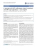
- World Journal of Surgical Oncology 2008, 6:60 http://www.wjso.com/content/6/1/60 between those two elements [1]. In Japan, MPC of the breast was added to the General Rules for Clinical and Pathological Recording of breast Cancer (16th Edi- tion)(The Japanese Breast Cancer Society) as a special form of carcinoma [2]. It was reported that MPC cells had the combined characteristics of both epithelial cells and mesenchymal cells [3-5]. Wargotz et al., suggested the tumor cells of MPC might be of epithelial-myoepithelial derivation depending on immunohistochemical analysis and electron microscopic results [1]. In addition, mouse model using Brca1 deficiency has suggested an important role for BRCA1 in basal-like breast carcinoma with meta- plastic elements [6]. There is thus no consensus in his- togenesis of MPC. We report our findings for a Japanese woman with MPC of the breast and discuss them together with the other 26 cases of MPC of the breast that have been reported in Japan to date. Case presentation The patient was a 42-year-old Japanese female with a chief complaint of a lump in her right breast. Her personal and Figure 1 Computed tomography scan imaging (CT) of the tumor family histories contained nothing of special note. Computed tomography scan imaging (CT) of the tumor. Contrast-enhanced CT revealed, in the right lateral-upper Examination revealed a hard tumor with a clear boundary quadrant, an irregularly shaped, 2,5 cm tumor showing and a diameter of about 2 cm in the lateral-upper quad- peripheral ring-shaped contrast enhancement. rant of the right breast. The axillary and supraclavicular lymph nodes could not be palpated. Mammography revealed focal asymmetric density in the right lateral- area consisted of cord-like and sheet-like structures of pro- upper quadrant, accompanied by amorphous microcalci- liferating carcinoma cells having a cuboidal or oval- fication. Breast echography indicated a tumor with shaped nucleus. The central myxoedematous and chon- dimensions of 2.3 × 1.8 × 1.5 cm and a slightly indistinct droid-looking matrix contained an extensive area of boundary, and the internal portion was heterogeneous necrosis, but no definite chondrocytes or osseous differ- and included a hyperechoic region. Contrast-enhanced entiation (Figs. 2, 3). Ductal carcinoma in situ (DCIS) CT revealed, in the right lateral-upper quadrant, an irreg- with comedo necrosis was found adjacent to the main ularly shaped, 2.5 cm tumor showing peripheral ring- tumor. Immunohistochemically, the tumor cells in both shaped contrast enhancement (Fig. 1). There was no evi- the peripheral epithelial area and the central myxoedema- dence of lymph node metastasis or clear distant metasta- tous area were negative for estrogen receptor (ER), proges- sis. Aspiration cytology showed many tumor cells, having terone receptor (PgR) and Her2. In addition, the tumor cuboidal to oval-shaped nucleus, were observed in the cells of both areas stained positively for both vimentin myxoedematous background containing much necrotic and S-100 protein (Fig. 4), and they also showed partial material, but without any sarcomatous spindle cells. The positive staining for each of cytokeratin AE1/AE3, CK7, myxoedematous matrix in the background stained pale CK8 and CK19 (Fig. 5). Conversely, the tumor cells of both areas stained negatively for each of α-smooth muscle gray with Papanicolau stain. The tumor cells were iso- antigen (α-SMA), p63 and glial fibrillary acidic protein lated, in loosely cohesive groups and in short chains. The nuclear to cytoplasmic ratio was high with coarsely gran- (GFAP), which were myoepithelial cell markers. The ular chromatin. The diagnosis was suspicious for malig- tumor cells were also negative for each of the basal mark- nancy. No particular abnormalities were noted on ers, i.e., CK5/6, CK14, CK17, and epidermal growth factor laboratory data, and tumor markers were all within their receptor (EGFR). Appropriate human breast cancers normal ranges: 1.6 ng/ml for CEA and 19 ng/ml for CA known to express ER, PgR and Her2 were included in each 15-3. Right breast cancer was suspected on the basis of the slide run. The luminal cells of non-tumorous adjacent above findings, and lumpectomy was performed. ducts were positive with ER and PgR as internal controls. The myoepithelial cells of adjacent non-tumorous ducts and acini were also positive with α-SMA, p63, CK5/6, Histopathological findings The tumor consisted of a peripheral epithelial area and a CK14 and CK17. Detailed information of the immunohis- central myxoedematous area. The peripheral epithelial tochemistry procedures and the antibodies used was listed Page 2 of 6 (page number not for citation purposes)
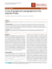
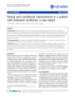
- World Journal of Surgical Oncology 2008, 6:60 http://www.wjso.com/content/6/1/60 adjuvant chemotherapy was recommended, but the patient refused this approach and it was thus not admin- istered. Ten months after the axillary surgery, multiple metastases and liver metastasis were diagnosed, and the patient died 8 months after recurrence of the disease. Discussion Epithelial-mesenchymal transition has been reported to be an etiological factor in metaplastic carcinoma [7]. The overt carcinoma cells of almost all of the MPC breast can- cer cases reported in Japan were positive for both epithe- lial cell markers and mesenchymal cell markers (Table 2). Electron microscope findings and the results of immuno- histochemical studies were reported to indicate that MPC is of myoepithelial cell origin [1,8]. On the other hand, Okuyama et al. examined specimens from 8 patients and reported that the overt carcinoma cells of all of those cases were negative for myoepithelial cell markers [3]. Moreo- Figure 2 Low-magnification view of the tumor ver, Only 4 of the 27 patients in Japan was positive for Low-magnification view of the tumor. The central myxoede- matous area contained an extensive area of necrosis at its myoepithelial cell markers. In the patient we have center (HE). described, as well, the overt carcinoma cells were positive for vimentin, S-100 protein and cytokeratins (AE1/AE3, CK7, CK8 and CK19). They showed negative staining for α-SMA, p63, CK5/6 and GFAP, which are myoepithelial in the Table 1. In special staining, the tumor stroma stained positive with alcian blue (pH2.5), which was par- cell markers. p63 has been reported to be useful as diag- tially eliminated by digestion with hyaluronidase. nostic marker for metaplastic carcinoma [9,10]. It was reported that the carcinoma cells with spindle and/or squamous differentiation in metaplastic carcinoma Clinical course Axillary lymph node dissection was performed on the showed positive staining for p63. The malignant compo- basis of a definitive diagnosis of MPC of the breast, but nent with no squamous or sarcomatous differentiation in there were no findings of metastasis. The remaining breast MPC of our patient might cause negative staining for p63. tissue was irradiated with a total of 50 Gray. Postoperative Figure 3 High-magnification view of the peripheral epithelial area (a) and the central area (b) of the tumor High-magnification view of the peripheral epithelial area (a) and the central area (b) of the tumor. The peripheral epithelial area consisted of cord-like and sheet-like structures of proliferating carcinoma cells having a cuboidal to oval-shaped nucleus. In the central areas of the tumor, sparse distribution of oval tumor cells was observed (HE). Page 3 of 6 (page number not for citation purposes)
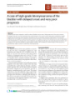
- World Journal of Surgical Oncology 2008, 6:60 http://www.wjso.com/content/6/1/60 Figure 4 Immunohistochemical staining for vimentin of the peripheral epithelial area (a) and the central myxoedematous area (b) Immunohistochemical staining for vimentin of the peripheral epithelial area (a) and the central myxoedematous area (b). The tumor cells of the both area stained positively for vimentin. Our patient's MPC exhibited the same cell marker profile and the central my edematous area showed no differences as that reported by Okuyama et al., showing the properties in their staining profiles. Recent molecular studies have of both epithelial cells and mesenchymal cells. It was shown the monoclonal origin of carcinosarcoma of the reported that the results of immunohistochemistry dif- breast, as the carcinomatous and sarcomatous elements fered between the peripheral epithelial area and the cen- share common genetic alterations [12,13]. These observa- tral myxoedematous area. In the central myxoedematous tions support the hypothesis that the matrix-producing area, which can be thought to be causing metaplasia, carcinoma may be derived from a single totipotent stem down-regulation of epithelial markers and up-regulation cell. of mesenchymal markers were observed [1,4,5,11]. On the contrary, for our patient, the peripheral epithelial area Immunohistochemical staining for cytokeratin AE1/AE3 of the peripheral epithelial area (a) and the central myxoedematous Figure area (b) 5 Immunohistochemical staining for cytokeratin AE1/AE3 of the peripheral epithelial area (a) and the central myxoedematous area (b). The tumor cells of the both area stained positively for cytokeratin AE1/AE3. Page 4 of 6 (page number not for citation purposes)
- World Journal of Surgical Oncology 2008, 6:60 http://www.wjso.com/content/6/1/60 Table 1: Characteristics of the overt carcinoma cells in matrix-producing carcinoma of the breast in Japan α-SMA ER PgR Her2 EMA AE1/AE3 Desmin GFAP p63 S-100 Vimentin (+) 0 1 0 16 20 0 4 1 0 15 17 (-) 23 22 14 0 1 11 15 0 1 0 3 ER: Estrogen receptor PgR: Progesterone receptor Her2: Human epidermal growth factor 2 EMA: epithelial membrane antigen GFAP: glial fibrillay acidic protein SMA: α-smooth muscle actin Our patient had triple-negative breast cancer with regard like cell type. BRCA1 has been shown to play an impor- to ER, PgR and Her2. In addition, it is interesting that tant role in mammary differentiation and the loss of almost all of the reported Japanese cases of MPC of the BRCA1 function resulted in the accumulation of cells breast were triple-negative. It is said that most cases of expressing the stem/progenitor marker ALDH-1 [19]. metaplastic carcinoma are also triple-negative breast can- Although the BRCA1 status of our patient has not been cer [14,15], and this is important in terms of elucidating identified, it was suggested the possibility that the tumor the etiology of the metaplastic change. Ninomiya et al., cells of our MPC might be blocked differentiation with reported that one of their two cases of MPC of the breast expansion of undifferentiated cell compartment. was the basal phenotype [5]. McCarthy et al., generated a conditional mouse model of BRCA1 deficiency. Mam- Okuyama et al., reported that the incidence of MPC of the mary tumors that developed in these mice had basal and breast was 0.05% in Japan [4]. Our search of the main Jap- metaplastic characteristics in the form of spindle cell and anese medical journals found a total of 27 cases of MPC squamous cell differentiation. Most of the tumors were of the breast reported in Japan to date, including our negative for ER, PgR and Her2 [6]. Additionally, a recent present patient [3-5,11]. Imaging diagnosis by contrast- report has shown that epithelial mesenchymal transition- enhanced CT and contrast-enhanced MRI have revealed like changes occurred preferentially in the basal-like sub- that this disease is characterized by a ring structure in its type of breast carcinomas [16]. Furthermore, subpopula- periphery. For that reason, it was concluded that it is nec- tions of cancer cells with stem cell properties are especially essary to consider the possibility of MPC of the breast frequent within basal-like breast cell lines [17]. Stem cell- when such image findings are obtained [3]. In our present like breast cell lines are also able to undergo epithelial patient, as well, contrast-enhanced CT revealed an irregu- mesenchymal transition [18]. These data suggest that larly shaped, 2.5 cm tumor showing peripheral ring- basal-like cancer cells may undergo epithelial mesenchy- shaped contrast enhancement. mal transition with intrinsic phenotype of cancer stem cells. Although most cases of triple-negative breast cancer Most MPC of the breast are triple-negative, and postoper- have the basal-like phenotype [6], MPC of our patient had ative adjuvant chemotherapy is often administered [13]. lack of any markers for myoepithelial cell type and basal- However, some studies have shown this therapy to have Table 2: Sources, dilution and pretreatment of antibodies used Antibody Clone Manufacturer Dilution Pretreatment ER 1D5 DakoCytomation, USA 1:50 boiling (pH9.0, 40 min) PgR PgR636 DakoCytomation, USA 1:800 boiling (pH9.0, 40 min) HER2 DakoCytomation, USA Prediluted (Hercep test) boiling (pH6.0, 40 min) CK5/6 D5/16B4 DakoCytomation, Denmark 1:100 autoclave (pH6.0, 10 min) CK14 LL002 NeoMarkers, USA 1:100 autoclave (pH6.0, 10 min) CK17 E3 DakoCytomation, Denmark 1:40 autoclave (pH6.0, 10 min) EGFR 2-18C9 DakoCytomation, USA Prediluted (pharmDX kit) proteinase K (room temperature, 5 min) AE1/AE3 AE/AE3 DakoCytomation, Denmark 1:50 pronase (37 C, 15 min) CK7 OV-TL12/30 DakoCytomation, Denmark 1:50 pronase (37 C, 15 min) 35βH11 CK8 DakoCytomation, USA 1:50 pronase (37 C, 15 min) CK19 RCK108 DakoCytomation, Denmark 1:50 autoclave (pH6.0, 10 min) α-SMA 1A4 DakoCytomation, Denmark 1:100 P63 4A4 DakoCytomation, Denmark 1:50 autoclave (pH6.0, 10 min) GFAP 6F2 DakoCytomation, Denmark 1:100 Page 5 of 6 (page number not for citation purposes)
- World Journal of Surgical Oncology 2008, 6:60 http://www.wjso.com/content/6/1/60 been ineffective, and further research on this issue is war- cer with cartilaginous and osseous metaplasia. Breast Cancer 2005, 12:52-56. ranted. 6. McCarthy A, Savage K, Gabriel A, Naceur C, Reis-Filho JS, Ashworth A: A mouse model of basal-like breast carcinoma with meta- plastic elements. J Pathol 2007, 211:389-398. The prognosis of MPC of the breast is said to be better 7. Lien HC, Hsiao YH, Lin YS, Yao YT, Juan HF, Kuo WH, Hung M-C, than that of other carcinomas that are accompanied by Chang KJ, Hsieh FJ: Molecular signature of metaplastc carci- osteocartilaginous metaplasia [20,21]. Wargotz et al., noma of the breast by large-scale transcriptional profiling: identification of genes potentially related to epithelial-mes- reported a 5-year survival rate of 68% for MPC of the enchymal transition. Oncogene 2007, 26:7859-7871. breast [1], but the number of reported cases has been 8. Kinkor Z, Baudova L, Ryska A, Kajo K, Svec A: Matrix-producing breast carcinoma with myoepithelial differentiation-descrip- small and the prognosis thus remains unclear. Our patient tion of 11 cases and review of literature aimed at histogene- refused postoperative adjuvant chemotherapy, and dis- sis and differential diagnosis. Ceska Gynekol 2004, tant metastasis was detected at 10 months after the partial 69(3):229-236. 9. Koker MM, Kleer CG: p63 expression in breast cancer: a highly mastectomy. In the future it will be necessary to study a sensitive and specific marker of metaplastic carcinoma. Am J larger number of patients with MPC of the breast and fur- Surg Pathol 2004, 28:1506-1512. ther elucidate the clinicopathological characteristics of 10. Tse GM, Tan PH, Chaiwun B, Putti TC, Lui PC, Tsang AK, Wong FC, Lo AW: p63 is useful in the diagnosis of mammary metaplas- this malignancy. tic carcinomas. Pathology 2006, 38:16-20. 11. Hama Y, Tsuda H, Sato K, Hiraide H, Mochizuki H, Kusano S: Inva- sive ductal carcinoma of the breast with a large central a cel- Conclusion lular zone associated with matrix-producing carcinoma. There have been reports that MPC of the breast is of Tumori 2004, 90:498-500. myoepithelial cell origin or basal cell origin. However, the 12. Manuel RT, Hanne Q, Per JB, Nikos P, Sverre H: Cytogenetic anal- ysis shows that carcinosarcomas of the breast are of mono- findings for our present patient suggested that MPC might colonal origin. Genes Chromosomes Cancer 1998, 22:145-151. be produced as a result of the undifferentiation process. 13. Wada H, Enomoto T, Tsujimoto M, Nomura T, Murata Y, Shroyer KR: Carcinosarcoma of the breast: molecular-biological study for analysis of histogenesis. Hum Pathol 1998, List of abbreviations 29:1324-1328. MPC: Matrix producing carcinoma; ER: Estrogen receptor; 14. Gibson GR, Qlan D, Ku JK, Lai LL: Metaplastic breast cancer: PgR: Progesterone receptor; Her2: Hercep test; α-SMA: α- clinical features and outcomes. Am Surgeon 2005, 71:725-730. 15. Livasy CA, Karaca G, Nanda R, Tretiakova MS, Olopade OI, Moore smooth muscle antigen; GEAP: Glial fibrillary acidic pro- DT, Perou CM: Phenotypic evaluation of the basal-like subtype tein; EGFR: Epidermal growth factor receptor of invasive breast carcinoma. Mod Pathol 2006, 19(2):264-271. 16. David S, Socorro MR-P, David H, Amparo C, Gema M-B, Jose P: Epi- thelial-mesenchymal transition in breast cancer relates to Competing interests the basal-like phenotype. Cancer Res 2008, 68:989-997. 17. Sheridan C, Kishimoto H, Fuchs RK, Mehrotra S, Bhat-Nakshatri P, The authors declare that they have no competing interests. Turner CH, Goulet R Jr, Badve S, Nakshatri H: CD44+/CD24- breast cancer cells exhibit enhanced invasive properties: an Authors' contributions early step necessary for metastasis. Breast Cancer Res 2006, 8:R59. HT, MS and NT took part in the care of the patient, YB 18. Hugo H, Ackland ML, Blick T, Lawrence MG, Clements JA, Williams examined surgical specimen and took photomicrographs ED, Thompson EW: Epithelial – mesenchymal and mesenchy- of the slides, JH and MS initiated and co-wrote the paper mal – epithelial transitions in carcinoma progression. J Cell Physiol 2007, 213:374-383. with TH and AT. All authors read approved the final man- 19. Liu S, Ginestier C, Charafe-Jauffret E, Foco H, Kleer CG, Merajver SD, uscript. Dontu G, Wicha MS: BRCA1 regulates human mammary stem/progenitor cell fate. Proc Natl Acad Sci USA 2008, 105:1680-1685. Acknowledgements 20. Beatty JD, Atwood M, Tickman R, Reiner M: Metaplastic breast Written consent was obtained from the husband of the patient or their rel- cancer: clinical significance. Am J Surg 2006, 191:657-664. 21. Tse GM, Tan PH, Putti TC, Lui PCW, Chaiwun B, Law BKB: Meta- ative for publication of this article. plastic carcinoma of the breast: a clinicopathological review. J Clin Pathol 2006, 59:1079-1083. References 1. Wargotz ES, Norris H: Matrix-producing carcinoma. Hum Pathol 1989, 20:628-635. Publish with Bio Med Central and every 2. Japanese Breast Cancer Society: General Rules for Clinical and scientist can read your work free of charge Pathological Recording of Breast Cancer. 15th edition. Kane- hara, Tokyo; 2005. "BioMed Central will be the most significant development for 3. Okuyama N, Sakamoto G, Sasaki T, Tokutome N, Sarumaru S, Hori disseminating the results of biomedical researc h in our lifetime." F, Horii R, Akiyama F, Kasumi F: Clinicopathological features of Sir Paul Nurse, Cancer Research UK matrix-producing carcinoma. pn J Breast Cancer 2004, 19:339-342. Your research papers will be: 4. Murata T, Ihara S, Kato H, Tanigawa K, Higashiguchi T, Imai T, Matsu- available free of charge to the entire biomedical community shita T, Nakamura K, Nakayama T, Shiraishi T, Moriya T: Matrix- producing carcinoma of the breast: Case report with radio- peer reviewed and published immediately upon acceptance graphical and cytopathological features. Pathol Int 1998, cited in PubMed and archived on PubMed Central 48:824-828. 5. Ninomiya J, Oyama T, Horiguchi J, Koibuchi Y, Yoshida T, Iijima K, yours — you keep the copyright Yoshida M, Takata D, Iino Y, Morishita Y: Two case of breast can- BioMedcentral Submit your manuscript here: http://www.biomedcentral.com/info/publishing_adv.asp Page 6 of 6 (page number not for citation purposes)
CÓ THỂ BẠN MUỐN DOWNLOAD

-
Báo cáo y học: " A neonate with left pulmonary artery thrombosis and left lung hypoplasia: a case report"
 5 p |
5 p |  57
|
57
|  5
5
-
Báo cáo khoa học: " A case of gangliocytic paraganglioma in the ampulla of Vater"
4 p | 68
| 5
-

báo cáo khoa học: "A case report of male breast cancer in a very young patient: What is changing"
5 p | 50
| 4
-

báo cáo khoa học: " A case of polyarteritis nodosa limited to the right calf muscles, fascia, and skin: a case report"
5 p | 49
| 4
-

báo cáo khoa học: " A case of limbic encephalitis presenting as a paraneoplastic manifestation of limited stage small cell lung cancer: a case report"
4 p | 39
| 4
-

Báo cáo khoa học: "A case of radiation-induced sternal malignant fibrous histiocytoma treated with neoadjuvant chemotherapy and surgical resection"
4 p | 54
| 4
-

báo cáo khoa học: "Peritoneal mesothelioma in a woman who has survived for seven years: a case report"
4 p | 105
| 4
-
báo cáo khoa học: " Dental and craniofacial characteristics in a patient with Dubowitz syndrome: a case report"
5 p | 133
| 4
-

Báo cáo khoa hoc:" A case study of new assessment and training of unilateral spatial neglect in stroke patients: effect of visual image transformation and visual stimulation by using a head mounted display system"
8 p | 72
| 4
-

báo cáo khoa học: " A rare case of giant leiomyosarcoma in a filarial scrotum: a case report"
5 p | 56
| 4
-

báo cáo khoa học: "A challenging case of gastric outlet obstruction (Bouveret's syndrome): a case report"
4 p | 42
| 4
-

Báo cáo khoa học: "A case for a CUG-initiated coding sequence overlapping torovirus ORF1a and encoding a novel 30 kDa product"
6 p | 35
| 3
-

Báo cáo khoa học: "A case of virilization induced by a Krukenberg tumor from gastric cancer"
5 p | 47
| 3
-

Báo cáo khoa học: "A case of Meigs syndrome mimicking metastatic breast carcinoma"
6 p | 42
| 3
-
Báo cáo khoa học: "A case of high-grade leiomyosarcoma of the bladder with delayed onset and very poor prognosis"
4 p | 49
| 2
-

báo cáo khoa học: "A male case of an undifferentiated carcinoma with osteoclast-like giant cells originating in an indeterminate mucin-producing cystic neoplasm of the pancreas. A case report and review of the literature"
6 p | 80
| 2
-

Báo cáo khoa học: " A case report of pseudoprogression followed by complete remission after proton-beam irradiation for a low-grade glioma in a teenager: the value of dynamic contrast-enhanced MRI"
5 p | 40
| 2


Chịu trách nhiệm nội dung:
Nguyễn Công Hà - Giám đốc Công ty TNHH TÀI LIỆU TRỰC TUYẾN VI NA
LIÊN HỆ
Địa chỉ: P402, 54A Nơ Trang Long, Phường 14, Q.Bình Thạnh, TP.HCM
Hotline: 093 303 0098
Email: support@tailieu.vn
Giấy phép Mạng Xã Hội số: 670/GP-BTTTT cấp ngày 30/11/2015 Copyright © 2022-2032 TaiLieu.VN. All rights reserved.

