


Báo cáo khoa học: "Adenocarcinoma of the appendix presenting as bilateral ureteric obstruction"
lượt xem 4
download
 Download
Vui lòng tải xuống để xem tài liệu đầy đủ
Download
Vui lòng tải xuống để xem tài liệu đầy đủ
Tuyển tập báo cáo các nghiên cứu khoa học quốc tế ngành y học dành cho các bạn tham khảo đề tài: Adenocarcinoma of the appendix presenting as bilateral ureteric obstruction
Bình luận(0) Đăng nhập để gửi bình luận!
Nội dung Text: Báo cáo khoa học: "Adenocarcinoma of the appendix presenting as bilateral ureteric obstruction"
- World Journal of Surgical Oncology BioMed Central Open Access Case report Adenocarcinoma of the appendix presenting as bilateral ureteric obstruction Kamran Ahmed*1, Robiol Hoque1, Sherif El-Tawil1, Mohammad S Khan2 and Mark L George1 Address: 1Department of General Surgery, St Thomas' Hospital, London, UK and 2Department of Urology, Guy's Hospital, London, UK Email: Kamran Ahmed* - kahmed198@yahoo.co.uk; Robiol Hoque - robiol.hoque@gstt.nhs.uk; Sherif El-Tawil - sharif.eltawil@gstt.nhs.uk; Mohammad S Khan - shamim.khan@gstt.nhs.uk; Mark L George - mark.george@gstt.nhs.uk * Corresponding author Published: 21 February 2008 Received: 14 October 2006 Accepted: 21 February 2008 World Journal of Surgical Oncology 2008, 6:23 doi:10.1186/1477-7819-6-23 This article is available from: http://www.wjso.com/content/6/1/23 © 2008 Ahmed et al; licensee BioMed Central Ltd. This is an Open Access article distributed under the terms of the Creative Commons Attribution License (http://creativecommons.org/licenses/by/2.0), which permits unrestricted use, distribution, and reproduction in any medium, provided the original work is properly cited. Abstract Background: Adenocarcinoma of the vermiform appendix is a rare neoplasm of the gastrointestinal tract. Presentation mimics acute appendicitis, but right iliac fossa mass and intestinal obstruction have also been reported. These presentations reflect various stages of a locally expanding tumour causing luminal obstruction of appendix. The investigation and subsequent management with a review of the literature is presented. Case presentation: We report a case of appendicular adenocarcinoma found unexpectedly in a 43 year old male who presented with urinary symptoms. Cystoscopy and uretero-renoscopy showed normal bladder but external compression of the ureters and therefore bilateral stents were inserted. CT scan showed a caecal mass. After colonoscopy, that showed external compression, and diagnostic laparoscopy the patient underwent right hemicolectomy. Histopathology revealed well differentiated adenocarcinoma with signet ring morphology with multiple lymph node involvement. The patient was referred for chemotherapy where he received infusional 5 fluorouracil but died 7 months after surgery. Conclusion: Patients with atypical manifestations related to right lower abdominal quadrant should be thoroughly investigated with an open mind. Every attempt should be made to make a precise diagnosis through all the available means to direct the treatment along correct lines. There are other clinical presentations and here we report a Background The appendix is an uncommon site of gastrointestinal case of appendicular signet ring cell adenocarcinoma malignancy. Presentation mimics acute appendicitis, but found unexpectedly in a patient who presented to the right iliac fossa mass and intestinal obstruction have been urologists with urinary symptoms. reported. These presentations reflect various stages of a locally expanding tumour causing luminal obstruction of Case presentation appendix. A 43 years old male presented to the emergency depart- ment with a two week history of right lower quadrant pain radiating to the right testis. Baseline blood tests were nor- Page 1 of 5 (page number not for citation purposes)
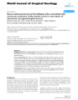
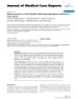
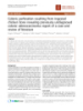
- World Journal of Surgical Oncology 2008, 6:23 http://www.wjso.com/content/6/1/23 Figure 1 (a&b) – CT Scan: Bilateral hydronephrosis with associated caecal mass (arrow showing hydronephrosis) (a&b) – CT Scan: Bilateral hydronephrosis with associated caecal mass (arrow showing hydronephrosis). mal apart from creatinine of 227 (umol/l) and blood in International Classification of Diseases for Oncology the urine. An intravenous urogram showed bilateral uret- (ICD-O) groups divides the adenocarcinoma of appendix eric obstruction with a standing column of contrast in the into three categories: colonic, mucinous and signet ring ureters extending up to the lower one third of the ureters. cell adenocarcinoma with a mean age of 60 years (Range The patient was transferred to a specialist unit. Re-exami- 17–90) at diagnosis [2,3] (Table 1). nation revealed a right iliac fossa mass and a clinically fro- zen pelvis on digital rectal examination. Cystoscopy and Most of the patients present as acute appendicits (37%), uretero-renoscopy showed normal bladder but external frequently with an appendiceal abscess [4]. Rarely is the compression of the ureters and therefore bilateral stents diagnosis made preoperatively. Unusual presentations were inserted. CT scan (Figure 1) showed a 5 × 4 cm caecal mass with no peritoneal or distal metastatic spread. The patient under- went colonoscopy (Figure 2) which showed extrinsic compression of the caecum, but with no intrinsic lesion. A diagnostic laparoscopy (Figure 3) was performed which confirmed a tumour of appendix with biopsies showing poorly differentiated adenocarcinoma. Due to on going pain the patient underwent a laparotomy. The pelvis was frozen secondary to peritoneal disease and a right hemi- colectomy was performed. Pathology showed a poorly differentiated adenocarcinoma (Figure 4) with signet ring morphology with multiple lymph node involvement (16/ 28). The patient was discharged on the third post opera- tive day and referred for chemotherapy. He received infu- sional 5 fluorouracil but died 7 months after surgery. Discussion Adenocarcinoma of the vermiform appendix is a rare neo- plasm of the gastrointestinal tract with an incidence of Figure caecal bulge in the caecal wall) showing2 Colonoscopy: Extrinsic compression of caecal pole (arrow about 0.01–0.2% [1]. Only about 250 cases of primary Colonoscopy: Extrinsic compression of caecal pole (arrow adenocarcinoma of appendix have been described since showing caecal bulge in the caecal wall). Berger first recognized the neoplasm in 1882. Page 2 of 5 (page number not for citation purposes)
- World Journal of Surgical Oncology 2008, 6:23 http://www.wjso.com/content/6/1/23 Figure 3 (a&b) Laparoscopy: appendicular tumour (a&b) Laparoscopy: appendicular tumour. have been reported before such as chronic renal failure type of tumour group is significantly worse than the other and a right renal mass and others have reported appendi- appendiceal carcinoma and some authors stress that this ceal carcinoma presenting as primary ovarian tumour type should be considered a separate type of appendiceal [5,6]. malignancy because of its poor prognosis [3,7]. Primary signet cell carcinoma of appendix is an extremely Right hemicolectomy is considered to be the treatment of rare entity and is notorious for its spread to adjacent choice for all lesions with invasion beyond the mucosa. organs (76%) at presentation compared with mucinous For in situ carcinoma some authors suggest there is no sur- (63%) and colonic type (37%) cancers [3]. Survival in this vival advantage in performing a right hemicolectomy over Figure 4 (a&b) Histopathology (H&E stain): Tumour invading bowel wall, breaching serosal surface (a&b) Histopathology (H&E stain): Tumour invading bowel wall, breaching serosal surface. Appearance of signet cells. (adeno- carcinoma). Page 3 of 5 (page number not for citation purposes)
- World Journal of Surgical Oncology 2008, 6:23 http://www.wjso.com/content/6/1/23 Table 1: Characteristics of patients presenting with appendiceal adenocarcinoma 3 Characteristics Mucinous adenocarcinoma Colonic type adnocarcinoma Signet ring cell carcinoma Age at diagnosis 60 (Range 17–99) 62 (Range 19–98) 58 (Range 25–90) Gender Male 49% Male 60% Male 46% Female 51% Female 40% Female 54% Race White 89% White 80%% White 93% Black 6% Black 13% Black 1% Other 5% Other 7% Other 4% appendicectomy alone. Varisco et al, in meta-analysis operative intraperitoneal chemotherapy, radical surgery involving 100 patients, supported the use of appendicec- with peritonectomy and combination of treatments. In tomy alone in localized cases of adenocarcinoids of the our case laparotomy also allowed assessment of the pelvis appendix with low tumour histology with no caecal to see if the patient needs peritonectomy and intraopera- involvement [8]. The role and safety of laparoscopic tive chemotherapy, but the pelvis was frozen and there- appendicectomy for management of incidentally discov- fore this option was not pursued. The treatment for ered appendiceal tumours has not yet been established. metastatic disease is standard post-operative adjuvant Laparoscopic approach has slightly higher rate of inade- chemotherapy. For management of metastatic peritoneal quate resection. However, it is not associated with a signif- disease hperthermic intraperitoneal chemotherapy or icantly worse patient prognosis than open peritonectomy can be considered in appropriate centers appendicectomy. The treatment options for metastatic [9,10]. The overall 5-year survival rate for appendicular disease include chemotherapy alone, hyperthermic intra- adenocarcinoma reported by Park et al is 20.5% [4]. Figure 5 Diagnostic and Management Approach Diagnostic and Management Approach. Page 4 of 5 (page number not for citation purposes)
- World Journal of Surgical Oncology 2008, 6:23 http://www.wjso.com/content/6/1/23 Studies have reported urological involvement due to the surveillance, epidemiology and end-results program, 1973–1998. Cancer 2002, 94:3307-3312. direct invasion of tumour into the bladder [11,12]. Previ- 4. Park IJ, Yu CS, Kim HC, Kim JC: Clinical features and prognostic ously unilateral ureteral obstruction due to appendiceal factors in primary adenocarcinoma of the appendix. Korean J Gastroenterol 2004, 43:29-34. carcinoma has been reported by a few authors [13,14]. A 5. Parsons JK, Freeswick PD, Jarrett TW: Appendiceal cystadenoma case of urinoma formation due to extravasation of urine mimicking a cystic renal mass. Urology 2004, 63(5):981-2. secondary to ureteral obstruction by metastatic squamous 6. Liapis A, Michailidis E, Bakas P, Kondi-Pafiti A, Creatasas G: Muci- nous tumours of the appendix presenting as primary cell carcinoma of the appendix has been described in lit- tumours of the ovary. Report of two cases. Eur J Gynaecol Oncol erature [15]. Risher et al reported an incidental finding of 2004, 25:113-115. 7. Thomas RM, Sobin LH: Gastrointestinal cancer. Cancer 1995, calcified mucocele of appendix that was discovered during 75:154-170. evaluation of ureteral obstruction [16]. The above men- 8. Varisco B, McAlvin B, Dias J, Franga D: Adenocarcinoid of the tioned case was unique in a sense that the patient pre- appendix: is right hemicolectomy necessary? A meta-analy- sis of retrospective chart reviews. Am Surg 2004, 70:593-599. sented with bilateral ureteric obstruction due to the 9. Smejkal P, Pazdro A, Smejkal M, Pafko P, Frantlova M: The cystade- metastatic spread of tumour resulting in a frozen pelvis. nocarcinoma of the appendix. Rozhl Chir 2005, 84:33-36. Adenocarcinoma of appendix is rare and often presents at 10. Vaira M, Scuderi S, Costamagna , Barone R, Aghemo B, Mioli PR, De Simone M: Cytoreductive surgery and intraperitoneal hyper- an advanced stage. Despite surgery and adjuvant treat- thermic antiblastic therapy (HAPP) in peritoneal carcino- ment, the prognosis remains poor. matosis. Minerva Chir 2001, 57(5):597-605. 11. Mori N, Noma M, Hara T, Yamaguchi S, Shibata K, Ishii T, Adachi S: A case of mucinous cystadenocarcinoma of the appendix Conclusion penetrating the urinary bladder. Hinyokika Kiyo 2002, Appendicular lesions, both inflammatory and neoplastic, 48:351-354. 12. Richie JP: Primary adenocarcinoma of the appendix masquer- are notorious for atypical presentation. It is thus not sur- ading as a bladder tumour. Arch Surg 1977, 112:666-667. prising that the rate of this misdiagnosis is quite high par- 13. VAira M, Scuderi S, Costamagna D, Barone R, Aghemo B, Mioli PR, ticularly if solely based on clinical grounds. In conclusion, De Simone M: [Cytoreductive surgery and intraperitoneal hyperthermic-antiblastic therapy (HAPP) in peritoneal car- patients with atypical manifestations related to right cinomatosis]. Minerva Chir 2001, 57(5):597-605. [Article in Italian] lower abdominal quadrant should be thoroughly investi- 14. Katsuno G, Kagawa S, Kokudo Y, Muraoka A, Tatemoto A, Sone Y, Tsumura M, Tsuruno M, Mizobuchi K: Ureteral metastasis from gated with an open mind (Figure 5). Every attempt should appendiceal cancer: report of a case. Surg Today 2005, be made to make a precise diagnosis through all the avail- 35:168-171. able means to direct the treatment along correct lines. 15. Angulo JC, Lopez JI, Lopez-Arregui E, Flores N: Urinoma forma- tion secondary to ureteral obstruction by metastatic squa- mous cell carcinoma of the appendix. Case report. Tumori Competing interests 1993, 79:447-449. 16. Risher WH, Ray JE, Hicks TC: Calcified mucocele of the appen- The author(s) declare that they have no competing inter- dix presenting as ureteral obstruction. J La State Med Soc 1991, ests. 143:29-31. Authors' contributions KA carried out the design of the study, acquired patient's records, and drafted the manuscript. RH participated in acquisition of data. SET participated in acquisition of data. MSK participated in acquisition of data and added his opinion about the urological aspect of this study. MG (Senior Author) carried out the design of the study, coor- dinated the study, and drafted the manuscript. All authors read and approved final manuscript. Acknowledgements Permission was obtained from National Research Ethics Service for publi- Publish with Bio Med Central and every cation of this case report. scientist can read your work free of charge This was presented at Clinical Skills Section Meeting, Royal Society of Med- "BioMed Central will be the most significant development for icine, London (Poster & Podium Presentation, 17th Nov. 2005) disseminating the results of biomedical researc h in our lifetime." Sir Paul Nurse, Cancer Research UK References Your research papers will be: 1. Rassu PC, Cassinelli G, Ronzitti F, Bronzino P, Stanizzi T, Casaccia M: available free of charge to the entire biomedical community Primary adenocarcinoma of the appendix. Case report and review of the literature. Minerva Chir 2002, 57:695-698. peer reviewed and published immediately upon acceptance 2. Percy C, Van Holten, Muir C, editors: International Classification cited in PubMed and archived on PubMed Central of Diseases for Oncology (ICD-O). 2nd edition. Geneva: World Health Organization; 1990. yours — you keep the copyright 3. McCusker ME, Cote TR, Clegg LX, Sobin LH: Primary malignant BioMedcentral Submit your manuscript here: neoplasms of the appendix: a population-based study from http://www.biomedcentral.com/info/publishing_adv.asp Page 5 of 5 (page number not for citation purposes)
CÓ THỂ BẠN MUỐN DOWNLOAD

-

Báo cáo khoa học: "Evaluation of adjuvant chemoradiation therapy for ampullary adenocarcinoma: the Johns Hopkins Hospital - Mayo Clinic collaborative study"
 31 p |
  53
|
53
|  6
6
-
báo cáo khoa học: "Concurrent insulinoma and pancreatic adenocarcinoma: report of a rare case and review of the literature"
3 p | 63
| 5
-
báo cáo khoa học: " Primary adenocarcinoma of the stomach in von Recklinghausen’s disease with high serum levels of multiple tumor markers: a case report"
5 p | 45
| 5
-
Báo cáo khoa học: "Serous adenocarcinoma of the fallopian tube, associated with verrocous carcinoma of the uterine cervix: a case report of synchronic rare gynecological tumors"
5 p | 61
| 5
-

Báo cáo y học: " Coexistence of primary adenocarcinoma of the lung and Tsukamurella infection: a case report and review of the literature"
4 p | 57
| 4
-
Báo cáo y học: " Adenocarcinoma of the bladder following nephrogenic adenoma: a case report"
4 p | 45
| 4
-
báo cáo khoa học: "Colonic perforation resulting from ingested chicken bone revealing previously undiagnosed colonic adenocarcinoma: report of a case and review of literature"
4 p | 66
| 4
-

báo cáo khoa học: "A role of 18F-fluorodeoxyglucose positron emission/computed tomography in a strategy for abdominal wall metastasis of colorectal mucinous adenocarcinoma developed after laparoscopic surgery"
5 p | 66
| 4
-

Báo cáo khoa học: "Intraductal and invasive adenocarcinoma of duct of Luschka, mimicking chronic cholecystitis and cholelithiasis"
4 p | 47
| 3
-

Báo cáo khoa học: "An unusual case of low-grade tubulopapillary adenocarcinoma of the sinonasal tract"
3 p | 59
| 3
-

Báo cáo khoa học: "Carbon ion radiotherapy for basal cell adenocarcinoma of the head and neck: preliminary report of six cases and review of the literature"
6 p | 63
| 3
-

báo cáo khoa học: " Incidence of high chromogranin A serum levels in patients with non metastatic prostate adenocarcinoma"
5 p | 68
| 3
-

Báo cáo khoa học: " Intensity Modulated Radiotherapy (IMRT) in the postoperative treatment of an adenocarcinoma of the endometrium complicated by a pelvic kidney"
5 p | 51
| 3
-

Báo cáo khoa học: "A rare case of isolated wound implantation of colorectal adenocarcinoma complicating an incisional hernia: case report and review of the literature"
6 p | 58
| 2
-

Báo cáo khoa học: "Toxicity report of once weekly radiation therapy for low-risk prostate adenocarcinoma: preliminary results of a phase I/II trial"
8 p | 39
| 2
-

Báo cáo khoa học: "Isolated recurrence of distal adenocarcinoma of the extrahepatic bile duct on a draining sinus scar after curative resection: Case Report and review of the literature"
4 p | 51
| 2
-

Báo cáo khoa học: "Adenocarcinoma of the third portion of the duodenum in a man with CREST syndrome"
4 p | 53
| 2


Chịu trách nhiệm nội dung:
Nguyễn Công Hà - Giám đốc Công ty TNHH TÀI LIỆU TRỰC TUYẾN VI NA
LIÊN HỆ
Địa chỉ: P402, 54A Nơ Trang Long, Phường 14, Q.Bình Thạnh, TP.HCM
Hotline: 093 303 0098
Email: support@tailieu.vn
Giấy phép Mạng Xã Hội số: 670/GP-BTTTT cấp ngày 30/11/2015 Copyright © 2022-2032 TaiLieu.VN. All rights reserved.

