


báo cáo khoa học: " Brain metastases from solid tumors: disease outcome according to type of treatment and therapeutic resources of the treating center"
lượt xem 4
download
 Download
Vui lòng tải xuống để xem tài liệu đầy đủ
Download
Vui lòng tải xuống để xem tài liệu đầy đủ
Tuyển tập báo cáo các nghiên cứu khoa học quốc tế ngành y học dành cho các bạn tham khảo đề tài: Brain metastases from solid tumors: disease outcome according to type of treatment and therapeutic resources of the treating center
Bình luận(0) Đăng nhập để gửi bình luận!
Nội dung Text: báo cáo khoa học: " Brain metastases from solid tumors: disease outcome according to type of treatment and therapeutic resources of the treating center"
- Fabi et al. Journal of Experimental & Clinical Cancer Research 2011, 30:10 http://www.jeccr.com/content/30/1/10 RESEARCH Open Access Brain metastases from solid tumors: disease outcome according to type of treatment and therapeutic resources of the treating center Alessandra Fabi1*, Alessandra Felici1, Giulio Metro1, Alessandra Mirri2, Emilio Bria1, Stefano Telera3, Luca Moscetti4, Michelangelo Russillo1, Gaetano Lanzetta5, Giovanni Mansueto6, Andrea Pace7, Marta Maschio7, Antonello Vidiri8, Isabella Sperduti9, Francesco Cognetti1, Carmine M Carapella3 Abstract Background: To evaluate the therapeutic strategies commonly employed in the clinic for the management of brain metastases (BMs) and to correlate disease outcome with type of treatment and therapeutic resources available at the treating center. Methods: Four Cancer centres participated to the survey. Data were collected through a questionnaire filled in by one physician for each centre. Results: Clinical data regarding 290 cancer patients with BMs from solid tumors were collected. Median age was 59 and 59% of patients had ≤ 3 brain metastases. A local approach (surgery and stereotactic radiosurgery) was adopted in 31% of patients. The local approach demonstrated to be superior in terms of survival compared to the regional/systemic approach (whole brain radiotherapy and chemotherapy, p =
- Fabi et al. Journal of Experimental & Clinical Cancer Research 2011, 30:10 Page 2 of 7 http://www.jeccr.com/content/30/1/10 in Rome, “I.N.I.” Hospital in Grottaferrata, “Umberto I” younger age (< 65 years), good control of primary tumor Hospital in Frosinone and “ Belcolle ” Hospital in and absence of extracranial disease are among factors predicting for better survival [6,7]. Other positive prog- Viterbo) were recruited for the survey. To be included, nostic factors include presence of a brain metastasis, patients had to have received at least one treatment for favorable tumor histology, response to steroid treatment brain metastases. The resources available at each institu- and no impairment of neurocognitive functions [7,8]. tion are described in Table 1. Local treatments (neuro- Using recursive partitioning analysis (RPA) derived from surgery and SRS) were available only in one center, a database of several Radiation Therapy Oncology while WBRT and chemotherapy were available in two Group (RTOG) trials, Gaspar et al. identified three prog- and three centers respectively. nostic categories of patients with a significant inter- For each patient the following clinical data were group variability of survival (from 7.1 months for RPA obtained through a questionnaire filled in by one physi- class I to 2.3 months for class III patients) [6]. cian per center: age, sex, primary tumor, date of initial Over the past few decades, whole brain radiotherapy diagnosis of primary cancer, date of radiographic diag- (WBRT) has been considered the standard treatment for nosis of BMs, number and location of BMs, neurologic brain metastases [9]. More recently, stereotactic radiosur- symptoms, presence/absence of extracranial disease, up- gery (SRS), namely the delivery of a single, high-dose frac- front treatment for BMs, date of progression of BMs, tion of external radiation to a target lesion in the brain, type of second treatment for BMs, death of the patient. has emerged as a promising therapeutic option for these Data were recorded in a central data base system at the patients. Surgery is another important treatment modality Regina Elena National Cancer Institute. For the aims of for BMs, although current evidence suggests that it should this study: be reserved to selected patients with single brain metasta- Chemotherapy: refers to the administration of any sis and favorable prognostic factors [10]. Regarding che- cytotoxic drugs currently approved for use in the meta- motherapy, its poor activity in cerebral metastases can static setting of each specific tumor. only be partially attributed to the blood-brain barrier SRS: indicates any single high fraction dose of focal (BBB), that limits the penetration of some chemothera- radiotherapy delivered from a linear accelerator (LINAC) or g-rays from Cobalt-60 sources in a gamma peutic agents into thecentral nervous system (CNS). How- ever, the mechanisms responsible for molecular knife. transportation across the BBB have been only partially elu- Surgical resection: refers to complete removal of the cidated. Moreover, the tumor-specific enhancing proper- tumor by any macroscopic excision procedure. ties of agents used in Computed Tomography (CT) and Whole brain radiotherapy: refers to entire brain radio- Magnetic Resonance Imaging (MRI) also suggest that BBB therapy to a total dose of 30 Gy. might be partially disrupted in patients with brain metas- tases. As a result, intracranial responses are observed in Statistical analysis chemosensitive tumors [11] and new chemotherapeutic The standard summary statistics was used for both con- and biologic agents show in the CNS an activity similar to tinuous and discrete variables. The objective response that exhibited at extracranial sites [12,13]. rate was reported with its 95% Confidence Interval (CI). In the context of a multidisciplinary approach involving Time to brain recurrence was the time in months different specialists, namely oncologists, radiotherapists between the diagnosis of primary cancer and the radio- and neurologic surgeons, thoughtful appropriate observa- graphic detection of brain metastases. Time to brain tional studies are helpful to guide clinical management. progression and overall survival were calculated accord- On behalf of the Neuro-Oncology Group Consortium for ing to the Kaplan-Meier method from the date of first Outcome Research, we carried out a survey on cancer treatment for BMs to the date of brain progression or patients treated for BMs derived from solid tumors. Four death, respectively [14]. If a patient had no progression different Italian institutions participated to the survey. or death, the time to progression or the survival was Our aims were a) to evaluate in an unselected population of patients the strategies commonly employed for the Table 1 Availability of resources at each Institution management of BMs b) to correlate the type of treatment Centre Neurosurgery SRS WBRT Chemotherapy Patients Cohort with clinical outcome c) to define whether the unavail- 1a Yes Yes Yes Yes 235 A ability of local approaches (neurosurgery and SRS) at the 2b referring centers would impact on disease outcome. No No Yes Yes 28 B 3c No No No Yes 16 Methods 4d No No No Yes 11 Cancer patients with BMs referring to four different Ita- a Regina Elena National Cancer Institute (Rome); bBelcolle Hospital (Viterbo); cI. lian institution ("Regina Elena” National Cancer Institute N.I. Hospital (Grottaferrata-Rome); dUmberto I Hospital (Frosinone).
- Fabi et al. Journal of Experimental & Clinical Cancer Research 2011, 30:10 Page 3 of 7 http://www.jeccr.com/content/30/1/10 censored at the time of the last visit. The differences in order of frequency by breast cancer (29.5%), colorectal survival were compared by long rank test. cancer (8.5%) and melanoma (6%). Nearly all patients had a KPS ≥ 70 and presented with extra-cranial disease. The Hazard risk and the confidence limits were esti- mated for each variable using the Cox univariate model Forty-one percent of patients had more than 3 brain and adopting the most suitable prognostic category as metastases. referent group. A multivariate Cox proportional hazard Tumor-specific time to brain recurrence was as fol- model was also adopted using stepwise regression (for- lows: 46 months (range 2-207) for breast cancer, ward selection) with predictive variables which were sig- 42 months (range 3-75) for colorectal cancer, 22 months nificant in the univariate analyses. Enter limit and (range 1-153) for melanoma and 9 months (range 1-105) remove limit were p = 0.10 and p = 0.15, respectively. for NSCLC. Overall, median time to brain recurrence The SPSS (11.0) statistical program was used for was 25 months (range 1-274). analysis. All 290 patients received at least one up-front treat- ment for BMs, while only half of them (n = 145) Results received also a second-line treatment (Table 3). Whole brain radiotherapy (WBRT) was the first chosen option From October 2004 to April 2007 clinical data from 290 in the majority of cases (n = 136, 47%), followed by che- patients with BMs from different solid tumors were col- motherapy (n = 66, 23%), surgery (n = 60, 21%) and lected. Characteristics of patients are reported in Table 2. SRS (n = 28, 10%) respectively. Among the 145 patients The most represented BMs were those from non-small receiving a second-line treatment for BMs, chemother- cell lung cancer (NSCLC) (44%), followed in decreasing apy and WBRT were the most used approach (51% and 36.5% respectively). Table 2 Demographic Among patients who underwent a local approach as first treatment, namely surgery or SRS, those with ≤ 3 Total patients 290 Age - years brain lesions were 92% (n = 55/60) and 100% (n = 28/ Median (range) 59 (20-88) 28) respectively. Among patients receiving WBRT and < 65 years 200 (69%) chemotherapy as up-front therapy, patients with > 3 ≥ 65 years 90 (31%) BMs were 62% (n = 84/136) and 41% (n = 27/66). Only patients with BMs from the four most frequent Gender (%) primary cancers (NSCLC, breast, colorectal cancer, and Male 133 (46) melanoma, n = 253) were considered for analyses of Female 157 (54) time to brain progression and survival. At a median fol- Neurocognitive impairment (%) low-up of 25 months (range 1-104) from detection of Yes 160 (55) BMs, time to brain progression was 26 months (C.I. No 130 (54) 95%: 23-29) and survival was 13 months (C.I. 95%: 10- Primary tumor (%) 16). At 1, 2 and 3 years, 52%, 26% and 12% of patients Lung (NSCLC) 126 (44) were still alive respectively. Breast 85 (29.5) Median time to brain tumor progression was Colon-rectum 24 (8.5) 11 months for either breast cancer (C.I. 95%: 7-14) and Melanoma 18 (6) melanoma (C.I. 95%: 6-17), 9 months for NSCLC (C.I. Others 37 (12) 95%: 7-10) and 5 months (C.I. 95%: 2-8) for colorectal RPA-RTOG classes (%) cancer (P = .03). The corresponding 1- and 2-year survi- I 80 (27.5) val rate were 58 % and 36% for breast cancer (median II 148 (51) survival 16 months, C.I. 95%: 11-20), 51% and 20% for III 62 (21.5) NSCLC (median survival 12 months, C.I.95%: 9-16), 40% Number of BMs (%) and 18% for melanoma (median survival 10 months, C.I. ≤3 180 (59) >3 120 (41) Table 3 Treatments for Brain Metastases Location of BMs (%) First-line treatment Second-line treatment Supratentorial 144 (50) (n = 290 pts) (n = 145 pts) Subtentorial 44 (15) Surgery 60 (20.5%) 10 (7%) Supra/Subtentorial 102 (35) Radiosurgery 28 (9.5%) 8 (5.5%) Extra-cranial disease (%) WBRT 136 (7%) 53 (36.5%) Yes 278 (96) Chemotherapy 66 (23%) 74 (51%) No 12 (4)
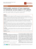
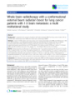
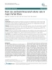
- Fabi et al. Journal of Experimental & Clinical Cancer Research 2011, 30:10 Page 4 of 7 http://www.jeccr.com/content/30/1/10 95%:9-14) and 18% and 9% for colorectal cancer (med- Discussion ian survival 6 months, C.I. 95%:1-12) respectively In this survey, we aimed at assessing the therapeutic (P = .01) (Figure 1). strategies currently adopted in the clinic for unselected Local approaches (surgery or SRS) demonstrated to be patients with BMs from solid tumors treated at four Ita- superior in terms of time to BM progression and survi- lian cancer institutions. The cure algorithm for patients val compared to either WBRT and chemotherapy with BMs is extremely variable and depends on several (P = .02 and P = .0001 respectively) (Table 4; Figure 2). factors such as primary histology and other clinical Multivariate analysis found that primary tumor, neurolo- characteristics of patients. Moreover, though a multidis- gic symptoms at diagnosis of brain involvement, number ciplinary strategy is needed when approaching such of BMs, and type of treatment were independent prog- complex patients, the lack of technical resources may nostic factors for survival (Table 5). influence the therapeutic decision of the treating physi- To assess whether the availability of resources for cian. In fact, in clinical practice, the treatment of BMs is local approach would impact on disease outcome of often planned on the basis of the resources available at patients with BMs, we analyzed the up-front strategy for each treating center. BMs on the basis of the treatment received at each insti- The incidence of BMs reported in our series of tution with respect to the number of brain lesions (≤ 3 patients for each tumor was similar to that reported in vs > 3). Group A included 235 patients referring to a other studies [2]. In our analysis, breast cancer was the comprehensive cancer center where resources for either tumor with the longest time to brain recurrence local (surgery and SRS) and regional/systemic (WBRT (46 months), probably reflecting the advantages of an and chemotherapy) approaches were available. Group B early diagnosis and the availability of effective treat- included 55 patients referring to 3 different institutions ments. In fact, anthracycline- and taxanes-including where only regional/systemic approaches were available regimens as well as new hormonal and biologic agents (WBRT in one center, chemotherapy in all centers) have significantly increased disease-free and overall sur- (Table 1). Patients with ≤ 3 brain lesions were 58% in vival in early breast cancer patients potentially leading both cohorts (n = 137/235 for group A and n = 32/55 to a higher incidence of BMs [15-17]. Regardless of the for group B). In subpopulation of patients with ≤ 3 treatment used for BMs, breast cancer showed the high- BMs, local treatment was delivered in 54% of cases for est 2-year survival rate (36%). The dramatic reduction of group A (75 out of 137 patients) but in only 18% for survival at 2 years observed for NSCLC and melanoma group B (6 out of 32 patients). No difference was found might be due to poor control of either cranial and extra- in terms of time to brain progression at 1 year between cranial disease usually achieved in both malignancies, group A and B (74.2% vs 71.6% respectively, P = .89). thus reflecting the intrinsic radio-resistance of their 2-yr OS 100 colon breast melanoma 80 lung 60 % 40 36 % (breast) 20 % (lung) 20 18 % (melanoma) p=. 01 9 % (colon) 0 0 2 4 6 8 10 12 14 16 18 20 22 24 months Figure 1 Kaplan-Meier survival curves at 2 years according to primary tumor.
- Fabi et al. Journal of Experimental & Clinical Cancer Research 2011, 30:10 Page 5 of 7 http://www.jeccr.com/content/30/1/10 Table 4 Time to brain progression (TTBP) and overall survival (OS) according to the type of treatment for brain metastases Surgery-SRS 88 pts WBRT 136 pts Chemotherapy 66 pts BPFa survival at 1 year 80 % 76 % 62 % BPF survival at 2 years 71 % 53.5 % 34 % median TTBP 27 months 25 months 14 months (C.I. 95%:16-21) (C.I. 95%:20-30) (C.I. 95%:11-17) 1 year OS 74.9 % 47.3 % 33.6 % 2 years OS 42.1 % 23 % 11.5 % median OS 18 months 10 months 8 months (C.I. 95%:26-28) (C.I. 95%:7-14) (C.I. 95%:7-10) a Brain Progression Free Survival. might have favored physicians ’ choice of using che- BMs [18] and the low systemic efficacy of medical thera- motherapy as up-front treatment for BMs along with pies [19,20]. Similarly to breast cancer, a long time to the fact that an oncology unit was available in each brain recurrence (42 months) was observed also for col- institution. Finally, the presence of uncontrolled extra- orectal cancer. Nevertheless, only 18% of patients with cranial disease might have played an important role in BMs from colorectal cancer survived at 1 year (in con- selecting chemotherapy as first treatment option for trast with a 1-year survival of 58% for breast cancer BMs, but the information about control rate on extra- patients with BMs), indicating that in colorectal cancer cranial sites could be retrieved only partially in our brain spread probably represents a final event in the patients, thus it was not considered for analysis. At the course of the disease. present, no prospective comparison has ever been In our series of patients, WBRT was the most used made between chemotherapy and WBRT as upfront up-front therapy for BMs (about 50% of patients) fol- treatment for brain metastases. Interestingly, a recent lowed by chemotherapy which was delivered in survey suggests that in patients with asymptomatic approximately one fourth of cases. The reason why BMs from NSCLC, platinum-based chemotherapy pro- many patients received chemotherapy as up-front vides equal benefit to WBRT as treatment of first treatment for BMs despite the fact that only 41% of choice [21]. In our study the multivariate analysis patients suffered from multiple (> 3) brain lesions, can showed no prognostic difference between chemother- be explained by several reasons. Firstly, nearly all apy and WBRT as up-front treatment for BMs, and patients of our series had active systemic disease at the noteworthy this finding was independent from neuro- time of diagnosis of brain metastases. Secondly, about logic status at diagnosis of brain metastases. half of patients had no neurological symptoms, which 2-yr OS 100 CT RT WB Surg+SRS 80 60 % 42 % of patients 40 23 % of patients 20 p
- Fabi et al. Journal of Experimental & Clinical Cancer Research 2011, 30:10 Page 6 of 7 http://www.jeccr.com/content/30/1/10 Table 5 Univariate and multivariate analysis of prognostic factors for overall survival Overall survival Univariate Analysis Multivariate Analysis HR (95% CI) p value HR (95% CI) p value Age (≤ 65 vs >65) 1.31 (0.93-1.87) 0.12 Sex (male vs female) 1.37 (0.99-1.91) 0.06 Primary Tumor NA 0.01 NA 0.017 Site NA 0.60 (subtentorial vs supratentorial) 0.72 (0.40-1.29) 0.28 (supratentorial and subtentorial vs 1.40 (0.96-2.05) 0.75 supratentorial ) (supratentorial and subtentorial vs 1.93 (1.1-2.53) 0.03 subtentorial Neurologic Symptom (yes vs no) 1.51 (1.06-2.14) 0.02 0.66 (0.44-0.99) 0.046 RPA-RTOG classes NA 0.21 (2 vs 1) 1.18 (0.77-1.70) 0.43 (3 vs 1) 1.78 (0.93-3.43) 0.08 (2 vs 3) 0.66 (0.36-1.19) 0.16 Type of treatment NA < 0.0001 0.02 (CT vs WBRT) 1.05 (0.72-1.53) 0.78 1.16 (0.76-1.76) 0.47 (Surgery/SRS vs WBRT) 0.37 (0.23-0.61) < 0.0001 0.47 (0.26-0.87) 0.02 (Surgery/SRS vs CT) 0.35 (0.21-0.60) < 0.0001 0.41 (0.21-0.77) 0.006 Number of brain metastases NA < 0.0001 0.013 (2-3 vs 1) 1.39 (0.86-2.24) 0.17 1.36 (0.79-2.34) 0.25 (>3 vs 1) 2.20 (1.48-3.27) < 0.0001 2.04 (1.26-3.33) 0.004 (2-3 vs >3) 0.63 (0.41-0.96) 0.03 0.66 (0.41-1.07) 0.10 the heterogeneous characteristics of our patients, which O f note, the multivariate analysis identified local reflects the scenario of clinical practice, where the choice approaches (surgery and SRS) as independent prognostic of front-line strategies for BMs are influenced not only factors for survival. In this survey, we observed that a by the experience of each single physician, but also by local approach was delivered as up-front treatment in the availability of resources. approximately 30% of patients, despite the fact that some data suggest that local treatment could be beneficial for many patients with ≤ 3 brain metastases (59% of patients Conclusions from our series). To this regard, historical data indicate Cancer patients with BMs who are deemed eligible for a that surgery might significantly prolong survival of local approach (SRS, surgery) on the basis of their clini- patients with single BMs [22,23], whereas more recently cal characteristics might obtain improved survival from it has been demonstrated that SRS alone might provide such treatment. Neverthless, in order to optimize the equal results in terms of survival and neurocognitive treatment of BMs, it becomes of crucial importance, to functioning to SRS plus WBRT in patients with ≤ 4 brain carefully select patients who should be offered local lesions [24]. The discrepancy we found between the treatments for BMs. number of patients with ≤ 3 brain metastases and those who received a local approach, can be explained at least Author details in part by the fact that neurosurgery and SRS were avail- 1 Department of Medical Oncology, Regina Elena National Cancer Institute, able only in one centre. In fact, when patients with ≤ 3 Rome - Italy. 2Division of Radiotherapy Regina Elena National Cancer Institute, Rome - Italy. 3Division of Neurosurgery, Regina Elena National BMs were analyzed on the basis of the resources available Cancer Institute, Rome - Italy. 4Belcolle Hospital, Division of Medical at each center, a higher percentage of patients referring Oncology, Viterbo, Italy. 5I.N.I Hospital, Grottaferrata (Rome), Italy. 6Umberto I to a comprehensive cancer center was preferentially trea- Hospital, Division of Medical Oncolog y (FR), Italy. 7Division of Neurology, Regina Elena National Cancer Institute, Rome - Italy. 8Diagnostic Imaging ted with either surgery or SRS (group A) compared to Unit, Regina Elena National Cancer Institute, Rome - Italy. 9Biostatistic Unit, that treated in cancer institutions with no local treat- Regina Elena National Cancer Institute, Rome, Italy. ments (group B). Surprisingly, time to brain progression Authors’ contributions for patients treated locally in each group versus those AF, AF, GM and CMC conceived the study and participated in its design, receiving regional/systemic treatments did not differ sig- coordination and they writed manuscript. AF, AF, GM, AM, EB, ST, LM, MR, nificantly. In our opinion, this finding can be ascribed to GL, GM, AP, MM, AV, IS, FC and CMC read and approved the manuscript-
- Fabi et al. Journal of Experimental & Clinical Cancer Research 2011, 30:10 Page 7 of 7 http://www.jeccr.com/content/30/1/10 long-term control and morbidity of patients surviving more than one Competing interests year after gamma knife radiosurgery for brain metastases. Int J Radiat The authors declare that they have no competing interests. Oncol Biol Phys 2005, 62:1125-1132. Carney DN: Lung cancer–time to move on from chemotherapy. N Engl J 19. Received: 4 December 2010 Accepted: 18 January 2011 Med 2002, 346:126-128. Published: 18 January 2011 20. La Porta CA: Drug resistance in melanoma: new perspectives. Curr Med Chem 2007, 14:387-391. References Moscetti L, Nelli F, Felici A, Rinaldi M, De Santis S, D’Auria G, Mansueto G, 21. 1. Posner JB: Brain metastases: 1995. A brief review. J Neurooncol 1996, Tonini G, Sperduti I, Pollera FC: Up-front chemotherapy and radiation 27:287-293. treatment in newly diagnosed nonsmall cell lung cancer with brain 2. Johnson JD, Young B: Demographics of brain metastases. Clin N Am 1996, metastases: survey by Outcome Research Network for Evaluation of 7:337-344. Treatment Results in Oncology. Cancer 2007, 109:274-281. 3. Sawaya R, Bindal RK, Lang FF, Abi-Said D: Metastatic brain tumors. In Brain 22. Patchell RA, Tibbs PA, Walsh JW, Dempsey RJ, Maruyama Y, Kryscio RJ, tumors. An encyclopedic approach. 2 edition. Edited by: Churchill Markesbery WR, Macdonald JS, Young B: A randomized trial of surgery in Livingstone. London: Kaye AH and Laws Jr ER; 2001:999-1026. the treatment of single metastases to the brain. N Engl J Med 1990, 4. Brem SS, Bierman PJ, Black P, Blumenthal DT, Brem H, Chamberlain MC, 322:494-500. Chiocca EA, DeAngelis LM, Fenstermaker RA, Fine HA, Friedman A, Glass J, 23. Vecht CJ, Haaxma-Reiche H, Noordijk EM, Padberg GW, Voormolen JH, Grossman SA, Heimberger AB, Junck L, Levin V, Loeffler JJ, Maor MH, Hoekstra FH, Tans JT, Lambooij N, Metsaars JA, Wattendorff AR, et al: Narayana A, Newton HB, Olivi A, Portnow J, Prados M, Raizer JJ, Treatment of single brain metastasis: radiotherapy alone or combined Rosenfeld SS, Shrieve DC, Sills AK Jr, Spence AM, Vrionis FD: Central with neurosurgery? Ann Neurol 1993, 33:583-590. nervous system cancers: Clinical Practice Guidelines in Oncology. J Natl 24. Aoyama H, Shirato H, Tago M, Nakagawa K, Toyoda T, Hatano K, Kenjyo M, Compr Canc Netv 2005, 3:644-690. Oya N, Hirota S, Shioura H, Kunieda E, Inomata T, Hayakawa K, Katoh N, 5. Langer CJ, Mehta MP: Current management of brain metastases, with a Kobashi G: Stereotactic radiosurgery plus whole-brain radiation therapy focus on systemic options. J Clin Oncol 2005, 23:6207-6219. vs stereotactic radiosurgery alone for treatment of brain metastases: a 6. Gaspar L, Scott C, Rotman M, Asbell S, Phillips T, Wasserman T, randomized controlled trial. JAMA 2006, 7:2483-2491. McKenna WG, Byhardt R: Recursive partitioning analsis (RPA) of prognostic factors in three Radiation Therapy Oncology Group (RTOG) doi:10.1186/1756-9966-30-10 brain metastases trials. Int J Radiat Oncol Biol Phys 1997, 37:745-751. Cite this article as: Fabi et al.: Brain metastases from solid tumors: 7. Lagerwaard FJ, Levendag PC, Nowak PJ, Eijkenboom WM, Hanssens PE, disease outcome according to type of treatment and therapeutic Schmitz PI: Identification of prognostic factors in patients with brain resources of the treating center. Journal of Experimental & Clinical Cancer metastases: a review of 1292 patients. Int J Radiat Oncol Biol Phys 1999, Research 2011 30:10. 43:795-803. 8. Meyers CA, Smith JA, Bezjak A, Mehta MP, Liebmann J, Illidge T, Kunkler I, Caudrelier JM, Eisenberg PD, Meerwaldt J, Siemers R, Carrie C, Gaspar LE, Curran W, Phan SC, Miller RA, Renschler MF: Neurocognitive function and progression in patients with brain metastases treated with whole-brain radiation and motexafin gadolinium: results of a randomized phase III trial. J Clin Oncol 2004, 22:157-165. 9. Bradley KA, Mehta MP: Management of brain metastases. Semin Oncol 2004, 31:693-701. 10. Soffietti R, Cornu P, Delattre JY, Grant R, Graus F, Grisold W, Heimans J, Hildebrand J, Hoskin P, Kalljo M, Krauseneck P, Marosi C, Siegal T, Vecht C: EFNS Guidelines on diagnosis and treatment of brain metastases: report of an EFNS Task Force. Eur J Neurol 2006, 13:674-681. 11. Langer CJ, Mehta MP: Current management of brain metastases, with a focus on systemic options. J Clin Oncol 2005, 23:6207-6219. 12. Fabi A, Vidiri A, Ferretti G, Felici A, Papaldo P, Carlini P, Mirri A, Nuzzo C, Cognetti F: Dramatic regression of multiple brain metastases from breast cancer with Capecitabine: another arrow at the bow? Cancer Invest 2006, 24:466-468. 13. Cappuzzo F, Ardizzoni A, Soto-Parra H, Gridelli C, Maione P, Tiseo M, Calandri C, Bartolini S, Santoro A, Crinò L: Epidermal growth factor receptor targeted therapy by ZD 1839 (Iressa) in patients with brain metastases from non-small cell lung cancer (NSCLC). Lung Cancer 2003, 41:227-231. 14. Kaplan EL, Meier P: Non parametric estimation from incomplete observations. J Am Stat Assoc 1958, 53:457-481. 15. Crivellari D, Pagani O, Veronesi A, Lombardi D, Nolè F, Thürlimann B, Hess D, Borner M, Bauer J, Martinelli G, Graffeo R, Sessa C, Goldhirsch A: Submit your next manuscript to BioMed Central High incidence of central nervous system involvement in patients with and take full advantage of: metastatic or locally advanced breast cancer treated with epirubicin and docetaxel. Ann Oncol 2001, 12:353-356. 16. Andre F, Slimane K, Bachelot T, Dunant A, Namer M, Barrelier A, Kabbaj O, • Convenient online submission Spano JP, Marsiglia H, Rouzier R, Delaloge S, Spielmann M: Breast cancer • Thorough peer review with synchronous metastases: trends in survival during a 14-year period. J Clin Oncol 2004, 22:3302-3308. • No space constraints or color figure charges 17. Clayton AJ, Danson S, Jolly S, Ryder WD, Burt PA, Stewart AL, Wilkinson PM, • Immediate publication on acceptance Welch RS, Magee B, Wilson G, Howell A, Wardley AM: Incidence of cerebral • Inclusion in PubMed, CAS, Scopus and Google Scholar metastases in patients treated with trastuzumab for metastatic breast cancer. Br J Cancer 2004, 91:639-643. • Research which is freely available for redistribution 18. Varlotto JM, Flickinger JC, Niranjan A, Bhatnagar A, Kondziolka D, Lunsford LD: The impact of whole-brain radiation therapy on the Submit your manuscript at www.biomedcentral.com/submit
CÓ THỂ BẠN MUỐN DOWNLOAD

-

Báo cáo khoa hoc:" A brain-computer interface with vibrotactile biofeedback for haptic information"
 12 p |
  50
|
50
|  6
6
-
Báo cáo khoa hoc:" Recovery of visual fields in brain-lesioned patients by reaction perimetry treatment"
16 p | 57
| 6
-

báo cáo khoa học: "Isolated angiitis of the central nervous system with tumor-like lesion, mimicking brain malignant glioma: a case report and review of the literature"
4 p | 74
| 4
-
Báo cáo khoa hoc:" Effectiveness of a Wii balance board-based system (eBaViR) for balance rehabilitation: a pilot randomized clinical trial in patients with acquired brain injury"
10 p | 66
| 4
-

Báo cáo khoa hoc:" Brain-computer interfacing using modulations of alpha activity induced by covert shifts of attention"
10 p | 34
| 4
-
Báo cáo khoa hoc:" Applying a brain-computer interface to support motor imagery practice in people with stroke for upper limb recovery: a feasibility study"
17 p | 54
| 4
-
báo cáo khoa học: "Multimodality treatment of brain metastases: an institutional survival analysis of 275 patients"
9 p | 63
| 3
-

Báo cáo khoa hoc:" Brain-Computer Interface Controlled Functional Electrical Stimulation System for Ankle Movement"
14 p | 36
| 3
-
Báo cáo khoa hoc:" Biased feedback in brain-computer interfaces"
4 p | 44
| 3
-
báo cáo khoa học: "Brain herniation in a patient with apparently normal intracranial pressure: a case report"
4 p | 44
| 3
-

Báo cáo y học: "Brain metabolism is significantly impaired at blood glucose below 6 mM and brain glucose below 1 mM in patients with severe traumatic brain injury"
13 p | 45
| 3
-

Báo cáo khoa học: " Rapid Brain Cooling in Intubated Pigs through Nasal Flushing with Oxygen: Prevention of Brain Hyperthermia"
6 p | 53
| 3
-

Báo cáo khoa học: " Whole brain radiation therapy in management of brain metastasis: results and prognostic factors"
7 p | 43
| 3
-

Báo cáo khoa học: "Stereotactic radiosurgery for brain metastases: analysis of outcome and risk of brain radionecrosis"
9 p | 43
| 3
-
Báo cáo khoa học: "SemiWhole brain radiotherapy with a conformational external beam radiation boost for lung cancer patients with 1-3 brain metastasis: a multi institutional study"
8 p | 66
| 3
-
Báo cáo y học: " Brain size and brain/intracranial volume ratio in major mental illness"
9 p | 49
| 3
-

Báo cáo khoa học: "Extrapulmonary small cell sarcinoma: involvement of the brain without evidence of extracranial malignancy by serial PET/CT scans"
5 p | 35
| 2


Chịu trách nhiệm nội dung:
Nguyễn Công Hà - Giám đốc Công ty TNHH TÀI LIỆU TRỰC TUYẾN VI NA
LIÊN HỆ
Địa chỉ: P402, 54A Nơ Trang Long, Phường 14, Q.Bình Thạnh, TP.HCM
Hotline: 093 303 0098
Email: support@tailieu.vn
Giấy phép Mạng Xã Hội số: 670/GP-BTTTT cấp ngày 30/11/2015 Copyright © 2022-2032 TaiLieu.VN. All rights reserved.

