


báo cáo khoa học: " Comparison of hyperthermia and adrenaline to enhance the intratumoral accumulation of cisplatin in a murin model of peritoneal carcinomatosis"
lượt xem 4
download
 Download
Vui lòng tải xuống để xem tài liệu đầy đủ
Download
Vui lòng tải xuống để xem tài liệu đầy đủ
Tuyển tập báo cáo các nghiên cứu khoa học quốc tế ngành y học dành cho các bạn tham khảo đề tài: Comparison of hyperthermia and adrenaline to enhance the intratumoral accumulation of cisplatin in a murin model of peritoneal carcinomatosis
Bình luận(0) Đăng nhập để gửi bình luận!
Nội dung Text: báo cáo khoa học: " Comparison of hyperthermia and adrenaline to enhance the intratumoral accumulation of cisplatin in a murin model of peritoneal carcinomatosis"
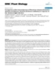
- Facy et al. Journal of Experimental & Clinical Cancer Research 2011, 30:4 http://www.jeccr.com/content/30/1/4 RESEARCH Open Access Comparison of hyperthermia and adrenaline to enhance the intratumoral accumulation of cisplatin in a murin model of peritoneal carcinomatosis Olivier Facy1,2, François Radais1, Sylvain Ladoire1, Delphine Delroeux3, Hervé Tixier1, François Ghiringhelli1, Patrick Rat1,2, Bruno Chauffert1,4, Pablo Ortega-Deballon1,2* Abstract Background: The best method to deliver intraperitoneal chemotherapy (IPC) for peritoneal carcinomatosis from ovarian cancer is not well defined. The aim of this study was to assess the ability of hyperthermia and adrenaline to enhance the intratumoral accumulation of cisplatin in a rat model of peritoneal carcinomatosis. Methods: Four groups of 5 BDIX rats with ovarian peritoneal carcinomatosis underwent IPC with 30 mg/l of cisplatin according to the following conditions: normothermia at 37° for 1 or 2 hours, hyperthermia at 42°C for 1 hour or normothermia at 37°C for 2 hours with 2 mg/l adrenaline. Tissue platinum content was measured by atomic absorption spectroscopy. The effect of hyperthermia, adrenaline and the duration of exposure to the drug was measured in vivo (tissue concentration of platinum in tumor, abdominal and extra abdominal tissues) and in vitro (cytotoxicity on human ovarian cancer cells). Results: In vitro, hyperthermia and longer exposure enhanced the accumulation and the cytotoxic effect of cisplatin on cancer cells. In vivo, only the 2 hours treatment with adrenaline resulted in increased platinum concentrations. The rats treated with adrenaline showed significantly lower concentrations of cisplatin in extra peritoneal tissues than those treated with hyperthermia. Conclusion: Adrenaline is more effective than hyperthermia in order to enhance the intratumoral concentration of cisplatin in rats with peritoneal carcinomatosis from ovarian origin. It may also decrease the systemic absorption of the drug. Introduction reduced quality of life in comparison with standard sys- temic chemotherapy [6]. Intraoperative IPC after cytore- Despite recent improvements, the prognosis of patients ductive surgery is a widely used alternative which with peritoneal carcinomatosis from digestive or ovarian achieves good results [7-9]. However, the best method origin treated with systemic chemotherapy remains poor for IPC has not yet been determined [10,11]. Heated [1,2]. Intraperitoneal chemotherapy (IPC) improves the intraperitoneal chemotherapy (HIPEC) with moderate control of regional disease in ovarian cancer and hyperthermia (41°C to 43°C) is a potentially curative increases survival in carcinomatosis of colorectal origin approach for peritoneal carcinomatosis [4]. Very [3,4]. Trials have shown a survival benefit with post- encouraging results have been recently obtained with operative IPC versus intravenous administration of cis- HIPEC using oxaliplatin at 43°C for 30 minutes in platin-based chemotherapy in ovarian cancer [5,6]. How- selected patients with carcinomatosis from colorectal ever, post-operative IPC showed poor tolerance and origin [9]. As cisplatin is currently the most active sys- temic drug against ovarian carcinoma, it has also been * Correspondence: pablo.ortega-deballon@chu-dijon.fr used for HIPEC [12-16]. This technique is feasible, but 1 INSERM 866, Equipe Avenir, Dijon, France Full list of author information is available at the end of the article © 2011 Facy et al; licensee BioMed Central Ltd. This is an Open Access article distributed under the terms of the Creative Commons Attribution License (http://creativecommons.org/licenses/by/2.0), which permits unrestricted use, distribution, and reproduction in any medium, provided the original work is properly cited.
- Facy et al. Journal of Experimental & Clinical Cancer Research 2011, 30:4 Page 2 of 8 http://www.jeccr.com/content/30/1/4 complete culture medium with fetal bovine serum to somewhat toxic, and most people limit HIPEC with cis- inhibit trypsin. The PROb cells were suspended in 3 ml platin to 1 hour at 42°C or 43°C. No randomized studies of serum-free Ham’s F10 medium and then injected into have compared heated with non-heated intraperitoneal the peritoneum of anesthetized rats (2 × 10 6 cells in cisplatin in ovarian carcinoma. In previous papers, we reported that intraperitoneal each rat). The size of the peritoneal tumor nodules adrenaline increased platinum uptake in rat peritoneal depended upon time. tumor nodules by a factor of 2 to 3 [17-19]. Adrenaline In vitro drug cytotoxicity assay acts through vasoconstriction by limiting drug wash out from the peritoneal cavity. Animals treated with intra- The PROb rat colon cancer cell line and the three peritoneal cisplatin and adrenaline were definitively human ovarian cancer cell lines (SKOV-3, OVCAR-3, and IGROV-1) were incubated in vitro with 30 mg/l cis- cured, whereas those treated with intraperitoneal cispla- tin alone had only a delay in tumor growth [18]. In two platin at 42°C for 1 hour, 37°C for 2 hours (in the pre- phase I studies, intraperitoneal cisplatin with adrenaline sence or not of 2 mg/l adrenaline), or 37°C for 1 hour was feasible in patients with refractory peritoneal carci- (control cells). In vitro cytotoxicity of cisplatin on cancer cells was nomatosis. We also established the maximal tolerated concentration of adrenaline (2 mg/l) in combination determined using a quantitative clonogenic assay. Cells (5 × 10⁴/well) were seeded and cultivated in 96-well tis- with 30 mg/l of cisplatin in two successive 1-hour peri- toneal baths at 37°C after complete cytoreductive sur- sue culture plates for 72 hours until confluence. Cell gery [20,21]. However, the ability of hyperthermia and incubation with cisplatin was performed in serum-free adrenaline to enhance the effect of cisplatin has never Ham culture medium at 37°C or 42°C. After rinsing, the been compared. This was the aim of this experimental cells were trypsinized and seeded again in 24-well tissue preclinical comparative study conducted in a rat model culture plates. After 6 days of culture, the cells were of peritoneal carcinomatosis. washed with phosphate buffered saline, fixed with pure ethanol for 10 min, and then stained with 1% crystal Methods violet in distilled water. After flushing the excess dye with water, the remaining dye was eluted with 33% Animals acetic acid. The optical density (OD) was read on an Female inbred BDIX strain rats, 3 months old, weighing automatic photometer at a wavelength of 540 nm. Cell 200-250 g, were bred in constant conditions of tempera- survival was determined as the ratio of OD in treated ture, hygrometry and exposure to artificial light. Experi- mental protocols followed the “ Guidelines on the wells to OD in control wells × 100. Experiments were protection of experimental animals ” published by the done twice in triplicate. Council of the European Community (1986). The Bur- gundy ’ s University Animal Care and Use Committee Treatment of animals approved all of the procedures. The rats were treated 21 days after intraperitoneal cell inoculation. Laparotomy was performed in anaesthetized rats (isoflurane inhalation as induction and then 100 Cancer cells and tumor model A previously described rat model of peritoneal carcino- mg/kg of intramuscular ketamine and 15 mg xylazine matosis was used. We previously reported the likeness of into the back leg for maintenance) to check the pre- this rat model to human ovarian carcinomatosis in terms sence of a peritoneal carcinomatosis (present in 95% of of peritoneal extension and chemo sensitivity to cisplatin animals). At day 21 after cell injection, the tumor [22]. The DHD/K12/TRb cell line originated from a nodules were confluent in the epiploic area and dimethylhydrazine-induced colonic carcinoma in BDIX extended partly to the peritoneum wall, including rats (ECACC N° 90062901). Its PROb clone was selected nodules in the area of the diaphragm. The abdomen was for its regular tumorigenicity when injected into syngenic then closed in such a way as to make it watertight. rats [23]. PROb cells were maintained in Ham’s F10 cul- Twenty rats were distributed into 4 groups of treatment ture medium supplemented with 10% fetal bovine serum. (5 rats per group), which are presented in Table 1. SKOV-3 (HTB-77) and OVCAR-3 (HTB-161) human The first group (control group) received 30 mg/l of intraperitoneal cisplatin (Sigma-Aldrich, L’Isle d’Abeau, ovarian carcinoma cells originated from ATCC (Mana- ssas, VA). IGROV-1 human ovarian carcinoma cells were France) in 50 ml of saline solution (9 g/l NaCl) at 37°C. a courtesy from Jean Benard, MD (Institut Gustave The second groupreceived HIPEC for 1 hour at 42°C Roussy, Villejuif, France). The human ovarian cells were with 30 mg/l of cisplatin. After laparotomy, an electro- cultured in RPMI medium with 10% fetal bovine serum. nic thermal probe was placed in the epiploic area, an The cells were detached from the culture flask using inward catheter above the right liver, and an outward trypsin and EDTA and centrifuged in the presence of catheter in the left splenic area. After watertight
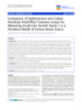
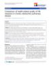
- Facy et al. Journal of Experimental & Clinical Cancer Research 2011, 30:4 Page 3 of 8 http://www.jeccr.com/content/30/1/4 hour HIPEC was impossible due to intolerance of the Table 1 Characteristics of treatment in each group of rats animals. Group Cisplatin Adrenaline Temperature Duration of treatment Atomic absorption spectrometry 1 30 mg/ No 37°C 1h ml The total concentration of platinum was measured by (1 30 mg/ 2 mg/l 37°C 1h atomic absorption spectrometry (AAS). Cultured cells bis*) ml were washed twice after cisplatin incubation, then tryp- 2 30 mg/ No 42°C 1h sinised and counted. Cell pellets were frozen at - 80°C ml until AAS assay. After weighing, the frozen rat tissues 3 30 mg/ 2 mg/ml 37°C 2 h (twice 1 hour) were digested in a microwave digester (MLS-1200 Mega, ml Milestone, Sorisole, Italy). Platinum concentration was 4 30 mg/ No 37°C 2 h (twice 1 hour) measured after dilution in distilled water, using a Zee- ml man atomic absorption spectrometer (Spectra-A; Varian, (*) In another experiment group 1 bis achieved the same tissue concentration Les Ulis, France). Platinum is 65.01% of the molecular of cisplatin as group 1 (unpublished data), thus this group was not repeated in the present study mass of cisplatin; to convert platinum concentrations into cisplatin concentrations, the first must be multi- plied by 1.54. abdomen closure, a closed circuit was established by an Statistical Analysis electric pump (Abbott-Gemstar, Crestline Medical, Plea- Because of the small sample size, nonparametric tests sant Grove, UT, USA) at a flow rate of 15 ml/min. were used to analyze the concentrations of platinum Total volume of the circuit was 500 ml of saline solution and the operative time. The Kruskal-Wallis test was per- which was pre-heated to 37°C. Starting time was defined formed to detect global statistically significant differ- as the moment the temperature reached 41.5°C and 30 ences in the extent of platinum accumulation in the mg/l cisplatin was added. The temperature was kept organs and tumors between the four groups. When a constant at 42°C for 1 hour in the peritoneal cavity by significant difference was found the Mann-Whitney test immersing an intermediate reservoir and about 1 meter was used for 2 × 2 comparisons between groups. A two- of the circuit tubing in a thermostat-regulated bath at tailed P value of\0.05 was considered significant for all an average temperature of 48°C. The third grouphad a tests. Data collection and statistical calculations were 2 hours treatment with 30 mg/l of cisplatin and 2 mg/l performed by SPSS (version 10.0) software (SPSS, Chi- of intraperitoneal adrenaline: after 1 hour the abdomen cago, IL, USA). was open to empty the peritoneal cavity and a second identical bath was then performed for 1 additional hour. Results A previous experiment showed that 1 hour of treatment In vitro accumulation and cytotoxicity of cisplatin on with 2 mg/ml adrenaline at 37°C did not increase the platinum content in peritoneal nodules and, thus, such a cancer cells A temperature of 42°C was toxic by itself. In compari- group was not planned in this study (unpublished data). son with the basal level, the number of residual adher- The fourth groupunderwent the same treatment as the ent cells in the wells was reduced after 1 hour third group, but without adrenaline. All animals from incubation at 42°C (decrease of percentage of 18%, 43%, the 4 groups were kept anesthetized, lying on the back, 51%, and 17% for the PROb, SKOV-3, OVCAR-3, and for the entire duration of the treatment, using repeated IGROV-1, respectively). This was not the case after 2 IM ketamine and xylazine injections as necessary. hours of treatment with cisplatin with or without adre- At the end of treatment, the rats were sacrificed; the naline at 37°C. Cellular platinum concentration was abdominal cavity was opened and abundantly washed increased by hyperthermia in all cells (Figure 1). Extend- with water. Epiploic tumor nodules (200 mg), the left ing the incubation to 2 hours also increased the plati- diaphragm, a piece of the muscle lining the abdominal num content in all cell lines, but there was no influence cavity measuring 5 × 5 × 1 mm thick, parietal thoracic of adrenaline. muscle (200 mg) in order to reflect the extra-abdominal Sensitivity to cisplatin depended on the cell lines (Fig- tissues, half of the left kidney, and about 200 mg of the ure 2). The most sensitive line was OVCAR-3 (IC 50 anterior edge of the liver were sampled and kept at -80° less than 2.5 mg/l after 1 hour incubation at 37°C), C until the platinum assay. whereas the least sensitive lines were SKOV-3 and The comparison of groups 1 and 2 should assess the IGROV-1 (IC 50 ranging between 5 and 10 mg/l). The effect of hyperthermia; that of groups 3 and 4 should rat PROb cell line had intermediate sensitivity to cispla- assess the effect of adrenaline; and that of groups 1 and tin (IC 50 2.5 mg/l). A concentration of 30 mg/l 4 should assess the effect of the duration of IPC. A 2-
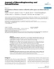
- Facy et al. Journal of Experimental & Clinical Cancer Research 2011, 30:4 Page 4 of 8 http://www.jeccr.com/content/30/1/4 35 Platinum (μg/million cells) 30 25 20 15 10 abcd abcd abcd 5 a bcd 0 PRO SKOV IGROV OVCAR Figure 1 In vitro platinum accumulation in cancer cells. Cells (1 × 10⁶ /well) were seeded in 12-well culture plates for 72 hours then incubated with 30 mg/l cisplatin in serum-free Ham medium. Incubation conditions were: 1 hour at 37°C (a), 1 hour at 42°C (b), and 2 hours at 37°C without (c) or with (d) 2 mg/l adrenaline. Mean and SD of 3 determinations are represented. treatments (Figure 3). Regarding the platinum content c isplatin was found to be almost complete cytotoxic (≥90%) for all cell lines. This concentration was chosen in peritoneal nodules, the difference between group 1 for the in vivo experiments. The cell toxicity of cisplatin (control, 1 hour IPC), and groups 4 (2 hours IPC) or 2 (HIPEC) did not reach significance (p = 0.06 and 0.19, was significantly enhanced by 1 hour of hyperthermia at respectively). In contrast, a 3-fold increase in tumor pla- 42°C for the resistant SKOV-3 and IGROV-1 cell lines, tinum content was found in group 3 (adrenaline) as but not for the sensitive OVCAR-3 and PROb cells. Cis- compared to groups 1 (control, p = 0.005) and 2 platin cytotoxicity was also enhanced by extending the (HIPEC, p = 0.005). Platinum concentration in the incubation time to 2 hours; the improvement in cyto- abdominal muscle lining the peritoneal cavity was also toxicity was of the same order as that achieved by 1 significantly greater in group 3 (adrenaline) as compared hour of hyperthermia. to group 4 (HIPEC) (p = 0.006), but did not reach sig- nificance in the diaphragm (p = 0.08). Platinum accumulation in rat peritoneal nodules and Out of the peritoneal cavity (kidney and thoracic mus- organs cle), the accumulation of platinum was lower in group 3 In the hyperthermia group, the closed circuit made it (adrenaline) than in groups 1 (control) and 4 (HIPEC) possible to reach a stable intra-abdominal temperature (p = 0.05 and p = 0.001, for the kidney and the thoracic (42.1°C ± 0.46°C) in a mean time of 15.5 minutes (range muscle, respectively). 4-21 minutes) with variations of less than 0.5°C along the procedure. Temperature was dependent on the flow Discussion rate and was unstable at a flow of less than 15 ml/min. Tolerance to HIPEC was poor. Only 3 out of 5 rats The present study reports the greater uptake of plati- survived until the end of the experiment. The others num in peritoneal nodules and in peritoneum lining presented an abnormal respiratory rhythm at about 45 muscle when adrenaline was used in combination with minutes and died before the end. This precluded the cisplatin, as compared to HIPEC. This underlines the performance of a 2-hour HIPEC. In contrast, all of the interest of adrenaline to increase the tissue concentra- animals that were treated at 37°C, for either 1 or 2 tion of chemotherapy and the fact that the best method hours, with or without adrenaline, were alive and well at to deliver of IPC remains to be defined [10,17,21]. the end of the experiment. The rats treated with adrenaline (group 3) received Platinum concentrations in rat organs and peritoneal this treatment for 2 hours, as compared to those under- nodules were measured according to the different going HIPEC (group 2) during only 1 hour. A 1-hour
- Facy et al. Journal of Experimental & Clinical Cancer Research 2011, 30:4 Page 5 of 8 http://www.jeccr.com/content/30/1/4 PROb SKOV-3 100 100 80 80 60 60 40 40 20 20 0 0 0 2,5 5 10 20 30 0 2,5 5 10 20 30 Cisplatin (mg/L) Cisplatin (mg/L) IGROV-1 OVCAR-3 100 100 80 80 60 60 40 40 20 20 0 0 0 2,5 5 10 20 30 0 2,5 5 10 20 30 Cisplatin (mg/L) Cisplatin (mg/L) Figure 2 In vitro cytotoxicity of cisplatin. Cells (5 × 10 /well) were seeded in 96-well culture plates for 72 hours, then treated with cisplatin in 4 serum-free Ham medium. Treatment conditions were: 1 hour at 37°C (dark triangles), 1 hour at 42°C (open triangles), 2 hours at 37°C without (dark squares) or with (clear squares) 2 mg/l adrenaline. Mean and SD of 4 determinations of cell survival (percent of control cells) are represented. adrenaline group was not performed because a previous the longer exposure explains the higher tissue uptake of unpublished experiment found no significant difference cisplatin. However, group 4 had a 2 hours IPC and did after this treatment as compared to the control group. not achieved significantly better concentrations than A 2-hour HIPEC was impossible due to intolerance of group 1 (1 hour IPC); the difference was close to signifi- the animals to such a procedure. It could be argued that cance (p = 0.06), but it can not explain a 3-fold increase
- Facy et al. Journal of Experimental & Clinical Cancer Research 2011, 30:4 Page 6 of 8 http://www.jeccr.com/content/30/1/4 30 25 Platinum (μg/g of tissue) 20 * * 15 10 5 a b cd a bc d abcd ab cd a bc d 0 TUMOR DIAPHR ABD/MU KIDNEY LIVER THOR/MU Figure 3 In vivo accumulation of platinum in peritoneal tumor and organs. Intraperitoneal chemotherapy was performed using 30 mg/l of cisplatin. Tumor and organs were sampled: after 1 hour cisplatin at 37°C (a), after 1 hour cisplatin at 42°C (b), after 2 hours cisplatin with (c) or without (d) 2 mg/l adrenaline. Mean and SD of 5 animals. Asterisk indicates a statistical difference (p < 0.01) between the 2 hours treatment at 37°C with 2 mg/L adrenaline, and the 1 hour treatment at 42°C. ABD/MU = abdominal muscle and THOR/MU = thoracic muscle. of adrenaline which prevented the systemic diffusion, in concentration. The effect of time probably exists, but and thus, the potential toxicity of cisplatin. At the oppo- is small. This is consistent with the results of a previous site, HIPEC has been shown to increase systemic pharmacokinetic study which showed that most of the absorption of chemotherapy drugs due to heat-induced uptake happens at the beginning of IPC, when the gradi- vasodilatation [11]. ent of concentrations is higher: a twice 1-hour bath (as Our results confirmed the well-known enhancing done in the present study) with a newly prepared identi- effect of hyperthermia on the platinum uptake, as well cal solution was more effective than a 2-hour bath [24]. in vitro as in vivo [25-28]. In vitro , the thermal Similar results have been obtained in HIPEC with oxali- enhanced ratio (TER) after 1 hour exposure at 42°C platin [11]. compared to 37°C ranged from 1.5 to 2.1, depending on Adrenaline also increased the drug content in the the cell line. The TER was lower than that found in muscle of the abdominal wall. We observed a ratio of 5 other studies (3.4 for 1 hour at 43°C in a different colon to 17 in drug uptake between an abdominal muscle and cancer cell line in rats; 2.2 or 3.9 for hamster kidney a distant thoracic muscle. This reflects the pharmacolo- cells and Chinese hamster fibroblasts, respectively) gical advantage of IPC to obtain high local drug concen- [26,27]. The reasons for these discrepancies (technical trations in the abdominal wall, peritoneum and muscle variations or true differences in membrane permeability lining, all of which are possibly infiltrated by malignant in different cell lines) are unknown. The increased accu- cells in peritoneal carcinomatosis. In previous studies mulation due to extending exposure to 2 hours (1.6 to we used a higher concentration of adrenaline (5 or 10 2.5) was of the same order as the TER recorded after 1 mg/L) [18,19]. In the present study it was reduced hour. Temperature is mainly thought to accelerate the according to a recent phase I clinical trial, which estab- passive diffusion of cisplatin by disturbing the phospho- lished the safety of 2 mg/l of adrenaline, whereas 3 mg/l lipid bilayer arrangement, even if other mechanisms, induced cardiovascular collateral effects (tachycardia, such as a direct apoptotic or necrotic effect, may be arterial hypertension or electric signs of cardiac ische- involved in cell death. mia) [21]. In vitro experiments on cancer cell lines alone cannot Despite their longer exposure, rats treated with adre- predict the in vivo effect of temperature or adrenaline. naline showed lower extraperitoneal concentrations of Tumor tissue penetration is the limiting factor for the platinum than both, the control and the HIPEC groups. activity of the chemotherapeutic agents [29]. It has been This is probably explained by the vasoconstrictor effect
- Facy et al. Journal of Experimental & Clinical Cancer Research 2011, 30:4 Page 7 of 8 http://www.jeccr.com/content/30/1/4 hypothesized that the depth of penetration of cisplatin Acknowledgements This paper was supported by grants from the French National League could be increased by hyperthermia through its effects against Cancer (Committees of Saône et Loire, Nièvre, and Côte d’Or). We on convection and diffusion in tissues, increasing cell thank Philip Bastable for the help in revising the manuscript. uptake of the drug, tumor blood flow and vascular per- We thank Pierre-Emmanuel Puig Ph.D., Laurent Benoit M.D., Sylvain Causeret M.D. and Bernard Royer M.D., Ph.D. for their help with the experiments and meability. Despite the clinical development of HIPEC their suggestions. We also thank Jean Luc Beltramo Ph.D. for the platinum with platinum compounds, only a few studies have been assays. done in order to establish the basis of this technique. Author details Two contradictory studies have been reported in rat 1 INSERM 866, Equipe Avenir, Dijon, France. 2Department of Digestive Surgical models of peritoneal carcinomatosis [27,30,31]. Differ- Oncology, University Hospital of Dijon, France. 3Department of Digestive Surgery, University Hospital of Besançon, France. 4Department of Medical ences in the hyperthermia technique could explain this Oncology, University Hospital of Amiens, France. discrepancy. Los et al. immersed the whole animal in a thermostatically controlled water bath, resulting in Authors’ contributions whole-body hyperthermia rather than locoregional OF, FR and DD carried out the in vivo experiments. SL and HT carried out the in vitro experiments. BC participated in the design of the study and hyperthermia [27]. This could have modified both blood performed the statistical analysis. POD, FG and PR conceived the study, and concentrations and vascular permeability, and may participated in its design and coordination. All authors read and approved explain why plasmatic cisplatin was about 3 times the final manuscript. greater at 41°5 than at 38°C and why platinum content Received: 2 December 2010 Accepted: 7 January 2011 was about twice as great in all organs, including the Published: 7 January 2011 extra-abdominal organs such as the lung. Our technique allowed us to heat only the abdominal cavity. Using this References 1. Gadducci A, Cosio S, Conte PF, Genazzani AR: Consolidation and method of heating, a 1-hour HIPEC at 42°C did not maintenance treatments for patients with advanced epithelial ovarian increase platinum content in the peritoneal tumor cancer in complete response after first-line chemotherapy: a review of nodules or in the peritoneal wall lining. Abdominal the literature. Crit Rev Oncol Hematol 2005, 55:153-66. 2. Jayne DG, Fook S, Loi C, Seow-Choen F: Peritoneal carcinomatosis from hyperthermia was poorly tolerated by the animals; some- colorectal cancer. Br J Surg 2002, 89:1545-50. times it was even necessary to stop the procedure before 3. Fujiwara K, Armstrong D, Morgan M, Markman M: Principles and practice 60 minutes. This poor tolerance made it impossible to of intraperitoneal chemotherapy for ovarian cancer. Int J Gynecol Cancer 2007, 17:1-20. compare the two methods in terms of survival. Our 4. Verwaal VJ, Bruin S, Boot H, van Slooten G, van Tinteren H: 8-Year follow- negative results on HIPEC with cisplatin are consistent up of randomized trial: cytoreduction and hyperthermic intraperitoneal with those obtained by other authors using similar chemotherapy versus systemic chemotherapy in patients with peritoneal carcinomatosis of colorectal cancer. Ann Surg Oncol 2009, methods [31,32]. An explanation of this negative result 15:2426-32. could be the temperature-related increase in blood flow 5. Armstrong DK, Bundy B, Wenzel L, Huang HQ, Baergen R, Lele S, et al: through the peritoneal nodules and the peritoneum due Intraperitoneal cisplatin and paclitaxel in ovarian cancer. N Engl J Med 2006, 354:34-43. to local vasodilatation and resulting in an increase in 6. Fung-Kee-Fung M, Provencher D, Rosen B, Hoskins P, Rambout L, Oliver T, the wash out of the cisplatin [33]. et al: Intraperitoneal chemotherapy for patients with advanced ovarian In contrast with heat, adrenaline at a concentration of cancer: a review of the evidence and standards for the delivery of care. Gynecol Oncol 2007, 105:747-56. 2 mg/l for 2 hour achieved a 2 to 3-fold increase in pla- 7. Bankhead C: Intraperitoneal therapy for advanced ovarian cancer: will it tinum content in the peritoneal tumor nodules. Such an become standard care? J Natl Cancer Inst 2006, 98:510-2. increase boosts the cytotoxic effect of cisplatin in vitro 8. Yan TD, Stuart OA, Yoo D, Sugarbaker PH: Perioperative intraperitoneal chemotherapy for peritoneal surface malignancy. J Transl Med 2006, 4:17. (Figure 2). Previous rat experiments have shown us that 9. Elias D, Lefevre JH, Chevalier J, et al: Complete cytoreductive surgery plus 2 hours of IPC are required to observe the enhancing intraperitoneal chemohyperthermia with oxaliplatin for peritoneal effect of adrenaline [17,19], and our following clinical carcinomatosis of colorectal origin. J Clin Oncol 2009, 27:681-5. 10. Esquivel J: Technology of hyperthermic intraperitoneal chemotherapy in trials have taken into account this parameter [20,21]. the United States, Europe, China, Japan, and Korea. Cancer J 2009, Experimental data show that adrenaline is more effec- 15:249-54. tive and better tolerated than hyperthermia in order to 11. Ortega-Deballon P, Facy O, Jambet S, Magnin G, Cotte E, Beltramo JL, et al: Which method to deliver heated intraperitoneal chemotherapy with enhance the penetration of cisplatin. It also minimizes oxaliplatin? An experimental comparison of open and closed the systemic absorption of cisplatin. Hyperthermia was techniques. Ann Surg Oncol 2010, 17:1957-63. not well tolerated in this rat model, but it is in humans. 12. Cotte E, Glehen O, Mohamed F, Lamy F, Falandry C, Golfier F, et al: Cytoreductive surgery and intraperitoneal chemo-hyperthermia for Future clinical trials performing IPC with cisplatin for chemo-resistant and recurrent advanced epithelial ovarian cancer: ovarian carcinoma should compare the effectiveness of prospective study of 81 patients. World J Surg 2007, 31:1813-20. adrenaline and hyperthermia in order to improve the 13. Helm CW, Randall-Whitis L, Martin RS, Metzinger DS, Gordinier ME, Parker LP, et al: Hyperthermic intraperitoneal chemotherapy in effect of intraperitoneal chemotherapy. conjunction with surgery for the treatment of recurrent ovarian The authors declare that they have no competing carcinoma. Gynecol Oncol 2007, 105:90-6. interests. 14. Kusamura S, Younan R, Baratti D, Costanzo P, Favaro M, Gavazzi C, et al: Cytoreductive surgery followed by intraperitoneal hyperthermic
- Facy et al. Journal of Experimental & Clinical Cancer Research 2011, 30:4 Page 8 of 8 http://www.jeccr.com/content/30/1/4 perfusion: analysis of morbidity and mortality in 209 peritoneal surface malignancies treated with closed abdomen technique. Cancer 2006, doi:10.1186/1756-9966-30-4 106:1144-53. Cite this article as: Facy et al.: Comparison of hyperthermia and 15. Piso P, Dahlke MH, Loss M, Schlitt HJ: Cytoreductive surgery and adrenaline to enhance the intratumoral accumulation of cisplatin in a hyperthermic intraperitoneal chemotherapy in peritoneal carcinomatosis murin model of peritoneal carcinomatosis. Journal of Experimental & from ovarian cancer. World J Surg Oncol 2004, 2:21. Clinical Cancer Research 2011 30:4. 16. Raspagliesi F, Kusamura S, Campos Torres JC, de Souza GA, Ditto A, Zanaboni F, et al: Cytoreduction combined with intraperitoneal hyperthermic perfusion chemotherapy in advanced/recurrent ovarian cancer patients: The experience of National Cancer Institute of Milan. Eur J Surg Oncol 2006, 32:671-5. 17. Chauffert B, Favoulet P, Polycarpe E, Duvillard C, Beltramo JL, Bichat F, et al: Rationale supporting the use of vasoconstrictors for intraperitoneal chemotherapy with platinum derivatives. Surg Oncol Clin N Am 2003, 12:835-48. 18. Duvillard C, Benoit L, Moretto P, Beltramo JL, Brunet-Lecomte P, Correia M, et al: Adrenaline enhances penetration and anti-cancer activity of local cisplatin on rat sub-cutaneous and peritoneal tumors. Int J Cancer 1999, 81:779-84. 19. Favoulet P, Magnin G, Guilland JC, Beltramo JL, Osmak L, Benoit L, et al: Pre-clinical study of the adrenaline-cisplatin association for the treatment of intraperitoneal carcinomatosis. Eur J Surg Oncol 2001, 27:59-64. 20. Molucon-Chabrot C, Isambert N, Benoit L, Zanetta S, Fraisse J, Guilland JC, et al: Feasibility of using intraperitoneal adrenaline and cisplatin in patients with advanced peritoneal carcinomatosis. Anticancer Drugs 2006, 17:1211-7. 21. Guardiola E, Chauffert B, Delroeux D, et al: Intraoperative intraperitoneal (IP) chemotherapy with cisplatin and epinephrine after cytoreductive surgery in patients with recurrent ovarian cancer: a phase I study. Anticancer Drugs 2010, 21:320-5. 22. Chauffert B, Dimanche-Boitrel MT, Genne P, Petit JM, Onier N, Jeannin JF: Experimental chemotherapy of peritoneal carcinomatosis of colonic origin in rats. Gastroenterol Clin Biol 1992, 16:215-9. 23. Martin F, Caignard A, Jeannin JF, et al: Selection by trypsin of two sublines of rat colon cancer cells forming progressive or regressive tumors. Int J Cancer 1983, 32:623-7. 24. Royer B, Delroeux D, Guardiola E, Combe M, Hoizey G, Montange D, et al: Improvement in intraperitoneal intraoperative cisplatin exposure based on pharmacokinetic analysis in patients with ovarian cancer. Cancer Chemother Pharmacol 2008, 61:415-21. 25. Barlogie B, Corry PM, Drewinko B: In vitro thermochemotherapy of human colon cancer cells with cis-dichlorodiammineplatinum (II) and mitomycin C. Cancer Res 1980, 40:1165-8. 26. Eichholtz-Wirth H, Hietel B: Heat sensitization to cisplatin in two cell lines with different drug sensitivities. Int J Hyperthermia 1990, 6:47-55. 27. Los G, Sminia P, Wondergem J, Mutsaers PH, Havemen J, ten Bokkel HD, et al: Optimisation of intraperitoneal cisplatin therapy with regional hyperthermia in rats. Eur J Cancer 1991, 27:472-7. 28. Meyn RE, Corry PM, Fletcher SE, Demetriades M: Thermal enhancement of DNA damage in mammalian cells treated with cis- diamminedichloroplatinum (II). Cancer Res 1980, 40:1136-9. 29. Conti M, De GU, Tazzari V, Bezzi F, Baccini C: Clinical pharmacology of intraperitoneal cisplatin-based chemotherapy. J Chemother 2004, 16(Suppl 5):23-5. 30. Los G, van Vugt MJ, Pinedo HM: Response of peritoneal solid tumours after intraperitoneal chemohyperthermia treatment with cisplatin or carboplatin. Br J Cancer 1994, 69:235-41. Submit your next manuscript to BioMed Central 31. Zeamari S, Floot B, van d, Stewart FA: Pharmacokinetics and pharmacodynamics of cisplatin after intraoperative hyperthermic and take full advantage of: intraperitoneal chemoperfusion (HIPEC). Anticancer Res 2003, 23:1643-8. 32. El-Kareh AW, Secomb TW: A theoretical model for intraperitoneal delivery • Convenient online submission of cisplatin and the effect of hyperthermia on drug penetration distance. Neoplasia 2004, 6:117-27. • Thorough peer review 33. Ausmus PL, Wilke AV, Frazier DL: Effects of hyperthermia on blood flow • No space constraints or color figure charges and cis-diamminedichloroplatinum (II) pharmacokinetics in murine • Immediate publication on acceptance mammary adenocarcinomas. Cancer Res 1992, 52:4965-8. • Inclusion in PubMed, CAS, Scopus and Google Scholar • Research which is freely available for redistribution Submit your manuscript at www.biomedcentral.com/submit
CÓ THỂ BẠN MUỐN DOWNLOAD

-
báo cáo khoa học: " Comparison of KRAS and EGFR gene status between primary non-small cell lung cancer and local lymph node metastases: implications for clinical practice"
 8 p |
8 p |  90
|
90
|  8
8
-

Báo cáo khoa học: "Comparison of immunohistochemistry (IHC) and fluorescence in situ hybridization (FISH) assessment for Her-2 status in breast cancer"
6 p | 72
| 6
-
Báo cáo khoa học: "Comparison of RBE values of high- LET a-particles for the induction of DNA-DSBs, chromosome aberrations and cell reproductive death"
8 p | 59
| 5
-
Báo cáo khoa học: "Comparison of coplanar and noncoplanar intensity-modulated radiation therapy and helical tomotherapy for hepatocellular carcinom"
8 p | 59
| 5
-
báo cáo khoa học:" Comparison of numerical and verbal rating scales to measure pain exacerbations in patients with chronic cancer pain"
8 p | 56
| 4
-
báo cáo khoa học: "Comparison of determinants of research knowledge utilization by practitioners and administrators in the field of child and family social services"
12 p | 81
| 4
-
báo cáo khoa học: " Comparison of Radioimmuno and Carbon Nanotube Field-Effect Transistor Assays for Measuring Insulin-Like Growth Factor-1 in a Preclinical Model of Human Breast Cancer"
6 p | 55
| 4
-
báo cáo khoa học:"Comparison of health-related quality of life measures in chronic obstructive pulmonary disease"
6 p | 69
| 4
-

báo cáo khoa học:" Comparison between the disease-specific Airways Questionnaire 20 and the generic 15D instruments in COPD"
9 p | 49
| 3
-
báo cáo khoa học: "Comparison of knee motion on Earth and in space: an observational study"
8 p | 73
| 3
-
Báo cáo khoa hoc:" Comparison of nanoparticle-mediated transfection methods for DNA expression plasmids: efficiency and cytotoxicity"
11 p | 50
| 3
-
Báo cáo khoa hoc: Comparison of regression models for estimation of isometric wrist joint torques using surface electromyography
12 p | 64
| 3
-
Báo cáo khoa hoc:" Comparison between the HCV IRES domain IV RNA structure and the Iron Responsive Element"
8 p | 71
| 3
-
Báo cáo khoa học: "Comparison of intraoperative frozen section analysis for sentinel lymph node biopsy during breast cancer surgery for invasive lobular carcinoma and invasive ductal carcinoma"
8 p | 81
| 3
-
Báo cáo khoa học: "Comparison of conformal and intensity modulated radiation therapy techniques for treatment of pelvic tumors. Analysis of acute toxicity"
7 p | 52
| 3
-
Báo cáo khoa học: "Comparison of simple and complex liver intensity modulated radiotherapy"
9 p | 47
| 3
-
báo cáo khoa học: " Comparison of the transcriptomes of American chestnut (Castanea dentata) and Chinese chestnut (Castanea mollissima) in response to the chestnut blight infection"
11 p | 57
| 3


Chịu trách nhiệm nội dung:
Nguyễn Công Hà - Giám đốc Công ty TNHH TÀI LIỆU TRỰC TUYẾN VI NA
LIÊN HỆ
Địa chỉ: P402, 54A Nơ Trang Long, Phường 14, Q.Bình Thạnh, TP.HCM
Hotline: 093 303 0098
Email: support@tailieu.vn
Giấy phép Mạng Xã Hội số: 670/GP-BTTTT cấp ngày 30/11/2015 Copyright © 2022-2032 TaiLieu.VN. All rights reserved.

