


báo cáo khoa học: " Effects of DNMT1 silencing on malignant phenotype and methylated gene expression in cervical cancer cells"
lượt xem 5
download
 Download
Vui lòng tải xuống để xem tài liệu đầy đủ
Download
Vui lòng tải xuống để xem tài liệu đầy đủ
Tuyển tập báo cáo các nghiên cứu khoa học quốc tế ngành y học dành cho các bạn tham khảo đề tài: Effects of DNMT1 silencing on malignant phenotype and methylated gene expression in cervical cancer cells
Bình luận(0) Đăng nhập để gửi bình luận!
Nội dung Text: báo cáo khoa học: " Effects of DNMT1 silencing on malignant phenotype and methylated gene expression in cervical cancer cells"
- Zhang et al. Journal of Experimental & Clinical Cancer Research 2011, 30:98 http://www.jeccr.com/content/30/1/98 RESEARCH Open Access Effects of DNMT1 silencing on malignant phenotype and methylated gene expression in cervical cancer cells Yi Zhang1,2†, Fu-qiang Chen1†, Ye-hong Sun1†, Shu-yan Zhou1†, Ti-yuan Li1* and Rui Chen1,2† Abstract Background: DNA methylation has been widely used in classification, early diagnosis, therapy and prediction of metastasis as well as recurrence of cervical cancer. DNMT methyltransferase 1 (DNMT1), which plays a significant role in maintaining DNA methylation status and regulating the expression of tumor suppressor genes. The aim of this research was to investigate the relationship between DNMT1 and abnormal methylation of tumor suppressor genes and malignant phenotype in cervical cancer. Methods: Levels of DNMT1 mRNA and protein were detected using qPCR and Western blot, respectively. Cell proliferation was analyzed by MTT and apoptosis was performed by Annexin V-FITC/PI double staining flow cytometry, respectively. MeDIP-qPCR and qPCR were performed to measure demethylation status and mRNA re- expression level of 7 tumor-suppressor genes (CCNA1, CHFR, FHIT, PAX1, PTEN, SFRP4, TSLC1) in Hela and Siha cells after silencing DNMT1. Results: The average expression levels of DNMT1 mRNA and protein in Hela and Siha cells were decreased significantly compared with control group. The flow cytometry and MTT results showed that Hela and Siha cells apoptosis rates and cell viabilities were 19.4 ± 2.90%, 25.7 ± 3.92% as well as 86.7 ± 3.12%, 84.16 ± 2.67% respectively 48 h after transfection (P < 0.01). Furthermore, the promoter methylation of five tumor suppressor genes was decreased with the increased mRNA expression after silencing DNMT1, whereas there were no significant changes in PTEN and FHIT genes in Hela cells, and CHFR and FHIT genes in Siha cells. Conclusions: Our experimental results demonstrate that methylation status of DNMT1 can influence several important tumor suppressor genes activity in cervical tumorigenesis and may have the potential to become an effective target for treatment of cervical cancer. Background Currently, the known repressor genes are related to cer- vical cancer including CCNA1, CHFR, FHIT, PAX1, Cervical cancer is the second most common cancer in PTEN, SFRP4, TSLC1 and etc [1]. All these genes men- women worldwide and the leading cause of cancer deaths tioned above have performed a wide variety of functions to in women in developing countries. It is obviously that regulate the transcription and expression, any of which many genetic and epigenetic alternations occur during down-regulation as well as promoter hypermethylation will cervical tumorigenesis. Among those changes, aberrant lead to the precursor lesions in cervical development and promoter methylation of tumor-suppressor genes gives malignant transformation. DNA methylation is catalyzed rise to its silencing functions and results in the significant by several DNA methyltransferases, including DNMT1, carcinogenesis of cervical cancer. DNMT3a, DNMT3b and etc. DNMT1 is responsible for precise duplicating and maintaining the pre-existing DNA methylation patterns after replication. As reported by Szyf * Correspondence: tiyuan_li@163.com † Contributed equally [2], DNMT1 inhibited the transcription of tumor suppres- 1 The Second Medical College, Jinan University, Shenzhen Clinical Medical sor genes and facilitated the formation of tumorigenesis, Research Center, Shenzhen People’s Hospital, 518020, Shenzhen, PR China which linked to the development of cervical cancer. Full list of author information is available at the end of the article © 2011 Zhang et al; licensee BioMed Central Ltd. This is an Open Access article distributed under the terms of the Creative Commons Attribution License (http://creativecommons.org/licenses/by/2.0), which permits unrestricted use, distribution, and reproduction in any medium, provided the original work is properly cited.
- Zhang et al. Journal of Experimental & Clinical Cancer Research 2011, 30:98 Page 2 of 8 http://www.jeccr.com/content/30/1/98 for DNMT1 were 5’-AACCTTCACCTAGCCCCAG-3 ’ Meanwhile, Inhibition of DNMT1 activity could reduce (forward) and 5 ’ -CTCATCCGATTTGGCTCTTCA- hypermethylation of repressive genes and promote its re- 3’(reverse); for GAPDH were 5’-CAGCCTCAAGATCAT- expression, and reverse phenotype of malignant tumor. CAGCA-3 ’ (forward) and 5 ’ -TGTGGTCATGAGTCC Thus, specific inhibition of DNMT1 could be one strategy TTCCA-3 ’ (reverse). QPCR was performed in a 20 μ l for cervical therapy. volume containing 1 μ l cDNA template, 10 μ l SYBR In our study, we detected the demethylation and re- Green Real-time PCR Master Mix and 1 μl of each primer. expression levels of seven cervical cancer suppressor genes with DNMT1 silencing in Hela and Siha cells. The aim Levels of seven tumor suppressor genes mRNA expression was to elucidate the relations between DNMT1 and were also assayed with qPCR. This cycle was defined at abnormal methylation of these genes’ promoter as well as 95°C for 5 min, followed by 35 cycles of denaturing at the malignant phenotype of tumor cells, which might con- 95°C for 45s, annealing at 59°C for 35 s and extension at tribute to the investigations of functions and regulation 72°C for 1 min, and followed by the final extension at roles of DNMT1 in cervical cancer. 72°C for 10 min. The primers were shown in Table 1 and Table 2. Materials and methods Cell culture and transfection Western blot analysis The Hela and Siha human cervical cancer cells lines were Cells were harvested and rinsed twice in ice-cold PBS, obtained from American Type Culture Collection (Mana- and kept on ice for 30 min in cell lysis buffer containing ssas, VA, USA). Lipofectamine TM2000 was purchased 1 mM PMSF while agitating constantly, and insoluble from Invitrogen Co. These cells grown in Dulbeco’s Modi- cell debris was discarded by centrifugation for 10 min at fied Eagle Medium (DMEM) supplemented with 10% fetal 12,000 rpm at 4°C. The protein samples were separated bovine serum and incubated at 37°C in a humidified with 12% SDS-PAGE and subsequently transferred to chamber with 5% CO2. The siRNA primer sequences for PVDF membranes (Millipore). Membranes were blocked DNMT1 were 5’-UUAUGUUGCUCACAAACUUCUU- with 5% nonfat dry milk solution either at room tem- GUC-3’ (forward) and 5’-GACAAGAAG UUUGUGAG- perature for 2 h, and incubated with Rabbit anti- CAACAUAA-3’ (reverse), which were custom synthesized DNMT1 and secondary antibody at 37°C for 2 h respec- tively. The Membranes were stained with an enhanced by Shanghai Sangon (Shanghai, China). After transfection, chemiluminescence solution. Band intensities are nor- the inhibition efficiency was examined using quantitative malized to b-actin as a loading control. polymerase chain reaction (qPCR). Transfections were performed with Lipfectamine TM2000 according to the protocol (Invitrogen Co.). Annexin V-FITC/PI staining and flow cytometry Cell cycle analysis: Cells were digested by typsin (0.25%) and fixed with cold 70% ethanol at 48 h after transfec- Real-time qPCR assay QPCR was used to analyze mRNA expression level tion. After washed in phosphate-buffered saline, samples were incubated with 100 μl RNase A at 37°C for 30 min of DNMT1. Total RNA was extracted using Trizol and stained with 400 μl propidium iodide (Sigma). Flow reagent and reversely transcribed into cDNA. The primers Table 1 Primers used in RNA expression gene Sequences Tm (°C) Product Size(bp) F:5’GGGAAACTGTGGCGTGAT3’ QPCR GAPDH 59 299 R:5’GAGTGGGTGTCGCTGTTGA3’ F:5’GGAGATCAGAGGAGGAAATGG3’ FHIT 59 233 R:5’GGGAGTTGGAGTGACCGAG3’ F:5’ACACGACGGGAAGACAAGTT3’ PTEN 59 157 R:5’CTGGTCCTGGTATGAAGAATG3’ F:5’GCGTAGAAATGCCCAAACC3’ CHFR 59 171 R:5’TCCATCCAGCCCGAGTAGC3’ F:5’GGCCTCTTGATGTTGACTGTAA3’ SFRP4 59 204 R:5’GAGGGATGGGTGATGAGGA3’ F:5’GGTAGGAGTAGGGAGCACAGG3’ PAX1 59 100 R:5’CAAGTGTTGCGAGTGGAGG3’ F:5’TTATTTCAGGGACTTCAGGC3’ TSLC1 59 223 R:5’TTCCACCGCAGTGTCTTTC3’ F:5’GCCTGGCAAACTATACTGTGAAC3’ CCNA1 59 295 R:5’GTGCAGAAGCCTATGACGATTA3’
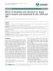
- Zhang et al. Journal of Experimental & Clinical Cancer Research 2011, 30:98 Page 3 of 8 http://www.jeccr.com/content/30/1/98 Table 2 Primers used in MeDIP-qPCR assay gene Sequences Tm (°C) Product Size(bp) F:5’GAAAGCCATAGTGACAGTAACCC3’ MSP FHIT 59 121 R:5’AAAGCCAAAGATTGTGCGATT3’ F:5’CTCCCGAGCCAGGGTTCT3’ CCNA1 59 76 R:5’CGTTCTCCCAACAGCCGC3’ F:5’GAGCGAATGCAGTCCACG3’ PTEN 59 232 R:5’AGGCAGGGTAGGCTGTTGT3’ F:5’TTGCCTCAGTATCTCACTTCTT3’ CHFR 59 118 R:5’TCGCCGTCTTTACTCCTCT3’ F:5’CCCCATTCTTTCCCACCTC3’ SFRP4 59 164 R:5’TCGCCTGAAGCCATCGTC3’ F:5’AGGAGACCCTGGCATCTTTG3’ PAX1 59 168 R:5’GACGGCGGCTGCTTACTT3’ F:5’GGGAGAACGGCGAGTTTAG3’ TSLC1 59 215 R:5’GGCTGAGGGCATCTGTGAG3’ was analyzed by qPCR on an Applied Biosystems 7500 cytometric analysis was performed at 488 nm to deter- Real-Time PCR System. Real-time PCR was performed in mine the DNA contents. a total 8 μl volume containing 1 μl of DNA template, 5 μl Apoptosis analysis: Cells were harvested as described of 2 × Master Mix, 1 μl ddH2O and 1 μl of each primer. above. After adding of 10 μl Binding reagent and 1.25 μl Annexin V-FITC, samples were suspended in 0.5 ml cold The relative changes in the extent of promoter methyla- 1 × Binding Buffer and stained with 10 μl PI. The samples tion were determined by measuring the amount of promo- were then analyzed for apoptosis by flow cytometry. ter in immunoprecipitated DNA after normalization to the input DNA: %(MeDNA-IP/Input) = 2^[(Ct(input)-Ct (MeDNA-IP)×100. MTT assay Cellular proliferation was measured using MTT assay. 104 cells were seeded in 96-well plates and cultured with Statistic analysis siRNA-DNMT1 at 37°C in a humid chamber with 5% Statistical analyses were performed with SPSS version CO2 for 24 h. 50 μl 1 × MTT was then added to each well 13.0(SPSS, Chicago, USA). Quantitative results were and incubated with cells at 37°C for 4 h. After removal of given as mean ± SD and statistical analysis was carried supernatant, 150 μl DMSO were added to each well. The out by t-test. P values less than 0.05 were considered as optical density (OD) was measured at 550 nm. The per- statistically significant. centage of viability was calculated according to the follow- Results ing formula: viability% = T/C×100%, where T and C refer to the absorbance of transfection group and cell control, Effects of siRNA on DNMT1 mRNA and protein level respectively. QPCR and western blot were performed to analyze the mRNA and protein expression levels of DNMT1 in Hela and Siha cells at 72 h after transfection. As shown in MeDIP-qPCR assay Transfections were performed as described above. MeDIP Figure 1A, Hela and Siha cells transfected with DNMT1- assay combined with qPCR were used to quantitatively siRNA (transfection group) displayed lower level of mRNA expression ( P < 0.01), with inhibitory ratios of assess the status of demethylation. Hela and Siha cells were transfected with siRNA and treated with 1.0 μM 5- 56.21% and 41.31% respectively compared with control az-dC (Sigma) respectively, and harvested at 72 h after group (negative siRNA). No significant change in DNMT1 incubation. Genomic DNA was extracted and randomly mRNA expression was found between control group and sheared to an average length of 0.2-1.0 kb by sonication. blank control (Lipo 2000). The transcript quantity of Dilution buffer and 60 μl Protein G Magnetic Bead sus- GAPDH in transfection group, control group and blank pension were added into the fragmented DNA and control did not change significantly. Figure 1B showed the allowed for more than 10 min of incubation. DNA was DNMT1 protein expression levels in Hela and Siha cells at then incubated overnight at 4°C with 8 μg antibody (Epi- 72 h after transfected with DNMT1-siRNA. The protein gentek) against 5-methylcytosine, followed by 2 h incuba- level of DNMT1 decreased significantly compared with control group and blank control (P < 0.01). The inhibitory tion with Mouse-IgG magnetic beads at 4°C. The methylated DNA/antibody complexes were then washed ratios of DNMT1 protein level in Hela and Siha cells were with 1 ml cold WB1, WB2 and WB3 buffer. Purified DNA 50.31% and 99.76%, respectively.
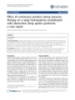
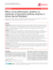
- Zhang et al. Journal of Experimental & Clinical Cancer Research 2011, 30:98 Page 4 of 8 http://www.jeccr.com/content/30/1/98 Figure 1 Effects of siRNA on DNMT1 mRNA and protein expression. (A): mRNA expression levels of DNMT1 in Hela and Siha cells were examined by qPCR. Compared with control group, Hela and Siha cells transfected with DNMT1-siRNA displayed lower level of mRNA expression (**P < 0.01). (B): DNMT1 protein levels in Hela and Siha cells were determined by western blot. The protein level of DNMT1 decreased significantly compared with control group and blank control. (1: transfection group (DNMT1-siRNA); 2: control group (negative siRNA); 3: blank group (Lipo2000), n = 3). 72 and 96 h after transfection, respectively (P < 0.05) com- Effects of DNMT1 silencing on cell cycle and apoptosis The G0/G1 ratio (74.72 ± 3.17%) of Hela cells in trans- pared with control group at each time point. fection group was higher than that in control group (65.88 ± 3.23%) ( P < 0.01), and cells at S phase were Effects of DNMT1 silencing on gene demethylation and fewer compared with control group. Meanwhile, The mRNA expression level in Hela cell G0/G1 ratio (76.43 ± 2.20%) of Siha cells in transfection Methylation status and mRNA expression level of seven group displayed significantly higher compared with con- repressive genes in Hela cells were performed with trol group (66.4 ± 1.99%) ( P < 0.01), while cells at S MeDIP-qPCR assay and Real-time PCR (Figure 4) com- phase were fewer than those in control group. No signif- pared with drug group(5-aza-dC, methylase inhibitors), icant changes in G0/G1 ratio or cells at S phase were control group and blank group. Specifically, PAX1, detected between the control group and blank control SFRP4 and TSLC1 possessed higher levels of methyla- (Figure 2A). Furthermore, as shown in Figure 2B, the tion, while CHFR and FHIT were relatively lower. apoptosis of Hela cells in transfection group was signifi- Except for FHIT and PTEN, the rest five suppressor cantly higher than that in control group ( P < 0.01). genes CCNA1, CHFR, PAX1, SFRP4 and TSLC1 in Similar results were observed in Siha cells. transfection group displayed lower level of methylation status compared with control group ( P < 0.01), which decreased to 34.42%, 15.57%, 22.36%, 52.09% and Effects of DNMT1 silencing on cell growth and 35.53%, respectively. The effects of DNMT1-siRNA and proliferation Cell growth and proliferation of Hela and Siha cells were 5-aza-dC treatment were performed the identical phe- examined using MTT assay. As shown in Figure 3, viabil- nomenon. The relative mRNA levels of seven repressive genes were detected by Real-time PCR. It ’ s clear that ities of Hela cells in transfection group were 91.47%, 86.74%, 78.92% and 48.98% at 24, 48, 72 and 96 h, respec- the expression of PTEN was higher than other genes. tively (P < 0.05) compared with control group at each time Except for FHIT and PTEN, the expression levels of point. We observed the similar results in Siha cells with CCNA1, CHFR, PAX1, SFRP4 and TSLC1 in transfec- viabilities of 90.45%, 84.16%, 71.09% and 60.47% at 24, 48, tion group were higher than those in control group,
- Zhang et al. Journal of Experimental & Clinical Cancer Research 2011, 30:98 Page 5 of 8 http://www.jeccr.com/content/30/1/98 Figure 2 Effects of DNMT1 silencing on cell cycle and apoptosis. (A): Phases of cell cycle of Hela and Siha cells were analyzed by flow cytometry assay at 48 h after transfection (**P < 0.01). (B): Apoptosis of Hela and Siha cells was analyzed by flow cytometry assay at 48 h after transfection (**P < 0.01). (1: transfection group (DNMT1-siRNA); 2: control group (negative siRNA); 3: blank group (Lipo2000), n = 3). Figure 3 Viability of Hela and Siha cells at different time after transfection determined by MTT assay. Viabilities of Hela and Siha cells in transfection group were 91.47%, 86.74%, 78.92%, 48.98% and 90.45%, 84.16%, 71.09%, 60.47% at 24, 48, 72 and 96 h, respectively. (n = 3, *P < 0.05, **P < 0.01, compared with control group).
- Zhang et al. Journal of Experimental & Clinical Cancer Research 2011, 30:98 Page 6 of 8 http://www.jeccr.com/content/30/1/98 Figure 4 Effects of DNMT1 silencing on gene methylation and mRNA expression of seven tumor suppressor genes in Hela cells assayed by MeDIP combined with Real-Time PCR. Except for FHIT and PTEN, the rest five suppressor genes CCNA1, CHFR, PAX1, SFRP4 and TSLC1 in transfected group displayed lower level of methylation with increased mRNA expression when compared with control group. (n = 3, **P < 0.01). PTEN, SFRP4 and TSLC1 in transfection group were with relative mRNA levels increased 6.13, 10.39, 4.98, higher than those in control group, with relative mRNA 4.87 and 3.51 folds, respectively. levels increased 7.22, 2.88, 2.32, 7.04 and 3.47 folds, respectively. Effects of DNMT1 silencing on gene demethylation and mRNA expression level in Siha cell Discussion Figure 5 showed the methylation status and mRNA levels in Siha cells were similar to those in Hell cells. PAX1, DNMT1 silencing in cervical cancer cells could induce SFRP4 and TSLC1 possessed higher level of methylation re-expression of most tumor suppressor genes by status, while PTEN and FHIT were relatively lower. demethylating its promoter region, and co-silencing of Except for FHIT and CHFR, the rest five repressor genes DNMT1 and DNMT3b might perform a greater inhibi- CCNA1, PAX1, PTEN, SFRP4 and TSLC1 in transfection tory effect on tumorigenesis [3]. Sowinska [4] demon- group displayed lower level of methylation compared strated that combined DNMT1 and DNMT3b siRNAs with control group (P
- Zhang et al. Journal of Experimental & Clinical Cancer Research 2011, 30:98 Page 7 of 8 http://www.jeccr.com/content/30/1/98 Hela cells and FHIT and CHFR in Siha cells in our [5] reported that DNMT3b deletion in a colorectal can- study, even though both of these two genes might cer cell line reduced global DNA methylation by less achieve high mRNA expression through low methyla- than 3%, but co-silencing of both DNMT1 and DNMT3b tion. It was previously reported that there was no PTEN nearly eliminated methyltransferase activity, and reduced mutation in 63 cases of squamous cervical carcinomas, genomic DNA methylation by greater than 95%. Thus, but 58% of the cases showed high methylation of PTEN DNMT1 and DNMT3b play the significant role in pro- promoter [11,12]. Wu et al [13] reported that FHIT was moter methylation of tumor suppressor genes and highly methylated in Hela, C33A and Siha cervical can- tumorigenesis in its early status. Currently, functions and cer cells, and that aberrant methylation of the FHIT mechanisms of DNMTs in cervical cancer cells remained gene might be a key mechanism for cervical tumorigen- unclear, and whether DNMT1 and DNMT3b act syner- esis, which could be reactivated and whose tumor sup- gistically or through other ways exploration efforts were pressing function could be restored by treatment of still required study. demethylating agent. Banno et al [14] reported that cer- In human bladder cancer cells, selective depletion of vical smears showed aberrant methylation of CHFR in DNMT1 with siRNA induced demethylation and reactiva- 12.3% of adenocarcinoma specimens, while aberrant tion of the silenced tumor-suppressor gene CDKN2A [6]. DNA methylation was not detected in normal cervical RNAi-mediated knockdown of DNMT1 resulted in signifi- cells. These researches demonstrated us that FHIT and cant reduction of promoter methylation and re-expression PTEN in Hela cells and FHIT and CHFR in Siha cells of RASSF1A, p16, and HPP1 in HCC1954 breast cancer might have the other regulation pathways for carcino- cells [7]. In ovarian cancer cell line CP70, DNMT1 siRNA genesis or transcription control, and which needs more treatment led to a partial removal of DNA methylation tests of cervical cancer cells and clinical specimens. from three inactive promoter CpG islands, TWIST, Apart from DNMT1 silencing, we treated Hela and RASSF1A, and HIN-1, and restored the expression of Siha cells with 5-aza-dC, which revealed the similar these genes [8]. Thus, RNAi-mediated DNMT1 depletion results with transfection group. Five repressor genes in different tumor cells could induce demethylation of var- were demethylated to various degrees and the mRNA ious tumor suppressor genes and enhance re-expression. expressions were also increased. These results are in However, contradictory results were reported even in the accordance with the findings of other reports [15-19], same cell line. Ting et al [9] found that hypermethylation which could be important in the development of new of CDKN2A, SFPR1, GATA4 and GATA5 were still main- and effective strategy in cervical treatment. tained in HCT116 colorectal cancer cells after transiently or stably depleted of DNMT1, and suggested that Conclusions DNMT1 might not play the dominant effect which caused hypermethylation of CpG islands in tumor suppressor In conclusion, our study demonstrates that DNMT1 genes. Knockout of DNMT1 in HCT116 cells by homolo- silencing could suppress proliferation and induce apop- gous recombination only reduced global DNA methylation tosis of Hela and Siha cells. DNMT1-siRNA induces by 20% and p16 maintained completely methylated status. demethylation of five tumor suppressor genes, including Besides, methylations of HMLH1, p16 and CDH1 in gas- CCNA1, CHFR, PAX1, SFRP4 and TSLC1 in Hela cells tric-cancer tissue samples at different progress periods do and CCNA1, PTEN, PAX1, SFRP4 and TSLC1 in Siha not correlate with the expression of DNMT1 directly [10]. cells, and enhances their mRNA expression. In a word, Therefore, whether over-expression of DNMT1 accounts DNMT1 represents an important potential diagnostic for the only or key causes of hypermethylation of tumor and therapeutic target for cervical cancer. suppressor genes remains to be confirmed. Currently, correlation between methlylation and mRNA Acknowledgements expression still remains unclear. In our study, methylation This study was supported by the Shenzhen major research projects of status of five suppressor genes (such as PAX1) in transfec- healthy department. tion group was significantly lower than that in control Author details group or blank control, and the mRNA expression levels 1 The Second Medical College, Jinan University, Shenzhen Clinical Medical Research Center, Shenzhen People’s Hospital, 518020, Shenzhen, PR China. were higher as compared to the two types of control, sug- 2 The Pharmacy College, Jinan University, 510632, Guangzhou, PR China. gesting that lower level of methylation facilitates mRNA expression. This trend was confirmed when CCNA1, Authors’ contributions SFRP4, TSLC1 and CHFR in Hela cells and CCNA1, YZ carried out the molecular genetic studies and wrote the manuscript, FQC and RC analyzed the dates and informations. YHS gave assistance with PTEN, SFRP4 and TSLC1 in Siha cells were analyzed. technical performance, SYZ contributed to the writing of the manuscript, Surprisingly, transfection did not affect the methyla- TYL designed the study and revised the manuscript. All authors read and tion status and mRNA expression of FHIT and PTEN in approved the final manuscript.
- Zhang et al. Journal of Experimental & Clinical Cancer Research 2011, 30:98 Page 8 of 8 http://www.jeccr.com/content/30/1/98 19. Steenbergen RD, Kramer D, Braakhuis BJ, Stern PL, Verheijen RH, Meijer CJ, Competing interests Snijders PJ: TSLC1 gene silencing in cervical cancer cell lines and cervical The authors declare that they have no competing interests. neoplasia. Journal of the National Cancer Institute 2004, 96(4):294-305. Received: 17 July 2011 Accepted: 17 October 2011 doi:10.1186/1756-9966-30-98 Published: 17 October 2011 Cite this article as: Zhang et al.: Effects of DNMT1 silencing on malignant phenotype and methylated gene expression in cervical References cancer cells. Journal of Experimental & Clinical Cancer Research 2011 30:98. 1. Ongenaert M, Wisman GB, Volders HH, Koning AJ, Zee AG, van Criekinge W, Schuuring E: Discovery of DNA methylation markers in cervical cancer using relaxation ranking. BMC Med Genomics 2008, 1:57. 2. Szyf M: The role of DNA methyltransferase 1 in growth control. Front Biosci 2001, 6:D599-609. 3. Peng DF, Kanai Y, Sawada M, Ushijima S, Hiraoka N, Kitazawa S, Hirohashi S: DNA methylation of multiple tumor-related genes in association with overexpression of DNA methyltransferase 1 (DNMT1) during multistage carcinogenesis of the pancreas. Carcinogenesis 2006, 27(6):1160-1168. 4. Sowinska A, Jagodzinski PP: RNA interference-mediated knockdown of DNMT1 and DNMT3B induces CXCL12 expression in MCF-7 breast cancer and AsPC1 pancreatic carcinoma cell lines. Cancer letters 2007, 255(1):153-159. 5. Rhee I, Bachman KE, Park BH, Jair KW, Yen RW, Schuebel KE, Cui H, Feinberg AP, Lengauer C, Kinzler KW, et al: DNMT1 and DNMT3b cooperate to silence genes in human cancer cells. Nature 2002, 416(6880):552-556. 6. Robert SM, Beaulieu Normand, Gauthier France: DNMT1 is required to maintain CpG methylation and aberrant gene silencing in human cancer cells. Nature genetics 2002, 33(9):61-65. 7. Suzuki M, Sunaga N, Shames DS, Toyooka S, Gazdar AF, Minna JD: RNA interference-mediated knockdown of DNA methyltransferase 1 leads to promoter demethylation and gene re-expression in human lung and breast cancer cells. Cancer research 2004, 64(9):3137-3143. 8. Leu YW, Rahmatpanah F, Shi H, Wei SH, Liu JC, Yan PS, Huang TH: Double RNA interference of DNMT3b and DNMT1 enhances DNA demethylation and gene reactivation. Cancer research 2003, 63(19):6110-6115. 9. Ting AH, Jair KW, Suzuki H, Yen RW, Baylin SB, Schuebel KE: CpG island hypermethylation is maintained in human colorectal cancer cells after RNAi-mediated depletion of DNMT1. Nature genetics 2004, 36(6):582-584. 10. Ye C, Shrubsole MJ, Cai Q, Ness R, Grady WM, Smalley W, Cai H, Washington K, Zheng W: Promoter methylation status of the MGMT, hMLH1, and CDKN2A/p16 genes in non-neoplastic mucosa of patients with and without colorectal adenomas. Oncology reports 2006, 16(2):429-435. 11. Hsieh SM, Maguire DJ, Lintell NA, McCabe M, Griffiths LR: PTEN and NDUFB8 aberrations in cervical cancer tissue. Advances in experimental medicine and biology 2007, 599:31-36. 12. Qi M, Anderson AE, Chen DZ, Sun S, Auborn KJ: Indole-3-carbinol prevents PTEN loss in cervical cancer in vivo. In Molecular medicine. Volume 11. Cambridge, Mass; 2005:(1-12):59-63. 13. Wu Y, Meng L, Wang H, Xu Q, Wang S, Wu S, Xi L, Zhao Y, Zhou J, Xu G, et al: Regulation of DNA methylation on the expression of the FHIT gene contributes to cervical carcinoma cell tumorigenesis. Oncology reports 2006, 16(3):625-629. 14. Banno K, Yanokura M, Kawaguchi M, Kuwabara Y, Akiyoshi J, Kobayashi Y, Iwata T, Hirasawa A, Fujii T, Susumu N, et al: Epigenetic inactivation of the CHFR gene in cervical cancer contributes to sensitivity to taxanes. International journal of oncology 2007, 31(4):713-720. 15. Cheung HW, Ching YP, Nicholls JM, Ling MT, Wong YC, Hui N, Cheung A, Tsao SW, Wang Q, Yeun PW, et al: Epigenetic inactivation of CHFR in Submit your next manuscript to BioMed Central nasopharyngeal carcinoma through promoter methylation. Molecular and take full advantage of: carcinogenesis 2005, 43(4):237-245. 16. Chung MT, Sytwu HK, Yan MD, Shih YL, Chang CC, Yu MH, Chu TY, Lai HC, Lin YW: Promoter methylation of SFRPs gene family in cervical cancer. • Convenient online submission Gynecologic oncology 2009, 112(2):301-306. • Thorough peer review 17. Kitkumthorn N, Yanatatsanajit P, Kiatpongsan S, Phokaew C, Triratanachat S, Trivijitsilp P, Termrungruanglert W, Tresukosol D, Niruthisard S, • No space constraints or color figure charges Mutirangura A: Cyclin A1 promoter hypermethylation in human • Immediate publication on acceptance papillomavirus-associated cervical cancer. BMC cancer 2006, 6:55. • Inclusion in PubMed, CAS, Scopus and Google Scholar 18. Lai HC, Lin YW, Huang TH, Yan P, Huang RL, Wang HC, Liu J, Chan MW, Chu TY, Sun CA, et al: Identification of novel DNA methylation markers in • Research which is freely available for redistribution cervical cancer. International journal of cancer 2008, 123(1):161-167. Submit your manuscript at www.biomedcentral.com/submit
CÓ THỂ BẠN MUỐN DOWNLOAD

-

Báo cáo khoa học: Nghiên cứu ảnh h-ởng của chế phẩm hữu cơ vi sinh MT đến
 6 p |
6 p |  313
|
313
|  59
59
-

Báo cáo khoa học: Nghiên cứu một số yếu tố ảnh hưởng đến kết quả ấp
7 p | 223
| 29
-
báo cáo khoa học: " Effects of ulinastatin and docataxel on breast tumor growth and expression of IL-6, IL-8, and TNF-a"
7 p | 86
| 5
-

báo cáo khoa học: " Effects of metastasis-associated in colon cancer 1 inhibition by small hairpin RNA on ovarian carcinoma OVCAR-3 cells"
12 p | 47
| 5
-
Báo cáo y học: "Effect of bladder volume on measured intravesical pressure:"
6 p | 121
| 4
-
báo cáo khoa học: " Effects of RNA interference-mediated gene silencing of JMJD2A on human breast cancer cell line MDA-MB-231 in vitro"
9 p | 76
| 4
-
báo cáo khoa học: "Effects of plasma concentrations of 5-fluorouracil on long-term survival after treatment with a definitive 5-fluorouracil/cisplatin-based chemoradiotherapy in Japanese patients with esophageal squamous cell carcinoma"
7 p | 66
| 3
-
báo cáo khoa học: " Effect of Chemokine Receptors CCR7 on Disseminated Behavior of Human T cell Lymphoma: clinical and experimental study"
9 p | 52
| 3
-

Báo cáo khoa học: " Mechanisms of the action of povidone-iodine against human and avian influenza A viruses: its effects on hemagglutination and sialidase activities"
10 p | 56
| 3
-

Báo cáo khoa học: "Effective suppression of Dengue fever virus in mosquito cell cultures using retroviral transduction of hammerhead ribozymes targeting the viral genome"
17 p | 86
| 3
-

Báo cáo y học: "Effectiveness of counseling for anxiety and depression in mothers of children ages 0-30 months by community workers in Karachi, Pakistan: a quasi experimental study"
9 p | 68
| 3
-

báo cáo khoa học: " Implementing evidence-based interventions in health care: application of the replicating effective programs framework"
10 p | 88
| 3
-

báo cáo khoa học: " An observational study of the effectiveness of practice guideline implementation strategies examined according to physicians' cognitive styles"
9 p | 131
| 3
-
Báo cáo y học: " Effect of continuous positive airway pressure therapy on a large hemangioma complicated with obstructive sleep apnea syndrome: a case report"
4 p | 66
| 3
-
báo cáo khoa học: "Effects of ulinastatin and docetaxel on breast cancer invasion and expression of uPA, uPAR and ERK"
7 p | 79
| 3
-

báo cáo khoa học: " Effect of mesoporous silica under Neisseria meningitidis transformation process: environmental effects under meningococci transformation"
8 p | 59
| 3
-
Báo cáo y học: "Effects of pro-inflammatory cytokines on expression of kynurenine pathway enzymes in human dermal fibroblasts"
7 p | 76
| 3


Chịu trách nhiệm nội dung:
Nguyễn Công Hà - Giám đốc Công ty TNHH TÀI LIỆU TRỰC TUYẾN VI NA
LIÊN HỆ
Địa chỉ: P402, 54A Nơ Trang Long, Phường 14, Q.Bình Thạnh, TP.HCM
Hotline: 093 303 0098
Email: support@tailieu.vn
Giấy phép Mạng Xã Hội số: 670/GP-BTTTT cấp ngày 30/11/2015 Copyright © 2022-2032 TaiLieu.VN. All rights reserved.

