


báo cáo khoa học: "Evaluation of HER2 and p53 expression in predicting response to docetaxel-based first-line chemotherapy in advanced breast cancer"
lượt xem 5
download
 Download
Vui lòng tải xuống để xem tài liệu đầy đủ
Download
Vui lòng tải xuống để xem tài liệu đầy đủ
Tuyển tập báo cáo các nghiên cứu khoa học quốc tế ngành y học dành cho các bạn tham khảo đề tài: Evaluation of HER2 and p53 expression in predicting response to docetaxel-based first-line chemotherapy in advanced breast cancer
Bình luận(0) Đăng nhập để gửi bình luận!
Nội dung Text: báo cáo khoa học: "Evaluation of HER2 and p53 expression in predicting response to docetaxel-based first-line chemotherapy in advanced breast cancer"
- Evaluation of HER2 and p53 expression in predicting response to docetaxel-based first-line chemotherapy in advanced breast cancer Camerini et al. Camerini et al. Journal of Experimental & Clinical Cancer Research 2011, 30:38 http://www.jeccr.com/content/30/1/38 (11 April 2011)
- Camerini et al. Journal of Experimental & Clinical Cancer Research 2011, 30:38 http://www.jeccr.com/content/30/1/38 RESEARCH Open Access Evaluation of HER2 and p53 expression in predicting response to docetaxel-based first-line chemotherapy in advanced breast cancer Andrea Camerini1*, Sara Donati1, Paolo Viacava2, Olimpia Siclari1, Cheti Puccetti1, Gianna Tartarelli1, Chiara Valsuani1, Filomena De Luca2, Leonardo Martini2, Andrea Cavazzana3 and Domenico Amoroso1 Abstract Background: The human epidermal growth factor receptor 2 (HER2) and p53 pathways may be involved in chemotherapy sensitivity and/or resistance. We explore the value of HER2 and p53 status to foretell docetaxel sensitivity in advanced breast cancer. Methods: HER2 and p53 expression was analysed in 36 (median age 55 yrs; range 37-87) metastatic breast cancer patients receiving docetaxel-based first-line chemotherapy. HER2 was determined by immunohistochemistry (IHC) and fluorescence in situ hybridization (FISH), p53 was tested by IHC. We correlate the expression of study parameters with pathologic parameters, RECIST response and survival. The standard cut-off value of 2 was used to determine HER2 overexpression while p53 mean expression level was used to divide low/high expressors tumors. Results: Median time to progression and overall survival were 9 (range 2 - 54) and 20 (range 3 - 101) months. Overall response rate was 41.6%. Nine cases showed HER2 overexpression. HER2 was more frequently overexpressed in less differentiated (p = 0.05) and higher stage (p = 0.003) disease. Mean FISH-HER2 values were significantly higher in responder than in non-responder pts (8.53 ± 10.21 vs 2.50 ± 4.12, p = 0.027). Moreover, HER2 overexpression correlates with treatment response at cross-tabulation analysis (p = 0.046). p53 expression was only associated with higher stage disease (p = 0.02) but lack of any significant association with HER status or docetaxel response. No significant relation with survival was observed for any parameter. Conclusion: Our data seem to indicate that FISH-determined HER2 status but not p53 is associated with docetaxel sensitivity in metastatic breast cancer. genome” [5] and the “cellular gatekeeper” [6], the p53 Background protein acts as cell modulator by driving lots of stress- Breast cancer (BC) is the leading cause of cancer and inducing signals to different antiproliferative cellular the second leading cause of cancer death in women in responses [7]. p53 can be activated in response to DNA the USA [1] and its incidence is increasing in many damage (such as cytotoxic agents), oncogene activation countries, including Italy [2] thus representing a major or hypoxia resulting to cellular outputs such as apopto- health problem. To date, the role of chemotherapy in sis, cell-cycle arrest, senescence, or modulation of autop- BC treatment is certain and taxanes are widely used in hagy [8-10]. Although about 50% of BCs harbours TP53 both early and advanced setting [3,4] but we have no gene mutations [11,12], the biological role and clinical validated sensitivity and/or resistance predictive factor importance of p53 alterations in BC are still unclear. and, hence, the search for a taxane-specific predictive marker is an hot topic. Colled as the “guardian of the This maybe related to the very complicated and exten- sive p53 network and to technical problems associated with surrogate markers to identify TP53 gene defects, as * Correspondence: andreacamerini@katamail.com most detection tests lack sensitivity and specificity. 1 Oncology Department, Medical Oncology Division, AUSL12 di Viareggio and Despite its limits, immunohistochemical p53 detection Istituto Toscano Tumori - Versilia Hospital, Lido di Camaiore, Italy Full list of author information is available at the end of the article © 2011 Camerini et al; licensee BioMed Central Ltd. This is an Open Access article distributed under the terms of the Creative Commons Attribution License (http://creativecommons.org/licenses/by/2.0), which permits unrestricted use, distribution, and reproduction in any medium, provided the original work is properly cited.
- Camerini et al. Journal of Experimental & Clinical Cancer Research 2011, 30:38 Page 2 of 8 http://www.jeccr.com/content/30/1/38 demonstrated in numerous studies to be a prognostic Table 1 Study population characteristics (n = 36) factor in BC [11-17] and that it may determine the sen- Median [range] age (yr) 55 [37-87] sitivity to specific therapeutic agents [18-22]. Some evi- Histotype# dences may indicate that abnormal p53 expression Invasive ductal carcinoma 28 (77.7%) could be associated with taxane sensitivity but its speci- Invasive lobular carcinoma 5 (13.8%) fic predictive role is unclear [22-24]. Mixed (ductal and lobular) 2 (5.5%) Another leading cell growth regulator in BC is the Undifferentiated 1 (3.0%) human epidermal growth factor receptor (HER) 2 Grading° (HER2; erbB2/neu). The HER2 oncogene encodes one of G2 21 (58.3%) four trans-membrane receptors within the erbB family. G3 15 (41.7%) Its over-expression, which occurs in approximately 25% ER status of all breast cancer tumors, is associated with a shor- Negative 14 (38.8%) tened disease-free interval and poor survival [25]. HER2 Positive 22 (61.2%) blockage in preclinical models of human BC and in pri- PgR status mary breast tumors from women treated with HER2- Negative 13 (36.1%) targeted therapies leads to the inhibition of survival Positive 23 (63.9%) pathways, which in turn induces tumor cell apoptosis HER2 status* [26]. The clinical benefit of HER2 inhibition by its speci- Negative 27 (75.0%) fic monoclonal antibody trastuzumab is meaningful in Positive 9 (25.0%) both early and advanced disease [27,28]. HER2 status Adjuvant chemotherapy^ may also influence chemotherapy sensitivity as proposed FEC 18 (52.9%) by Gennari et al [29] that focused on the adjuvant set- EC 11 (32.4%) ting showing that the added benefits of adjuvant che- CMF 5 (14.7%) motherapy with anthracyclines seems to be reserved to Mean ± SD time to first relapse (months) 15.8 ± 6.5 breast cancer harboring HER2 overexpression or Metastatis sites amplification. Bone 21 (58.3%) On this grounds, we analysed the relationship between Liver 21 (58.3%) HER2 and p53 expression and response to first-line doc- Lung 16 (44.4%) etaxel based chemotherapy in advanced BC finding that Lymphnodes 14 (38.8%) FISH-determined HER2 status but not p53 could predict Local 3 (8.3%) docetaxel sensitivity. Chemotherapy” TXT75 14 (38.8%) Methods TXT25 8 (22.2%) Patient characteristics and tissue samples TXT75+C 5 (13.8%) Tumor samples were obtained from breast cancer TXT75+T 9 (25.2%) patients who underwent surgery at Versilia Hospital in Treatment best response Lido di Camaiore (Italy) from 2000 to 2004. A total of Complete response 1 (2.7%) 36 breast cancer patients (median age 55 yrs; range 37- Partial response 14 (38.8%) 87) receiving between 2001 and 2005 a docetaxel-based Stable disease 12 (33.3%) first-line chemotherapeutic regimen for metastatic dis- Disease progression 9 (25.2%) ease were retrospectively selected for the study. Study Time to disease progression (months) population characteristics are shown in table 1. Mean Median [range] 9 [2-54] time from initial diagnosis to first relapse was 15.8 ± Overall survival (months) 6.5 months. Location of metastatic deposits includes Median [range] 20 [3-101] bone (21/36), liver (21/36), lung (16/36), lymphnodes # According to WHO hystological typing of breast tumor (Ref. 32). °According (14/36) and local recurrence (3/36) with 27 out of 36 to Elston and Ellis classification (Ref. 31). *Pre-study determination. “See text for regimen details. ^on 34 pts. patients presenting with multiple disease sites; remain- ing 9 patients with single-site metastasis presented with measurable non-bone disease. Patients receiving pre- received an association of 5-fluorouracil (5-FU), epiru- operative chemotherapy, having a family history of bucin and cyclophosphamides (FEC) for 6 cycles, 11 breast cancer or receiving docetaxel as part of adjuvant patients received an association of epirubucin and cyc- treatment were excluded as well as those for whom fol- lophosphamides (EC) for 4 cycles, and remaining low-up data were missing. Adjuvant treatment was per- 5 patients received an association of cyclophosphamides, formed in all patients but two as follow: 18 patients methotrexate and 5-FU (CMF) for 6 cycles.
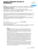
- Camerini et al. Journal of Experimental & Clinical Cancer Research 2011, 30:38 Page 3 of 8 http://www.jeccr.com/content/30/1/38 After endogenous peroxidase blocking sections were A ll patients received docetaxel-based first-line che- incubated for 45 min at 37°C with a 1:50 dilution of pri- motherapy for metastatic disease. In particular, 14 out mary mouse anti-human p53 monoclonal antibody of 36 patients received six cycles docetaxel (75 mg/m2) (clone: DO-7, isotype IgG2b) (Dako), then immunos- every 3 weeks (TXT75), 8 patients received docetaxel (25 mg/m 2 ) on a weekly basis (TXT25), 5 patients tained with secondary antibodies and finally counter- received a combination of docetaxel (75 mg/m2) on day stained with hematoxylin. Sections of known positive 1 plus capecitabine (1000 mg/m 2 bid day 1-14) every mammary carcinoma were used as positive controls. Negative controls were obtained by omitting the primary 3 weeks (TXT75+C) and the remaining 9 patients with antibodies. For p53 only a clear nuclear staining in the HER2-positive disease received a combination of doce- taxel (75 mg/m 2 ) and trastuzumab (8 mg/kg loading absence of cytoplasmic background coloration was con- sidered positive. A minimum of 1.000 cells were dose then 6 mg/kg) both on day 1 every 3 weeks counted for each tumor and immunoreactivity was (TXT75+T) (Table 1). Every treatment was planned for expressed as a percentage of positive cells on the total up to 6-9 months. Causes for early treatment stop were number of tumor cells. A value of 11% of positive cells, unacceptable toxicity, disease progression or patient corresponding to the mean value of p53 expressing refusal. Trastuzumab was administered alone after doce- tumor cells, was used as cut-off to distinguish high and taxel discontinuance as maintenance therapy until dis- low expressing tumors. ease progression in 6 responder patients. Tumor HER2 IHC evaluation was realized by the streptavidin- assessment was performed every 3 months by CT-scan biotin-peroxidase complex technique (StreptABC, and/or chest X-ray coupled with abdomen ultrasound DAKO) as standard for the time of analysis. Tissue sec- depending on those used at baseline. Time to progres- tions were deparaffinized and underwent antigenic sion (TTP) was calculated from the date of treatment retrieval and endogenous peroxidase blocking. Sections start to the date of first-documented progression. Over- were first incubated with polyclonal primary antibodies all survival (OS) was defined as the time interval against c-erbB-2 (A0485, DAKO) with a 1:500 dilution, between the start of treatment and death or last follow- then incubated in secondary biotinylated antibody and up contact. Treatment response was assessed according finally counterstained with Hematoxylin. Immunohisto- to RECIST criteria and we consider as responder a chemical analyses of c-erbB-2 expression describe the patient achieving a complete (CR) or partial (PR) intensity and staining pattern of tumor cells. The FDA- response to treatment. Patients achieving disease stabili- recognized test, the Herceptest ™ (DAKO), describes zation (SD) or disease progression (PD) were considered four categories: no staining, or weak staining in fewer as not-responders. Anyway, we planned a secondary than 10% of the tumor cells (0); weak staining in part of analysis considering as responders even patients achiev- the membrane in more than 10% of the tumor cells ing disease stabilization as best result. Median TTP was (1+); complete staining of the membrane with weak or 9 (range 2 - 54) months and overall response rate moderate intensity in more than 10% of the neoplastic (ORR) was 41.6% (14 out of 36) with 11 and 8 pts cells (2+); and strong staining in more than 10% (3+). experiencing disease stabilization and progression Cases with 0 or 1+ score were regarded as negative; the respectively. Median OS was 20 (range 3 - 101) months. ones with 3+ score were regarded as positive while 2+ Being a retrospective analysis patients were not asked to cases underwent FISH and categorized accordingly. All sign any informed consent; anyway samples were coded immunostained specimens were evaluated by two obser- and the names of the patients were not revealed. All vers independently (PV and AC) without knowledge of available clinico-pathological data were collected and clinical characteristics and/or follow-up information and stored in an appropriate database. Age, tumor grade and the discrepant cases were jointly re-evaluated and agree- stage [30,31], size, histotype,(32) estrogen receptor (ER) ment was met. and progesterone receptor (PgR) status were considered. Dual-color Fluorescence in situ Hybridization Immunoistochemistry P53 expression was evaluated by immunohistochemistry HER2 amplification was analyzed on microdissected (IHC) while HER2 expression was evaluated both by tumor samples using FISH HER2 PharmDx (Dako, IHC and fluorescence in situ hybridization (FISH - see K5331), which contains both fluorescently-labeled next paragraph). All IHC analyses were performed on HER2/neu gene and chromosome 17 centromere probes. routinely processed, formalin-fixed and paraffin- Microdissection was performed by an expert pathologist embedded tissue samples obtained from primary tumor. different from ones that performed IHC evaluation. For p53 IHC analysis, representative tumor sections In brief, sections were deparaffinized, heat-pretreated in (3 μm) were deparaffinized, rehydrated and immunos- citrate buffer at 80°C for near 1 hour, digested with tained using antigen retrieval by microwave technique. pepsin at room temperature for few minutes and
- Camerini et al. Journal of Experimental & Clinical Cancer Research 2011, 30:38 Page 4 of 8 http://www.jeccr.com/content/30/1/38 dehydrated in graded ethanol. After the HER2/CEN17 probe mix was applied to the dry slides. The slides were then incubated in hybridizer (Hybridizer Instru- ment for in situ hybridization, DAKO, S2450) for denaturation at 82°C for 5 minutes and hybridization at 45°C for about 18 hours. The slides were re- dehydrated in graded ethanol. FISH analyses were per- formed according to the HER2 FISH PharmDx (Dako) criteria. In each case, 100 non-overlapped, intact inter- phase tumor nuclei identified by DAPI staining were evaluated, and gene (red signal) and CEN17 (green sig- nal) copy numbers in each nucleus were assessed. The cases were considered to be amplified when the aver- age copy number ratio, HER2/CEN17, was ≥ 2.0 in all nuclei evaluated or when the HER2 signals formed a tight gene cluster. Among the cases in which the gene Figure 1 Immunohistochemical positive staining of p53 in a representative case of high-grade (G3) ductal carcinoma. was not amplified, samples showing more than four Immunostaining shows a clear and wide nuclear staining in an high copies of the HER2 gene and more than four CEN17 grade (G3) invasive ductal carcinoma. Original magnifications: ×100 in more than 10% of the tumor cells were considered (inset ×250). to be polysomic for chromosome 17. Conversely, HER2 positive breast tumors appear to be, Statistical analysis Correlation between p53, HER2 and other molecular as expected, less differentiated and of higher stage more frequently than negative ones (Table 3). In accordance and clinical parameters were assessed by contingency with literature data, 6 out of 9 (66.6%) HER2 positive table methods and tested for significance using the Pear- son’s chi-square test. Mean values were compared using while only 9 out 27 (33.3%) HER2 negative patients respectively responded to docetaxel treatment and this the student-T test. Survival curves were calculated using difference was significant (Table 3). Confirmatory results the Kaplan-Meier method and tested for significance were obtained by student-T test on mean FISH values using the log-rank test. Univariate and multivariate rela- between responders vs not-responders patients. In fact, tive risks were calculated using Cox proportional responder group showed significantly higher mean FISH hazards regression. Statistical analyses were performed values than not-responder (8.53 ± 10.21 vs 2.50 ± 4.12, using NCSS software. All tests were two-tailed, and p < p = 0.027). All HER2-positive patients received trastuzu- 0.05 was considered to be significant. mab in combination with docetaxel while HER2-negative Results Expression levels of p53 ranged from 0% to 70% of immu- nostained nuclei with a mean expression value of 11% (median = 5%) (Figure 1 and 2). Using this mean value as cut-off to distinguish high and low expressing tumors, staining was considered high in 11 (30.5%) out of 36 tumors in our series (similar results were obtained using as cut-off the median value). P53 expression levels were only related to disease stage with higher p53 levels in higher stage disease (p = 0.02) but lack of any significant association with HER2 status, other clinic-pathologic para- meters (age, ER and PgR status, Ki67 and tumor grading) or docetaxel response (Table 2). Even comparing mean p53 expression levels between responders vs not-respon- ders patients we did not find any significant difference (not shown) and mean TTP (8.6 ± 7.0 vs 9.2 ± 11.9 months; p = ns) and OS (21.6 ± 13.0 vs 19.8 ± 10.2 Figure 2 p53 immunohistochemical negative staining in a months; p = ns) did not differ between low and high p53 grade 2 ductal carcinoma. The wide majority of nuclei showed no staining with the exception of one clear positive nucleus (arrow) in groups. Morever, no significant relation with survival para- the upper left corner. Original magnifications: ×100 (inset ×250). meters was observed for p53 at Kaplan-Meier analysis.
- Camerini et al. Journal of Experimental & Clinical Cancer Research 2011, 30:38 Page 5 of 8 http://www.jeccr.com/content/30/1/38 Table 2 p53 expression in relation to main tumor Table 3 HER2 expression in relation to main tumor characteristics and treatment response cheracteristics and treatment response HER2 expression” p53 expression p value p value Total Low High Total Low High Age Age < 55 yrs 18 13 5 n.s. < 55 yrs 18 13 5 n.s. ≥55 yrs ≥55 yrs 18 12 6 18 14 4 ER expression ER expression Negative 14 8 6 n.s. Negative 14 10 4 n.s. Positive 22 17 5 Positive 22 17 5 PgR expression PgR expression Negative 13 9 4 n.s. Negative 13 9 4 n.s. Positive 23 16 7 Positive 23 18 5 Grading# Grading# G2 21 17 4 n.s. G2 21 18 3 0.05 G3 15 8 7 G3 15 8 7 Stage*° Stage*° I-IIA 17 15 2 0.02 I-IIA 17 16 1 0.003 IIB-III 16 7 9 IIB-III 16 8 8 HER2 Ki67 Negative” 27 21 6 n.s. Negative 22 18 4 n.s. Positive” 9 4 5 Positive 14 9 5 Ki67 Treatment response Negative 22 15 7 n.s. CR+PR 15 9 6 0.046 Positive 14 10 4 SD+PD 21 18 3 Treatment response “IHC 0, 1+ and 2+ FISH negative were regarded as negative while IHC 3+ or 2+ FISH positive were regarded as positive. #According to Elston and Ellis CR+PR 15 11 4 n.s. classification (see text for complete reference). *According to UICC-TNM SD+PD 21 14 7 classification of malignant tumours, sixth edition 2002. °At initial diagnosis time. n.s. = not significant; CR = complete response; PR = partial response; n.s. = not significant; CR = complete response; PR = partial response; SD = SD = stable disease; PD = disease progression. stable disease; PD = disease progression. #According to Elston and Ellis classification (Ref. 31). *According to UICC-TNM classification of malignant tumours, sixth edition 2002 (Ref. 30). °At initial diagnosis time. “IHC 0,1+ and survival analysis did not show a significant separation 2+ FISH negative were regarded as negative while IHC 3+ or 2+ FISH positive between HER2 positive and negative groups (Figure 3 were regarded as positive. for OS curves). The same results for both study mole- cules were obtained even incorporating in responders ones were treated with docetaxel with a known influence group patients achieving SD (not shown). Neither HER2 on and response rate and outcome. To shrink the possi- expression nor p53 status were independent predictors ble treatment-related bias we test the FISH value differ- of OS and TTS at Cox regression analysis. ence between docetaxel responders and not-responder in Lastly, we also observed at cross-tabulation analysis a HER2-negative subgroup (n = 27) so removing trastuzu- clear correlation between HER2 testing with IHC and mab treatment-related bias. Taking into account the FISH (p = 0.001). Mean ± SD FISH values in negative smaller sample size and the lower FISH values (< 2), we and positive groups were 1.51 ± 0.223 and 13.09 ± 9.98 found a non-statistically significant difference in mean respectively. FISH value with responders patients having higher values (1.64 ± 0.157 vs 1.38 ± 0.146; p = ns). We also performed Discussion the same analysis in FISH-positive group (11 pts all receiving docetaxel plus trastuzumab) and we observed Some preliminary comments about study limitations will also in this small subgroup a similar behaviour (16.86 ± facilitate the discussion of the results. First, presented 9.78 vs 9.85 ± 10.53; responders vs not-responders; p = data originate from a retrospective analysis that is natu- 0.18 ns). rally exposed to selection bias. Second, the relative small Mean TTP (positive vs negative: 7.9 ± 8.1 vs 9.8 ± 9.4 sample size could reduce the strength of statistical asso- months; p = 0.18 ns) and OS (positive vs negative: ciations and dramatically affects survival analyses. Third, 18.1 ± 11.7 vs 21.2 ± 12.1 months; p = 0.12 ns) showed all patients did not receive the same chemotherapy regi- a only modest trend towards significance with HER2 men both in term of schedule (weekly or every 3 weeks positive patients having worse prognosis. Kaplan-Meier administrations) and in term of associated drug (5 patient
- Camerini et al. Journal of Experimental & Clinical Cancer Research 2011, 30:38 Page 6 of 8 http://www.jeccr.com/content/30/1/38 100 100 p53 negative HER2 negative p53 positive HER2 positive 75 75 Percent alive 50 50 25 25 p = n.s. p = n.s. 0 0 30 60 90 0 30 60 90 Time (months) Time (months) Figure 3 Kaplan-Meier curves for overall survival according to p53 or HER2 status. Kaplan-Meier curves for overall survival showed no- significant separation between high vs low-espressors group for both p53 (left panel) and HER2 (right panel). Similar results were obtained for disease-free survival (not shown). Conversely, p53 mainly (but not exclusively) acts in received an association of docetaxel plus capecitabine). early phases of cell cycle inducing, after DNA damage, a Lastly, according to guidelines all HER2 positive patients G1 arrest by transactivation of p21Waf1/Cip1, a cyclin- (both patients that achieve a response and patients who dependent kinase inhibitor [37-39]. Therefore, the sub- did not) received trastuzumab while negative-ones were cellular localization of docetaxel molecular target and treated with docetaxel (alone or in combination). The dif- the timing of docetaxel action during cell cycle do not ference in treatment received and, notably, in the under- overlap with those of p53 and this could explain, at least lying cancer biology makes HER2 positive and negative in part, our negative results. Some opposite data were groups as different populations so affecting our data published some years ago about a possible predictive interpretation. role of TP53 mutation on paclitaxel sensitivity in breast Within that specific experimental context, IHC-assessed cancer [22,23]; Johnson et al [23] proposed a model in nuclear p53 status failed to show any significant associa- which the loss of p53 function reduced the G1 block tion with outcome and survival parameters. In fact, thus enhancing the efficacy of paclitaxel during mitosis. nuclear expression level of p53 did not differ between Our data do not support this hypothesis even account- responders and not-responders patients. Reasons for this ing for docetaxel over paclitaxel differences. phenomenon cannot be limited to the above mentioned Lastly, the correlation between p53 nuclear storage study limitations, probably, should be seek in the mechan- measured by IHC and p53 mutation detected by sequen- isms of action (MoA) of docetaxel and, to a lesser extent, cing has been estimated to be less than 75% in breast in technical limitations of p53 determination by IHC. carcinomas [40]. Indeed, not all mutations yield a stable Docetaxel, a semi-synthetic analogue of paclitaxel, is a protein, and some mutations lead to an abnormal pro- promoter of microtubule stabilization by direct binding tein not detected by IHC. On the other hand, wild-type leading to cell cycle arrest at G2/M and apoptosis [33-35]. The b-subunit of the tubulin heterodimer, the p53 may accumulate in some tumors as a result of the response to DNA damage, giving a positive IHC result key component of cellular microtubules, represent the not accounting for TP53 mutation [41]. molecular target of docetaxel [36]. This unique MoA On the other hand, we observed a clear predictive could offer a putative explanation for the lack of asso- value for HER2 status. Patients with HER2-positive ciation between p53 status and docetaxel sensitivity. In tumors were more likely to respond to docetaxel treat- fact, docetaxel is not a direct DNA-damaging drug and ment even taking into account the small sample size. docetaxel-induced cell cycle arrest occurs in a late phase This observation seems to be true independently of of cell cycle (G2/M transition).
- Camerini et al. Journal of Experimental & Clinical Cancer Research 2011, 30:38 Page 7 of 8 http://www.jeccr.com/content/30/1/38 patient category (HER2-positive or negative); in fact, in interpretation and paper writing; CP: study design and statistical analysis; GT: data collection and interpretation; CV: data interpretation and paper writing; both the whole population and in HER2 subgroups it FDL: data collection, immunohistochemistry performance and interpretation; seems that the higher is the FISH value the higher is LM: data collection, immunohistochemistry performance and interpretation; the probability to respond to docetaxel. In our opinion, AC: FISH performance and interpretation, data collection; DA: study design, data interpretation and paper writing. All authors read and approved the the most likely explanation of our data may resides in final manuscript. the higher proliferation rate of this subset of cancers [25]. Docetaxel, as near-all chemotherapeutic agents, Competing interests The authors declare that they have no competing interests. works better in tumors with an higher proliferation index because cancer growth-rate it’s “per se” the main Received: 30 November 2010 Accepted: 11 April 2011 determinant of cell sensitivity to non-target chemoter- Published: 11 April 2011 apy. Moreover, rapid growth cancers (as HER2 positive References breast cancer) have a greater percentage of cells in the 1. Jemal A, Siegel R, Ward E, Hao Y, Xu J, Murray T, Thun MJ: Cancer statistics, M phase of cell cycle and this could represent another 2008. CA Cancer J Clin 2008, 58:71-96. element to take into account. 2. Grande E, Inghelmann R, Francisci S, Verdecchia A, Micheli A, Baili P, Capocaccia R, De Angelis R: Regional estimates of breast cancer burden More specific molecular mechanisms, i.e. as for topoi- in Italy. Tumori 2007, 93:374-9. somerase II alpha, are unlikely. In fact, b-tubulin con- 3. Sledge GW, Neuberg D, Bernardo P, Ingle JN, Martino S, Rowinsky EK, sists of six isotypes, all of which have related aminoacid Wood WC: Phase III trial of doxorubicin, paclitaxel, and the combination of doxorubicin and paclitaxel as front-line chemotherapy sequences and are well conserved between species. Class for metastatic breast cancer: an Intergroup trial (E1193). J Clin Oncol I-btubulin is the most commonly expressed isotype in 2003, 21:588-592. 4. De Laurentiis M, Cancello G, D’Agostino G, Giuliano M, Giordano A, human beings, and the most common isotype in cancer Montagna E, Lauria R, Forestieri V, Esposito A, Silvestro L, Pennacchio R, cells [42]. The class-I isotype is encoded by the TUBB Criscitiello C, Montanino A, Limite G, Bianco AR, De Placido S: Taxane- gene located at 6p2513 far from HER2 gene located on based combinations as adjuvant chemotherapy of early breast cancer: a chromosome 17. Thus a co-amplification phenomenon meta-analysis of randomized trials. J Clin Oncol 2008, 26:44-53. 5. Lane DP: p53, guardian of the genome. Nature 1992, 358:15-16. is difficult to propose [42]. 6. Levine AJ: p53, the cellular gatekeeper for growth and division. Cell 1997, 88:323-331. Conclusions 7. Zilfou JT, Lowe SW: Tumor Suppressive Functions of p53. Cold Spring Harb Perspect Biol 2009, 1:a001883. FISH-determined HER2 status may predict docetaxel 8. Yee KS, Vousden KH: Complicating the complexity of p53. Carcinogenesis sensitivity in metastatic breast cancer and could be an 2005, 26:1317-1322. element to evaluate in the pre-treatment work-up. 9. Riley T, Sontag E, Chen P, Levine A: Transcriptional control of human p53- regulated genes. Nat Rev Mol Cell Biol 2008, 9:402-412. Obviously, a further prospective validation on a larger 10. Green DR, Kroemer G: Cytoplasmic functions of the tumour suppressor sample size is warranted before any possible clinical p53. Nature 2009, 458:1127-1130. application. Interestingly, HER2 is a well-known predic- 11. Petitjean A, Mathe E, Kato S, Ishioka C, Tavtigian SV, Hainaut P, Olivier M: Impact of mutant p53 functional properties on TP53 mutation patterns tor of trastuzumab efficacy and the association of trastu- and tumor phenotype: lessons from recent developments in the IARC zumab plus taxanes can be considered as a standard of TP53 database. Hum Mutat 2007, 28:622-629. care in the first-line setting, so the possibility to predict 12. Soussi T, Ishioka C, Claustres M, Béroud C: Locus-specific mutation databases: pitfalls and good practice based on the p53 experience. Nat treatment response by analysing one parameter (HER2) Rev Cancer 2006, 6:83-90. could be an attractive option. 13. Vousden KH, Prives C: p53 and prognosis: new insights and further Conversely, p53 status lack of any significant associa- complexity. Cell 2005, 120:7-10. 14. Lønning PE, Knappskog S, Staalesen V, Chrisanthar R, Lillehaug JR: Breast tion with docetaxel sensitivity in the same setting. Prob- cancer prognostication and prediction in the postgenomic era. Ann ably, TP53 gene mutational analysis could be more Oncol 2007, 18:1293-1306. informative that IHC, even if a simplistic association 15. Lacroix M, Toillon RA, Leclercq G: p53 and breast cancer, an update. Endocr Relat Cancer 2006, 13:293-325. between TP53 gene status and taxane treatment 16. Geisler S, Lønning PE, Aas T, Johnsen H, Fluge O, Haugen DF, Lillehaug JR, response seem to be unlikely given the wide and very Akslen LA, Børresen-Dale AL: Influence of TP53 gene alterations and c- complicated molecular pathway related to p53. erbB-2 expression on the response to treatment with doxorubicin in locally advanced breast cancer. Cancer Res 2001, 61:2505-2512. 17. Abdel-Fatah TM, Powe DG, Agboola J, Adamowicz-Brice M, Blamey RW, Lopez-Garcia MA, Green AR, Reis-Filho JS, Ellis IO: The biological, clinical Author details and prognostic implications of p53 transcriptional pathways in breast 1 Oncology Department, Medical Oncology Division, AUSL12 di Viareggio and cancers. J Pathol 2010, 220:419-434. Istituto Toscano Tumori - Versilia Hospital, Lido di Camaiore, Italy. 2Oncology 18. Sayeed A, Konduri SD, Liu W, Bansal S, Li F, Das GM: Estrogen receptor α Department, Pathology Division, AUSL12 di Viareggio and Istituto Toscano inhibits p53 mediated transcriptional repression: implications for the Tumori - Versilia Hospital, Lido di Camaiore, Italy. 3Pathology Division, AUSL 1 regulation of apoptosis. Cancer Res 2007, 67:7746-7755. Massa-Carrara and Istituto Toscano Tumori, Carrara, Italy. 19. Veerakumarasivam A, Scott HE, Chin SF, Warren A, Wallard MJ, Grimmer D, Ichimura K, Caldas C, Collins VP, Neal DE, Kelly JD: High resolution array- Authors’ contributions based comparative genomic hybridization of bladder cancers identifies AC: study design, statistical analysis, data interpretation and paper writing; mouse double minute 4 (MDM4) as an amplification target exclusive of SD: data collection and interpretation; PV: data collection, MDM2 and PT53. Clin Cancer Res 2008, 14:2527-2534. immunohistochemistry performance and interpretation; OS: data
- Camerini et al. Journal of Experimental & Clinical Cancer Research 2011, 30:38 Page 8 of 8 http://www.jeccr.com/content/30/1/38 20. Ding SL, Sheu LF, Yu JC, Yang TL, Chen BF, Leu FJ, Shen CY: Abnormality doi:10.1186/1756-9966-30-38 of the DNA double strand-break checkpoint/repair genes, ATM, BRCA1 Cite this article as: Camerini et al.: Evaluation of HER2 and p53 and TP53, in breast cancer is related to tumour grade. Br J Cancer 2004, expression in predicting response to docetaxel-based first-line 90:1995-2001. chemotherapy in advanced breast cancer. Journal of Experimental & 21. Rahko E, Blanco G, Bloigu R, Soini Y, Talvensaari-Mattila A, Jukkola A: Clinical Cancer Research 2011 30:38. Adverse outcome and resistance to adjuvant antiestrogen therapy in node-positive postmenopausal breast cancer patients – the role of p53. Breast 2006, 15:69-75. 22. Kandioler-Eckersberger D, Ludwig C, Rudas M, Kappel S, Janschek E, Wenzel C, Schlagbauer-Wadl H, Mittlböck M, Gnant M, Steger G, Jakesz R: TP53 mutation and p53 overexpression for prediction of response to neo-adjuvant treatment in breast cancer patients. Clin Cancer Res 2000, 6:50-56. 23. Johnson KR, Fan W: Reduced expression of p53 and p21WAF1/CIP1 sensitizes human breast cancer cells to paclitaxel and its combination with 5-fluorouracil. Anticancer Res 2002, 22:3197-3204. 24. Noguchi S: Predictive factors for response to docetaxel in human breast cancers. Cancer Sci 2006, 97:813-820. 25. Slamon DJ, Clark GM, Wong SG, Levin WJ, Ullrich A, McGuire WL: Human breast cancer: correlation of relapse and survival with amplification of the HER-2/neu oncogene. Science 1987, 235:177-182. 26. Yakes FM, Chinratanalab W, Ritter CA, King W, Seelig S, Arteaga CL: Herceptin-induced inhibition of phosphatidylinositol-3 kinase and Akt is required for antibody-mediated effects on p27, cyclin D1, and antitumor action. Cancer Res 2002, 62:4132-4141. 27. Morrow PKH, Zambrana F, Esteval FJ: Advances in systemic therapy for HER2-positive metastatic breast cancer. Breast Cancer Res 2009, 11:207. 28. Garnock-Jones KP, Keating GM, Scott LJ: Trastuzumab: a review of its use as adjuvant treatment in human epidermal growth factor receptor 2 (HER2)-positive early breast cancer. Drugs 2010, 70:215-39. 29. Gennari A, Sormani MP, Pronzato P, Puntoni M, Colozza M, Pfeffer U, Bruzzi P: HER2 status and efficacy of adjuvant anthracyclines in early breast cancer: A pooled analysis of randomized trials. J Natl Cancer Inst 2008, 100:14-20. 30. Sobin LH, Wittekind C: UICC TNM Classification of Malignant Tumours. New York: Wiley-Liss;, 6 2002. 31. Elston C, Ellis I: Pathological prognostic factors in breast cancer. I. The value of histologic grade in breast cancer: experience from a large study with long-term follow-up. Histopatology 1991, 19:403-10. 32. The World Health Organization: Histological typing of breast tumors. Neoplasma 1983, 30:113-23. 33. Clarke SJ, Rivory LP: Clinical pharmacokinetics of docetaxel. Clin Pharmacokinet 1999, 36:99-114. 34. Schiff PB, Fant J, Horwitz SB: Promotion of microtubule assembly in vitro by taxol. Nature 1979, 277:665-67. 35. Ganansia-Leymarie V, Bischoff P, Bergerat JP, Holl V: Signal transduction pathways of taxanes-induced apoptosis. Curr Med Chem Anti-Canc Agents 2003, 3:291-306. 36. Verweij JM, Clavel M, Chevalier B: Paclitaxel (Taxol™) and docetaxel (Taxotere™): Not simply two of a kind. Ann Oncol 1994, 5:495-505. 37. Brugarolas J, Chandrasekaran C, Gordon JI, Beach D, Jacks T, Hannon GJ: Radiation-induced cell cycle arrest compromised by p21 deficiency. Nature 1995, 377:552-557. 38. St Clair S, Manfredi JJ: The dual specificity phosphatase Cdc25C is a direct target for transcriptional repression by the tumor suppressor p53. Cell Cycle 2006, 5:709-713. 39. Deng C, Zhang P, Harper JW, Elledge SJ, Leder P: Mice lacking p21CIP1/ WAF1 undergo normal development, but are defective in G1 checkpoint Submit your next manuscript to BioMed Central control. Cell 1995, 82:675-684. 40. Norberg T, Lennerstrand J, Inganas M, Bergh J: Comparison between p53 and take full advantage of: protein measurements using the luminometric immunoassay and immunohistochemistry with detection of p53 gene mutations using • Convenient online submission cDNA sequencing in human breast tumors. Int J Cancer 1998, 79:376-383. 41. Bertheau P, Espiè M, Turpin E, Lehmann J, Plassa LF, Varna M, Janin A, de • Thorough peer review Thè H: TP53 status and response to chemotherapy in breast cancer. • No space constraints or color figure charges Pathobiology 2008, 75:132-139. • Immediate publication on acceptance 42. Berrieman HK, Lind MJ, Cawkwell L: Do β-tubulin mutations have a role in resistance to chemotherapy? Lancet Oncol 2004, 5:158-64. • Inclusion in PubMed, CAS, Scopus and Google Scholar • Research which is freely available for redistribution Submit your manuscript at www.biomedcentral.com/submit
CÓ THỂ BẠN MUỐN DOWNLOAD

-

báo cáo khoa học: "Evaluation of the new AJCC staging system for resectable hepatocellular carcinoma"
 26 p |
  66
|
66
|  6
6
-

báo cáo khoa học: " Part I, Patient perspective: activating patients to engage their providers in the use of evidencebased medicine: a qualitative evaluation of the VA Project to Implement Diuretics (VAPID)"
11 p | 143
| 5
-

Báo cáo khoa hoc:" Evaluation of triblock copolymeric micelles of δvalerolactone and poly (ethylene glycol) as a competent vector for doxorubicin delivery against cancer"
14 p | 60
| 5
-
Báo cáo khoa hoc:" Evaluation of vardenafil for the treatment of subjective tinnitus: a controlled pilot study"
12 p | 51
| 5
-

báo cáo khoa học: " Evaluation of protein pattern changes in roots and leaves of Zea mays plants in response to nitrate availability by two-dimensional gel electrophoresis analysis"
17 p | 40
| 5
-

Báo cáo khoa học: "Evaluation of a commercial Erns-capture ELISA for detection of BVDV in routine diagnostic cattle serum samples"
7 p | 43
| 5
-

Báo cáo khoa học: " Evaluation of three commercial bovine ELISA kits for detection of antibodies against Alphaherpesviruses in reindeer (Rangifer tarandus tarandus)"
10 p | 67
| 4
-

báo cáo khoa học:" Evaluation of oral health-related quality of life among Sudanese schoolchildren using Child-OIDP inventory"
12 p | 41
| 4
-

báo cáo khoa học:" Evaluation of internal reliability in the presence of inconsistent responses"
10 p | 39
| 4
-

báo cáo khoa học: "Evaluation of methods for extraction of the volitional EMG in dynamic hybrid muscle activation"
11 p | 41
| 4
-

báo cáo khoa học: "Evaluation of the microbial growth response to inorganic nanoparticles"
8 p | 46
| 4
-

Báo cáo khoa hoc:" Evaluation of upper extremity robot-assistances in subacute and chronic stroke subjects"
9 p | 39
| 4
-

Báo cáo khoa học: " Evaluation of Copper Supplementation to Control Haemonchus contortus Infections of Sheep in Sweden"
12 p | 48
| 4
-

báo cáo khoa học: " Evaluation of a clinical decision support tool for osteoporosis disease management: protocol for an interrupted time series design"
7 p | 54
| 4
-

Báo cáo khoa học: "Evaluation of a blocking ELISA for the detection of antibodies against Lawsonia intracellularis in pig sera"
6 p | 60
| 4
-

Báo cáo khoa học: "Evaluation of clinical, laboratory and morphologic prognostic factors in colon cancer"
7 p | 55
| 3
-

Báo cáo khoa học: "Evaluation of the Inheritance of the Complex Vertebral Malformation Syndrome by Breeding Studies"
5 p | 49
| 3


Chịu trách nhiệm nội dung:
Nguyễn Công Hà - Giám đốc Công ty TNHH TÀI LIỆU TRỰC TUYẾN VI NA
LIÊN HỆ
Địa chỉ: P402, 54A Nơ Trang Long, Phường 14, Q.Bình Thạnh, TP.HCM
Hotline: 093 303 0098
Email: support@tailieu.vn
Giấy phép Mạng Xã Hội số: 670/GP-BTTTT cấp ngày 30/11/2015 Copyright © 2022-2032 TaiLieu.VN. All rights reserved.

