


báo cáo khoa học: "Reduced expression of SMAD4 in gliomas correlates with progression and survival of patients"
lượt xem 3
download
 Download
Vui lòng tải xuống để xem tài liệu đầy đủ
Download
Vui lòng tải xuống để xem tài liệu đầy đủ
Tuyển tập báo cáo các nghiên cứu khoa học quốc tế ngành y học dành cho các bạn tham khảo đề tài: Reduced expression of SMAD4 in gliomas correlates with progression and survival of patients
Bình luận(0) Đăng nhập để gửi bình luận!
Nội dung Text: báo cáo khoa học: "Reduced expression of SMAD4 in gliomas correlates with progression and survival of patients"
- He et al. Journal of Experimental & Clinical Cancer Research 2011, 30:70 http://www.jeccr.com/content/30/1/70 RESEARCH Open Access Reduced expression of SMAD4 in gliomas correlates with progression and survival of patients Shi-ming He, Zhen-wei Zhao, Yuan Wang, Ji-pei Zhao, Liang Wang, Fang Hou and Guo-dong Gao* Abstract Background: To examine the expression of SMAD4 at gene and protein levels in glioma samples with different WHO grades and its association with survival. Methods: Two hundreds fifty-two glioma specimens and 42 normal control tissues were collected. Immunochemistry assay, quantitative real-time PCR and Western blot analysis were carried out to investigate the expression of SMAD4. Kaplan-Meier method and Cox’s proportional hazards model were used in survival analysis. Results: Immunohistochemistry showed that SMAD4 expression was decreased in glioma. SMAD4 mRNA and protein levels were both lower in glioma compared to control on real-time PCR and Western blot analysis (both P < 0.001). In addition, its expression levels decrease from grade I to grade IV glioma according to the results of real- time PCR, immunohistochemistry analysis and Western blot. Moreover, the survival rate of SMAD4-positive patients was higher than that of SMAD4-negative patients. We further confirmed that the loss of SMAD4 was a significant and independent prognostic indicator in glioma by multivariate analysis. Conclusions: Our data provides convincing evidence for the first time that the reduced expression of SMAD4 at gene and protein levels is correlated with poor outcome in patients with glioma. SMAD4 may play an inhibitive role during the development of glioma and may be a potential prognosis predictor of glioma. Keywords: glioma, SMAD4, Immunochemistry assay, Quantitative real-time PCR, Western blot analysis, prognosis 1. Introduction alterations that underlie glioma pathogenesis and their marked resistance to therapy [2]. So elucidation of these Human gliomas are the most common primary intracra- critical molecular events will improve therapy and indi- nial tumors in adults. A grading scheme proposed by vidualize therapeutic interventions for patients with the WHO distinguishes four different grades of gliomas, gliomas. of which glioblastoma multiforme (GBM) WHO grade Mothers against decapentaplegic homologue 4 IV is the most malignant variant with a median survival (SMAD4), expressed ubiquitously in different human time of 1 year [1]. Many aggressive treatment organ systems, was initially isolated as a tumor suppres- approaches, such as postoperative radiation therapy and sor gene on chromosome 18q21.1 in pancreatic ductal chemotherapy, have been used clinically. However, these adenocarcinomas [3]. The SMAD4 protein is the down- approaches do not benefit all patients equally. Adverse stream mediator of transforming growth factor beta effects of these approaches even dramatically deteriorate (TGF-b), which is an important multifunctional cytokine the quality-of-life of some patients. Therefore, individua- that regulates cell proliferation, differentiation and extra- lized therapy should be considered as a valuable cellular matrix production [4]. Conflicting data exist approach for patients with high-grade gliomas. Molecu- about the influence of SMAD4 on the development and lar profiling of gliomas may define the critical genetic progression of various human tumors. Papageorgis et al. reported that SMAD4 inactivation promotes malignancy * Correspondence: gguodong@fmmu.edu.cn and drug resistance of colon cancer [5]. The study of Department of Neurosurgery, Institute for functional neurosurgery P.L.A, TangDu Hospital, Fourth Military Medical University, Xi’an, 710038, PR China © 2011 He et al; licensee BioMed Central Ltd. This is an Open Access article distributed under the terms of the Creative Commons Attribution License (http://creativecommons.org/licenses/by/2.0), which permits unrestricted use, distribution, and reproduction in any medium, provided the original work is properly cited.
- He et al. Journal of Experimental & Clinical Cancer Research 2011, 30:70 Page 2 of 7 http://www.jeccr.com/content/30/1/70 included age, sex, date and type of initial operation, Sakellariou et al. found that SMAD4 may behave as a and details of the follow-up. Clinical information was tumor promoter in low grade gastric cancer and the sur- obtained by reviewing the medical records on radio- vival rates were significantly higher for patients with graphic images, by telephone or written correspon- reduced SMAD4 expression, in cases of well- or moder- dence, and by review of death certificate. A patient ately differentiated tumors [6]. In pancreatic cancer, was considered to have recurrent disease if this was inactivation of the SMAD4 gene through mutation revealed either by magnetic resonance imaging or the occurs frequently in association with malignant progres- occurrence of new neurologic symptoms. Parts of the sion [7]. In non-small-cell lung carcinoma, immunohis- specimens were fixed in 10% formaldehyde and tochemistry revealed that SMAD4 was expressed at high imbedded in paraffin for histological sections. Other level in normal broncho-tracheal epithelium, but at low parts were put into liquid N2 for 10 min, then into a level in tumor tissues, and closely correlated with tumor -70°C ultra-freezer for mRNA and protein isolation. In lymph node metastasis [8]. Lv et al. also demonstrated the follow-up period, overall survival was measured that the hypo-expression level of SMAD4 was associated from diagnosis to death or last follow-up. with the pathological stage, and lymph node metastasis of the patients with esophageal squamous cell carci- noma, however, it might not be the independent prog- 2.2 Immunohistochemistry assay nostic factor [9]. On the other hand, Sheehan et al. Immunohistochemical assay was performed using the indicated that SMAD4 protein expression persists in conventional immunoperoxidase technique according to prostatic adenocarcinomas compared with benign the protocol of the Department of Neurosurgery, Insti- glands, with both nuclear and cytoplasmic overexpres- tute for functional neurosurgery P.L.A, TangDu Hospi- tal, Fourth Military Medical University, Xi ’ an, P.R. sion correlating with prognostic variables indicative of aggressive tumor behavior [10]. Hiwatashi et al. also China. Briefly, following peroxidase blocking with 0.3% concluded that strong SMAD4 expression in hepatocel- H2O2/methanol for 30 min, specimens were blocked lular carcinoma is likely to suggest poor prognosis of with phosphate-buffered saline (PBS) containing 5% nor- patients [11]. However, little is known about the expres- mal horse serum (Vector Laboratories Inc., Burlingame, sion level of SMAD4 or its prognostic significance in CA, USA). All incubations with anti-SMAD4 antibody human gliomas. (clone B-8, Santa Cruz Biotechnology Inc, Heidelberg, In order to gain further insight into the status of Germany) at 1:50 dilution were carried out overnight at SMAD4 in the progression of glioma, we used immuno- 4°C. Then the specimens were briefly washed in PBS chemistry assay, quantitative real-time PCR and Western and incubated at room temperature with the anti-mouse blot analysis to investigate the expression pattern of antibody and avidin-biotin peroxidase (Vector Labora- SMAD4 in glioma specimens and normal control brain tories Inc., Burlingame, CA, USA). The specimens were tissues. Next, we analyzed the relationship between then washed in PBS and color-developed by diamino- SMAD4 expression and the glioma stage as well as the benzidine solution (Dako Corporation, Carpinteria, CA, survival of patients. USA). After washing with water, specimens were coun- terstained with Meyer’s hematoxylin (Sigma Chemical 2. Materials and methods Co., St Louis, MO, USA). Normal brain tissues were used as control tissues and non-immune IgG was also 2.1 Patients and Tissue Samples used as negative control antibody for immunohisto- This study was approved by the Research Ethics Com- chemical staining. mittee of the Institute for functional neurosurgery P.L.A, Stained sections were observed under a microscope. TangDu Hospital, Fourth Military Medical University, Xi ’ an, P.R. China. Written informed consent was Immunostaining was scored by two independent experi- enced pathologists, who were blinded to the clinico- obtained from all of the patients. All specimens were pathologic parameters and clinical outcomes of the handled and made anonymous according to the ethical patients. An immunoreactivity score system was applied and legal standards. as described previously [12]. The extensional standard Fresh glioma specimens were obtained from 252 was: (1) the number of positively stained cells 75% scored 4; (2) intensity of stain: colorless radiotherapy or chemotherapy prior to surgery. About scored 0; pallide-flavens scored 1; yellow scored 2; 42 normal brain tissue samples were taken from brown scored 3. Multiply (1) and (2). The staining score patients who underwent surgery for reasons other than was stratified as - (0 score, absent), + (1-4 score, weak), malignancy such as cerebral trauma. This served as the ++ (5-8 score, moderate) and +++ (9-12 score, strong) control. Tumors were histopathologically classified according to the proportion and intensity of positively according to the WHO classification. Patient data
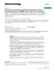
- He et al. Journal of Experimental & Clinical Cancer Research 2011, 30:70 Page 3 of 7 http://www.jeccr.com/content/30/1/70 b-mercaptoethanol for 5 min, separated by size on 15% stained cancer cells. Specimens were rescored if differ- polyacrylamide gel under SDS denaturing conditions, ence of scores from two pathologists was >3. and transferred to a nitrocellucose membrane at 90 V for 2 h. The nitrocellulose membranes were stained 2.3 Quantitative real-time PCR with ponceau S to assess the efficiency of transfer. Non- Total RNA purified from all 252 glioma tissues and 42 specifi c binding was blocked by incubation in block control brain tissues was prepared and reverse tran- buffer (5% non-fat dry milk, 0.05% Tween-20, 1 × tris- scribed. Real-time monitoring of polymerase chain reac- Cl-buffered saline) overnight at 4°C, The membranes tions (PCRs) was performed using the ABI 7900HT were hybridized with mouse monoclonal antibody (Idaho Technology, Idaho Falls, ID, USA) and the SYBR recognizing SMAD4 (sc-7966, Santa Cruz Biotechnol- green I dye (Biogene), which binds preferentially to dou- ogy, Inc., Santa Cruz, CA), then incubated with a horse- ble-stranded DNA. Fluorescence signals, which are pro- radish peroxidase-labeled goat anti-mouse IgG (1: 500). portional to the concentration of the PCR product, are The bound secondary antibody was detected by measured at the end of each cycle and immediately dis- enhanced chemiluminescence (Amersham Life Science, played on a computer screen, permitting realtime moni- Little Chalfont, UK). Housekeeping protein b-actin was toring of the PCR. The reaction is characterized by the used as a loading control. Positive immunoreactive point during cycling when amplification of PCR pro- bands were quantified densitometrically (Leica Q500IW ducts is first detected, rather than the amount of PCR image analysis system) and expressed as ratio of product accumulated after a fixed number of cycles. SMAD4 to b-actin in optical density units. The higher the starting quantity of the template, the earlier a significant increase in fluorescence is observed. The threshold cycle is defined as the fractional cycle 2.5 Statistical analysis number at which fluorescence passes a fixed threshold All computations were carried out using the software of above the baseline. The primers 5 ’ - TAT TAA GCA SPSS version13.0 for Windows (SPSS Inc, IL, USA). The TGC TAT ACA ATC TG -3’ and 5’- CTT CCA CCC rank sum test was used to analyze the ranked data. The AGA TTT CAA TTC -3’ were used to amplify 332-bp measurement data were analyzed by one-way ANOVA. transcripts of SMAD4 and the primers 5 ’ - GGT GGC Randomized block design ANOVA was used to analyze TTT TAG GAT GGC AAG -3’ and 5’- ACT GGA ACG the statistical difference among different tissue types. In GTG AAG GTG ACA G -3’ were used to amplify 161- the analysis of glioma morbidity for all patients, we used bp transcripts of b-actin. All primers were synthesized the Kaplan-Meier estimator and univariate Cox regres- by Sangon Co. (Shanghai, China). The PCR profile con- sion analysis to assess the marginal effect of each factor. sisted of an initial melting step of 1 min at 94°C, fol- The differences between groups were tested by log-rank lowed by 38 cycles of 15 s at 94°C, 15 s at 56°C and 45 analyses. The joint effect of different factors was s at 72°C, and a final elongation step of 10 min at 72°C. assessed using multivariate Cox regression. A Spear- man’s analysis was carried out to analyze the correlation Fluorescence data were converted into cycle threshold measurements using the SDS system software and between SMAD4 mRNA and protein expression levels. exported to Microsoft Excel. SMAD4 mRNA levels were Differences were considered statistically significant when compared to b-actin. Thermal dissociation plots were p was less than 0.05. examined for biphasic melting curves, indicative of 3. Results whether primer-dimers or other nonspecific products could be contributing to the amplification signal. 3.1 SMAD4 protein levels in glioma tissues by immunohistochemistry assay and survival analysis SMAD4 expression was studied in a total of 252 glioma 2.4. Western blot analysis Glioma and normal brain tissues were homogenized in specimens of which 113 were low grade glioma (grade I lysis buffer [PBS, 1% nonidet P-40 (NP-40), 0.5% sodium and II) and 139 were high grade (grade III and IV). deoxycholate, 0.1% sodium dodecyl sulfate (SDS), 100 About 42 specimens taken from normal brain tissue ug/ml aprotinin, 100 μg/ml phenylmethylsulfonyl fluor- served as control group. Based on immunohistochemis- ide (PMSF), Sodium orthovanadate] at 4°C throughout try analysis, positive staining for SMAD4 was mainly all procedures, and sonicated for 70 s, then add 300 μg observed in the cytoplasm and to a lesser degree in the PMSF per gram of tissue and incubate on ice for 30 nuclei of cancer cells. The representative photographs min, followed by centrifugation at 15,000 rpm for 20 were shown in Figure 1. Among the glioma specimens, min at 4°C. The protein content was determined accord- 138 (54.8%) glioma specimens were positively stained, ing to Bradford’s method (Bradford 1976), with bovine and 114 (45.2%) glioma specimens were negatively serum albumin used as a standard. Protein samples (30 stained. Among the control specimens, 34 (81.0%) were μg) were boiled with 2 × sample buffer containing 5% positively stained, and 8 (19.0%) were negatively stained.
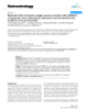
- He et al. Journal of Experimental & Clinical Cancer Research 2011, 30:70 Page 4 of 7 http://www.jeccr.com/content/30/1/70 Figure 1 Immunohistochemical staining of SMAD4 protein in tumor cells of GBM (A) and astrocytoma (B) (Original magnification ×400). Staining for this antigen is described in Materials and Methods. Positive staining of SMAD4 is seen in the cytoplasm and/or nuclei of tumors cells and is more abundant in the low- (B) than the high-grade (A) tumors. Intensively positive expression of SMAD4 (C) was observed in normal brain tissues. SMAD4 positive staining (P < 0.001; Figure 2A). The We also found a significant decrease of SMAD4 expres- median survival time of patients with strong positive (+ sion in glioma compared with normal brain tissues (P < ++) expression of SMAD4 could not be estimated by 0.001). statistical analysis because all patients survived better In addition, SMAD4 expression was not significantly than the overall median level, and those patients with affected by the gender and age (both P > 0.05) of the moderate positive (++), weak positive (+) and negative patients. In contrast, the SMAD4 expression was the expression of SMAD4 were 22.8 ± 1.3 months, 13.2 ± closely correlated with WHO grade (Table 1; P = 0.008), 1.6 months and 8.0 ± 0.5 months (log-rank test: P < as well as Karnofsky performance Status (KPS) (Table 1; 0.001). P < 0.001). Furthermore, Figure 2B shows the post-operative sur- Moreover, we reviewed clinical information of these vival curve of patients with glioma and SMAD4 expres- SMAD4-positive or -negative glioma patients. During sion after adjusting for age, gender, WHO grade and the follow-up period, 197 of the 252 glioma patients KPS. By multivariate analysis, the loss of SMAD4 (78.2%) had died (108 from the SMAD4-negative group expression was a significant and independent prognostic and 142 from the SMAD4-positive group). As deter- indicator for patients with glioma besides age, WHO mined by the log-rank test, the survival rate of patients grade and KPS. The Cox proportional hazards model without SMAD4 staining was lower than those showing showed that lower SMAD4 expression was associated with poor overall survival. Table 1 SMAD4 expression in human glioma tissues with different clinical-pathological features 3.2 Quantitative analysis of SMAD4 protein expression Clinicopathological No. of SMAD4 (n) P based on WHO grade in gliomas features cases As the results of Western blot analysis, we found that - + ++ +++ SMAD4 protein expression tended to increase from the WHO grade 114 60 51 27 glioma to the normal tissue (Figure 3A, C). We also I 53 12 16 13 12 0.008 investigated whether the expression of SMAD4 corre- II 60 17 21 15 7 lated with the WHO grade. SMAD4 expression was III 62 34 12 11 5 highest in grade I and lowest in grade IV (Figure 3B, C). IV 77 51 11 12 3 This result agreed with the findings of the immunohis- Age tochemistry analysis and indicated a close correlation of
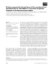
- He et al. Journal of Experimental & Clinical Cancer Research 2011, 30:70 Page 5 of 7 http://www.jeccr.com/content/30/1/70 Figure 2 Postoperative survival curves for patterns of patients with glioma and SMAD4 expression. (A) Kaplan-Meier postoperative survival curve for patterns of patients with glioma and SMAD4 expression. Unadjusted RR of SMAD4-negative (-), weak positive (+), moderate positive (++) and strong positive (+++) groups were 1.0, 0.4, 0.08 and 0.02, respectively (P < 0.001). (B) Cox proportional hazards model after adjusting for age, gender and grade. SMAD4 might be an independent predictor of survival, without consideration of age, gender or grade. Adjusted RR of SMAD4-negative (-), weak positive (+), moderate positive (++) and strong positive (+++) groups were 1.0, 0.4, 0.2 and 0.04, respectively (P < 0.001). altered in various tumors [13]. They consistently trans- tissues (P < 0.001). We further analyzed the expression mit the TGF- b signal from the cell membrane to the of SMAD4 mRNA based on KPS and WHO grade. nucleus. The mammalian SMAD family consists of eight Interestingly, SMAD4 mRNA expression decreased in members, which can be divided into three groups patients whose KPS lower than 80 (P < 0.001) and also according to their function: receptor-activated SMADs, decreased with advancement of WHO grade I to grade commonmediated SMADs, and inhibitory SMADs [14]. IV (P < 0.01). There was a significant positive correla- SMAD4 is one of the commonmediated SMADs and, in tion between the expression of SMAD4 mRNA and pro- general, SMAD4 is a central component of the TGF-b/ tein expression levels from the same glioma tissues (rs = SMAD pathway and is expressed in different human 0.886, P < 0.001). organ systems. TGF- b binds to homodimers of the TGF-b type II receptor (TbRII) which recruits and acti- 4. Discussion vates homodimers of TGF-b type I receptor (TbRI) ser- In the current study, we investigated the expression of ine/threonine kinase. Activated T b RI phosphorylates SMAD4 in 252 cases of human glioma and compared SMAD2 or SMAD3 which heterodimerize with SMAD4. the expression with tumor grade and survival rates of These heterocomplexes translocate into the nucleus patients. Our data demonstrated that SMAD4 protein where they bind DNA and regulate TGF- b dependent was decreased in glioma compared to normal brain tis- gene expression [15]. Deletion or degradation of sue. SMAD4 mRNA expression was also reduced in SMAD4 in tumors could specifically inhibit the tumor glioma compared with control normal brain tissue. We suppressor effect of TGF-b. SMAD4 alteration has been found a decreased trend of both SMAD4 protein level associated with specific loss of TGF-b induced growth and mRNA level from WHO grade I to WHO grade IV resulting in increased angiogenesis and loss of epithelial glioma. These results suggest that the transcriptional integrity [16]. Recent studies have shown that SMAD4 repression of human SMAD4 might participate in the inactivation is associated with the advanced disease state carcinogenesis and progression of glioma. SMAD4 may of various human tumors, including pancreatic carci- have an important role during the genesis or progres- noma, esophageal carcinoma, colorectal carcinoma, sion of glioma. renal cell carcinoma, as well as breast carcinoma SMAD proteins are the key intracellular mediators of transcriptional responses to TGF-b signaling which is [17-20]. Our results confirm that SMAD4 is
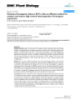
- He et al. Journal of Experimental & Clinical Cancer Research 2011, 30:70 Page 6 of 7 http://www.jeccr.com/content/30/1/70 downregulated during tumor progression. Kjellman et al. [21] analyzed the mRNA expression of TGF-b1, TGF- b 2, TGF- b 3, the TGF- b receptors type I (T b R-I) and type II (TbR-II), SMAD2, SMAD3, and SMAD4. Their data suggested that TGF- b normally up-regulates the TGF-b receptors, and TbR-I and TbR-II showed stron- ger expression in all gliomas when compared to normal tissues. However, the mRNA expression of SMAD2, SMAD3, and SMAD4 was decreased in GBM, which was consistent with the results of our study. We further analyzed the correlation of SMAD4 expression and survival rates of patients. Our data indi- cated that nearly 55% of glioma cases showed positive staining for SMAD4. The survival rate of patients with- out SMAD4 staining was lower than those showing SMAD4-positive staining. Kaplan-Meier analysis of the survival curves showed a significantly worse overall sur- vival for patients whose tumors had low SMAD4 levels, indicating that low SMAD4 protein level is a marker of poor prognosis for patients with glioma. Moreover, mul- tivariate analysis showed low SMAD4 expression to be a marker of worse outcome independent of the known clinical prognostic indicators such as age, KPS and grade. These data suggest that low expression of SMAD4 is correlated with a worse outcome of patients with glioma. Thus, SMAD4 might be an independent predictor of survival for glioma patients. In our study, which consisted of a large sample (n = 252), SMAD4 expression was analyzed by immunohistochemistry, real- time PCR and Western blot analysis. Thus, a large sam- Figure 3 Expression of SMAD4 protein in glioma and normal brain tissues by Western blot analysis. (A) SMAD4 expression ple size, a good methodology and a detailed clinical fol- levels in glioma and normal brain tissues. (B) SMAD4 expression low-up in our study make it reliable. levels in glioma with different WHO grades. (C) SMAD4 expression In conclusion, our data provides convincing evidence levels in normal brain tissues and glioma with different WHO for the first time that the reduced expression of SMAD4 grades. ‘N’ refers to normal brain tissues; ‘Ca’ refers to glioma tissues; at gene and protein levels is correlated with poor out- ‘Ca_ I’~’ Ca_ IV’ refer to glioma tissues with WHO grade I~ IV. b- actin was used as a control for equal protein loading. Values are come in patients with glioma. SMAD4 may play an inhi- means ± SD. ‘*’, p < 0.05, comparison with normal brain tissues; ‘**’, bitive role during the development of glioma and may p < 0.001, comparison with normal brain tissues. be a potential prognosis predictor of glioma. Authors’ contributions S-MH and Z-WZ carried out the Immunochemistry assay and Quantitative Table 2 Statistics of SMAD4 mRNA levels in glioma real-time PCR. SMH also drafted the manuscript. YW carried out the Western No. of cases SMAD mean (SD) P blot analysis and drafted the manuscript. J-PZ, LW and FH participated in the survival analysis. G-DG conceived of the study, and participated in its Tissue type design and coordination. All authors read and approved the final Control 42 2.096 (0.338)
- He et al. Journal of Experimental & Clinical Cancer Research 2011, 30:70 Page 7 of 7 http://www.jeccr.com/content/30/1/70 2. Sun B, Chu D, Li W, Chu X, Li Y, Wei D, Li H: Decreased expression of 21. Kjellman C, Olofsson SP, Hansson O, Von Schantz T, Lindvall M, Nilsson I, NDRG1 in glioma is related to tumor progression and survival of Salford LG, Sjögren HO, Widegren B: Expression of TGF-beta isoforms, patients. J Neurooncol 2009, 94:213-219. TGF-beta receptors, and SMAD molecules at different stages of human 3. Ding Z, Wu CJ, Chu GC, Xiao Y, Ho D, Zhang J, Perry SR, Labrot ES, Wu X, glioma. Int J Cancer 2000, 89:251-258. Lis R, Hoshida Y, Hiller D, Hu B, Jiang S, Zheng H, Stegh AH, Scott KL, doi:10.1186/1756-9966-30-70 Signoretti S, Bardeesy N, Wang YA, Hill DE, Golub TR, Stampfer MJ, Cite this article as: He et al.: Reduced expression of SMAD4 in gliomas Wong WH, Loda M, Mucci L, Chin L, DePinho RA: SMAD4-dependent correlates with progression and survival of patients. Journal of barrier constrains prostate cancer growth and metastatic progression. Experimental & Clinical Cancer Research 2011 30:70. Nature 2011, 470:269-273. 4. Ali NA, McKay MJ, Molloy MP: Proteomics of Smad4 regulated transforming growth factor-beta signalling in colon cancer cells. Mol Biosyst 2010, 6:2332-2338. 5. Papageorgis P, Cheng K, Ozturk S, Gong Y, Lambert AW, Abdolmaleky HM, Zhou JR, Thiagalingam S: Smad4 inactivation promotes malignancy and drug resistance of colon cancer. Cancer Res 2011, 71:998-1008. 6. Sakellariou S, Liakakos T, Ghiconti I, Hadjikokolis S, Nakopoulou L, Pavlakis K: Immunohistochemical expression of P15 (INK4B) and SMAD4 in advanced gastric cancer. Anticancer Res 2008, 28:1079-1083. 7. Blackford A, Serrano OK, Wolfgang CL, Parmigiani G, Jones S, Zhang X, Parsons DW, Lin JC, Leary RJ, Eshleman JR, Goggins M, Jaffee EM, Iacobuzio- Donahue CA, Maitra A, Cameron JL, Olino K, Schulick R, Winter J, Herman JM, Laheru D, Klein AP, Vogelstein B, Kinzler KW, Velculescu VE, Hruban RH: SMAD4 gene mutations are associated with poor prognosis in pancreatic cancer. Clin Cancer Res 2009, 15:4674-4679. 8. Ke Z, Zhang X, Ma L, Wang L: Deleted in pancreatic carcinoma locus 4/ Smad4 participates in the regulation of apoptosis by affecting the Bcl-2/ Bax balance in non-small cell lung cancer. Hum Pathol 2008, 39:1438-1445. Lv J, Cao XF, Ji L, Zhu B, Wang DD, Tao L, Li SQ: Association of β-catenin, 9. Wnt1, Smad4, Hoxa9, and Bmi-1 with the prognosis of esophageal squamous cell carcinoma. Med Oncol 2011. 10. Sheehan GM, Kallakury BV, Sheehan CE, Fisher HA, Kaufman RP Jr, Ross JS: Smad4 protein expression correlates with grade, stage, and DNA ploidy in prostatic adenocarcinomas. Hum Pathol 2005, 36:1204-1209. 11. Hiwatashi K, Ueno S, Sakoda M, Kubo F, Tateno T, Kurahara H, Mataki Y, Maemura K, Ishigami S, Shinchi H, Natsugoe S: Strong Smad4 expression correlates with poor prognosis after surgery in patients with hepatocellular carcinoma. Ann Surg Oncol 2009, 16:3176-3182. 12. Brown RS, Wahl RL: Overexpression of Glut-1 glucose transporter in human breast cancer: an immunohistochemical study. Cancer 1993, 72:2979-2985. 13. Mesker WE, Liefers GJ, Junggeburt JM, van Pelt GW, Alberici P, Kuppen PJ, Miranda NF, van Leeuwen KA, Morreau H, Szuhai K, Tollenaar RA, Tanke HJ: Presence of a high amount of stroma and downregulation of SMAD4 predict for worse survival for stage I-II colon cancer patients. Cell Oncol 2009, 31:169-178. 14. Koinuma D, Tsutsumi S, Kamimura N, Imamura T, Aburatani H, Miyazono K: Promoter-wide analysis of Smad4 binding sites in human epithelial cells. Cancer Sci 2009, 100:2133-2142. 15. Bornstein S, White R, Malkoski S, Oka M, Han G, Cleaver T, Reh D, Andersen P, Gross N, Olson S, Deng C, Lu SL, Wang XJ: Smad4 loss in mice causes spontaneous head and neck cancer with increased genomic instability and inflammation. J Clin Invest 2009, 119:3408-3419. 16. Korc M: Smad4: gatekeeper gene in head and neck squamous cell carcinoma. J Clin Invest 2009, 119:3208-3211. 17. Wilentz RE, Su GH, Dai JL, Sparks AB, Argani P, Sohn TA, Yeo CJ, Kern SE, Hruban RH: Immunohistochemical labeling for dpc4 mirrors genetic status in pancreatic adenocarcinomas: a new marker of DPC4 Submit your next manuscript to BioMed Central inactivation. Am J Pathol 2000, 156:37-43. 18. Wilentz RE, Iacobuzio-Donahue CA, Argani P, McCarthy DM, Parsons JL, and take full advantage of: Yeo CJ, Kern SE, Hruban RH: Loss of expression of Dpc4 in pancreatic intraepithelial neoplasia: evidence that DPC4 inactivation occurs late in • Convenient online submission neoplastic progression. Cancer Res 2000, 60:2002-2006. 19. Natsugoe S, Xiangming C, Matsumoto M, Okumura H, Nakashima S, • Thorough peer review Sakita H, Ishigami S, Baba M, Takao S, Aikou T: Smad4 and Transforming • No space constraints or color figure charges Growth Factor beta1 Expression in Patients with Squamous Cell • Immediate publication on acceptance Carcinoma of the Esophagus. Clin Cancer Res 2002, 8:1838-1842. 20. Cardillo MR, Lazzereschi D, Gandini O, Di Silverio F, Colletta G: • Inclusion in PubMed, CAS, Scopus and Google Scholar Transforming growth factor-beta pathway in human renal cell • Research which is freely available for redistribution carcinoma and surrounding normal-appearing renal parenchyma. Anal Quant Cytol Histol 2001, 23:109-117. Submit your manuscript at www.biomedcentral.com/submit
CÓ THỂ BẠN MUỐN DOWNLOAD

-

Báo cáo khoa học: Mechanism of the reaction catalyzed by dehydroascorbate reductase from spinach chloroplasts
 8 p |
8 p |  49
|
49
|  6
6
-
Báo cáo khoa học: Differential involvement of protein kinase C alpha and epsilon in the regulated secretion of soluble amyloid precursor protein
8 p | 48
| 5
-
Báo cáo khoa học: Reducing expression of NAD+ synthesizing enzyme NMNAT1 does not affect the rate of Wallerian degeneration
14 p | 36
| 5
-

Báo cáo y học: "Association of reduced heme oxygenase-1 with excessive Toll-like receptor 4 expression in peripheral blood mononuclear cells in Behçet's disease"
10 p | 47
| 5
-
Báo cáo y học: "Atorvastatin reduces lipopolysaccharide-induced expression of cyclooxygenase-2 in human pulmonary epithelial cells"
6 p | 50
| 5
-
Báo cáo khoa học: Kinetics of electron transfer from NADH to the Escherichia coli nitric oxide reductase flavorubredoxin
10 p | 29
| 4
-

Báo cáo khoa học: Expression of heme oxygenase-1 is repressed by interferon-c and induced by hypoxia in human retinal pigment epithelial cells
9 p | 43
| 4
-

Báo cáo khóa học: Suppression of b1,3galactosyltransferase b3Gal-T5 in cancer cells reduces sialyl-Lewis a and enhances poly N-acetyllactosamines and sialyl-Lewis x on O-glycans Lydia Mare and Marco Trinchera
9 p | 40
| 4
-
báo cáo khoa học: " Ectopic expression of MdSPDS1 in sweet orange (Citrus sinensis Osbeck) reduces canker susceptibility: involvement of H2O2 production and transcriptional alteration"
15 p | 55
| 4
-

Báo cáo y học: " Open Access No evidence of altered alveolar macrophage polarization, but reduced expression of TLR2, in bronchoalveolar lavage cells in sarcoidosis"
13 p | 60
| 4
-
Báo cáo khoa học: Hypoxia reduces the expression of heme oxygenase-2 in various types of human cell lines A possible strategy for the maintenance of intracellular heme level
12 p | 35
| 3
-
Báo cáo y học: " Reduced levels of reactive oxygen species correlate with inhibition of apoptosis, rise in thioredoxin expression and increased bovine leukemia virus proviral loads"
10 p | 58
| 3
-

Báo cáo y học: "Mild hypothermia alone or in combination with anesthetic post-conditioning reduces expression of inflammatory cytokines in the cerebral cortex of pigs after cardiopulmonary resuscitation"
11 p | 46
| 3
-
báo cáo khoa học: " Cloning of transgenic tobacco BY-2 cells; an efficient method to analyse and reduce high natural heterogeneity of transgene expression"
11 p | 48
| 3
-

Báo cáo y học: " Reduced Fas ligand-expressing splenic CD5+ B lymphocytes in severe collagen-induced arthritis"
12 p | 38
| 2
-
Báo cáo y học: "Alteration of viral lipid composition by expression of the phospholipid floppase ABCB4 reduces HIV vector infectivity"
9 p | 33
| 2
-
Báo cáo khoa học: Protein expressed by the ho2 gene of the cyanobacterium Synechocystis sp. PCC 6803 is a true heme oxygenase
0 p | 44
| 2


Chịu trách nhiệm nội dung:
Nguyễn Công Hà - Giám đốc Công ty TNHH TÀI LIỆU TRỰC TUYẾN VI NA
LIÊN HỆ
Địa chỉ: P402, 54A Nơ Trang Long, Phường 14, Q.Bình Thạnh, TP.HCM
Hotline: 093 303 0098
Email: support@tailieu.vn
Giấy phép Mạng Xã Hội số: 670/GP-BTTTT cấp ngày 30/11/2015 Copyright © 2022-2032 TaiLieu.VN. All rights reserved.

