


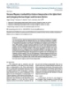
Báo cáo khoa học: "Spinal cord compression by a solitary metastasis from a low grade leydig cell tumour: a case report and review of the literature"
lượt xem 3
download
 Download
Vui lòng tải xuống để xem tài liệu đầy đủ
Download
Vui lòng tải xuống để xem tài liệu đầy đủ
Tuyển tập báo cáo các nghiên cứu khoa học quốc tế ngành y học dành cho các bạn tham khảo đề tài: Spinal cord compression by a solitary metastasis from a low grade leydig cell tumour: a case report and review of the literature
Bình luận(0) Đăng nhập để gửi bình luận!
Nội dung Text: Báo cáo khoa học: "Spinal cord compression by a solitary metastasis from a low grade leydig cell tumour: a case report and review of the literature"
- World Journal of Surgical Oncology BioMed Central Open Access Case report Spinal cord compression by a solitary metastasis from a low grade leydig cell tumour: a case report and review of the literature Efthimios P Samoladas*1, Ashraf S Anbar1, Jonathan D Lucas1, Hlias Fotiadis2 and Byron E Chalidis3 Address: 1Spinal Unit, Guy's Hospital, London, UK, 2Department of Orthopaedics, Veria General Hospital, Greece and 3Department of orthopaedics, USCF Hospital, San Francisco, USA Email: Efthimios P Samoladas* - msamolad@gmail.com; Ashraf S Anbar - ashraf_anbar@hotmail.com; Jonathan D Lucas - Jonathan.Lucas@gstt.sthames.nhs.uk; Hlias Fotiadis - fotiad-h@otenet.gr; Byron E Chalidis - byronchalidis@gmail.com * Corresponding author Published: 10 July 2008 Received: 21 November 2007 Accepted: 10 July 2008 World Journal of Surgical Oncology 2008, 6:75 doi:10.1186/1477-7819-6-75 This article is available from: http://www.wjso.com/content/6/1/75 © 2008 Samoladas et al; licensee BioMed Central Ltd. This is an Open Access article distributed under the terms of the Creative Commons Attribution License (http://creativecommons.org/licenses/by/2.0), which permits unrestricted use, distribution, and reproduction in any medium, provided the original work is properly cited. Abstract Background: Leydig tumour is rare and there are only three cases with metastatic disease reported. Case presentation: A 52 year-old Caucasian male was admitted, on emergency basis to the Orthopaedic Department with six weeks history of increasing midthoracic back pain, change in gait, poor balance, subjective weakness and numbness of the lower trunk and legs. MRI scan showed change in the signal intensity of T4 and T5 vertebral body but their height were maintained. Urgent T4 and T5 corpectomies, decompression of the spinal cord and reconstruction of the vertebral bodies were performed followed by radiotherapy. Neurological status significantly improved with a mild residual numbness over the dorsum of the right foot. The histology of the excised tumour was identical to the primary. At 2 years follow-up visit the patient is neurologically stable and disease free without other organs metastases. Conclusion: This is the first case in English literature, which shows that spinal metastases could occur even in the early stage of Leydig cell tumour, without other organs involvement. Aggressive surgical management of spinal metastases combined with post operative radiotherapy can give a better chance for long survivorship. mas and GIT adenocarcinomas. Among those, metastases Background Secondary tumours are the most common tumours from the first 3 tumours are the commonest [1,3]. involving the spine [1] and their incidence may be increased as further advances in cancer therapy prolong Leydig cell (interstitial cell) tumour of the testis was first the life expectancy of afflicted patients [2]. Malignant pri- described by Sacchi [2] in 1895. The interstitial cells of the mary tumours most frequently metastasizing to the spine testis, located between the seminiferrous tubules are des- are: bronchogenic carcinoma, breast carcinoma, prostatic ignated by the surname of the German anatomist who adenocarcinoma, renal cell carcinoma, thyroid carcino- first described them, Franz von Leydig. They primarily Page 1 of 5 (page number not for citation purposes)
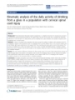
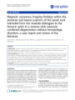
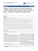
- World Journal of Surgical Oncology 2008, 6:75 http://www.wjso.com/content/6/1/75 secrete testosterone [4] and it is an exceedingly rare Nine months later the patient admitted to the Spinal Unit tumour [2]. in an emergency base complaining of increasing mid tho- racic pain, change in gait, poor balance, subjective weak- Only 7–10% of Leydig cell tumours shows malignant ness, numbness of the lower trunk and legs. Examination activity exclusively in adults [4-7] and metastasise. More- revealed a broad base gait, able to walk in toes and heel, over, it seldom metastasizes to the spine [8-10]. absence of tenderness or masses over T4 level, hypo aes- thesia below T5 level more pronounced over the left side, The tumour is generally refractory to radiotherapy and exaggerated tendon reflexes in the lower limbs, un-sus- chemotherapy. The natural course of patients with meta- tained ankle clonus bilaterally and normal plantar static variety of Leydig cell tumour is one of progression at reflexes. No objective motor weakness detected and intact an unpredictable pace. The median survival of these perianal sensations were recorded. patients with metastatic disease is less than 2 years [4,11- 15]. X-rays of the thoracic spine revealed a sclerotic appearance of the T4 vertebral body (Figure 1 &2) and an urgent Mag- We present the fourth case in English literature of malig- netic Resonance Imaging (MRI) showed quite dramatic nant Leydig cell tumour with spinal metastases and the change in the appearance of T4 compare to the previous first in the early stage of the disease. Surgical treatment in CT despite the maintenance of vertebral body height. Fur- combination with post-operative radiotherapy resulted in thermore, T5 vertebral body was also involved, but to a a very satisfactory outcome. This is the first case reported lesser extent. There was a soft tissue expansion into the with such a long disease free period. Case presentation A 52 year-old Caucasian male was admitted, on emer- gency basis to the orthopaedic department with six weeks history of increasing mid thoracic pain, change in gait, poor balance, subjective weakness, numbness of the lower trunk and legs. He didn't report any neurogenic bladder or bowel disturbances and he was otherwise fit and well. The patient had a right sided orchidectomy 3 years ago, for stage one well differentiated Leydig cell tumour. He was diagnosed having an enlarged right testis. No adju- vant therapy was given perioperatively. Afterwards, he fol- lowed up periodically and Computer Tomography (CT) scans of the chest, abdomen and pelvis were performed on the basis of evaluation and potential metastasizing of the neoplasm. Two years following the primary operation the patient complained of back pain. Plain films of the spine showed an "ivory" vertebra at T4. CT scan depicted a definite abnormality in the body of T4 with no evidence of general metastatic disease. There was no soft tissue extension and no vertebral body collapse. None of the visceral organs was involved and this was the only detectable pathologi- cal sign. The bones scan showed intense uptake in T4 and no other sights of increasing radioisotope uptake. The blood tests, including tumour markers, didn't show any abnormality. At that stage, the oncologists decided against biopsy as they felt it was potentially hazardous and the patient will have little to gain from it. Accordingly, in absence of symptoms and tenderness, a "wait and see" Figure 1 AP X ray of Thoracic spine policy was adopted. AP X ray of Thoracic spine. Page 2 of 5 (page number not for citation purposes)
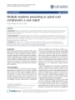
- World Journal of Surgical Oncology 2008, 6:75 http://www.wjso.com/content/6/1/75 Figure 2 Lateral X ray of Thoracic spine Lateral X ray of Thoracic spine. Figure 3 T2W MRI of Thoracic spine extradural space causing spinal cord compression (Figure T2W MRI of Thoracic spine. 3). Blood test, including inflammatory and tumour markers, were within normal values and a dose of 16 mg Dexame- sion was coming only from the front and the tension band thasone daily was started. A CT guided biopsy was per- of the posterior elements, at the involved level, were formed and the histological appearance of the lesion was intact. identical to the primary tumour. Surgery was performed through a right subscapular 3rd rib After discussion with the patient, a decision was made to thoracotomy, and the cord function was monitored by perform urgent T4 and T5 corpectomies, decompression Somatosensory Evoked Potentials (SSEPs) throughout the of the spinal cord and reconstruction of the vertebral bod- procedure. After complete canal decompression, recon- ies. Anterior surgery was contemplated as the compres- struction was achieved by a Synmesh packed with bone Page 3 of 5 (page number not for citation purposes)
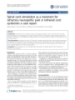
- World Journal of Surgical Oncology 2008, 6:75 http://www.wjso.com/content/6/1/75 of repeated scans. He complained only for a minimal numbness of the right foot and slight winging of the right scapula. Discussion The most common sites of metastatic involvement in Ley- dig cell tumour are the regional lymph nodes and then the lung, liver, and bone. The spine is very rarely involved and there are only three cases reported in the English literature with spine involvement [4,5,12]. In the reported cases, spinal involvement occurred late in the course of the dis- ease and other organs metastases had already occurred. Neurological deficit developed only as a pre-terminal event and the thoracic spine was involved in all three patients. In our case the spine was the first metastatic area without any other detectable metastases, which hasn't described before. Traditionally, spinal metastases treatment involves radia- tion therapy, either alone or in conjunction with chemo- therapy and/or surgical decompression. "Prophylactic" irradiation had not prevented local recurrence or meta- static spread within the radiation ports [15]. Several chemotherapeutic agents have been used in the treatment of metastatic Leydig cell tumour, with uniformly poor results [4,5,12]. Recently [16], a randomised study Figure AP post op 4 & Lat X rays of Thoracic spine showed that direct decompressive surgery plus postopera- post op AP & Lat X rays of Thoracic spine. tive radiotherapy is superior to treatment with radiother- apy alone for patients with spinal cord compression caused by metastatic cancer. graft obtained from the excised rib. Anterior Universal Spine System (USS) II system was added to augment the In the reported three previous cases none treated opera- construct (Figure 4). tively. One patient received spinal irradiation (2000 cGy) without improvement in neurological deficit but with The patient was then transferred to Intensive Therapy Unit some amelioration of back pain [4]. The second one (ITU) and had an uneventful recovery. He discharged one received Mitotane(1,1-dichloro-2 [o-chlorophenyl]-2-[p- week following operation with clear neurological chlorophenyl]ethane or o,p'-DDD) chemotherapy with- improvement. Three weeks postoperatively, he developed out clear benefit [5]. The third patient received no therapy a right-sided pneumonia, which resolved with antibiotics. and developed progressive neurological dysfunction [12]. The histological evaluation of the excised tumour was identical to the primary. A combination of operative treatment and radiotherapy was adopted in our case with a satisfactory result, Because it was a solitary metastasis and eradication of the although the diagnosis of the metastatic disease was made tumour wasn't feasible by surgical excision alone as it had at earlier stage. Our patient remained disease free at the already extended beyond the bony limits, postoperative last follow up visit two years and six months postopera- radiotherapy with radical intent was applied. The dura- tively. tion of the radiotherapy was a daily 5 weeks course and the dose was 50 Gy in 25 fractions to the area of T4/T5. It is well recognised that decompression surgery alone, Neither side effect from radiotherapy nor skin reactions whether anterior or posterior, might actually contribute to was reported. mechanical instability of the spine. This can lead to the spinal cord compression by creating post surgical deform- The patient was followed up periodically by Technetium ity. Therefore, we believe that reconstruction should bone scans and CT scans of chest, abdomen and pelvis. At always be added. There is no consensus on whether stabi- the last follow-up visit-two years and six months postop- lisation should be performed through an anterior or pos- eratively he was disease-free based according to the results terior approach since deformity and instability can be Page 4 of 5 (page number not for citation purposes)
- World Journal of Surgical Oncology 2008, 6:75 http://www.wjso.com/content/6/1/75 improved by either. It is frequently stated that anterior 9. Hall AJ, Mackay NNS: The results of laminectomy for compres- sion of the cord and cauda equina by extradural malignant procedures give better results, but this is probably a func- tumour. J Bone Joint Surg Br 1973, 55(3):497-505. tion of patient selection [17]. If the posterior elements are 10. White WA, Patterson RH, Bergland RM: Role of surgery in the treatment of spinal cord compression by metastatic neo- not involved by the tumour, it is recommended to avoid plasm. Cancer 1971, 27:558-561. disrupting the remaining intact posterior tension band. 11. Abelson D, Bulaschenko H, Trommer PR, Valdes-Dapena : A Malig- nant interstitial-cell tumor of the testis treated with o,p'- DDD. Metabolism 1966, 15:242-256. However, if the patient's general condition can't allow an 12. Azer PC, Braunstein GD: Malignant Leydig cell tumor: Objec- anterior approach, posterior decompression should tive tumor response to o,p'-DDD. Cancer 1981, 47:1251-1255. 13. Davis S, DiMartino NA, Schneider G: Malignant interstitial cell always be augmented by at least posterior internal fixation carcinoma of the testis: Report of two cases with steroid syn- and reconstruction of the anterior column also, via a lat- thetic profiles, response to therapy, and review of the litera- eral extracavitary approach (LECA). In our case, as the ture. Cancer 1981, 47:425-431. 14. Feldman PS, Kovacs K, Horvath E, Adelson GL: Malignant Leydig compression was mainly at the front an anterior approach cell tumor: Clinical, histologic and electron microscopic fea- with decompression and reconstruction was selected. tures. Cancer 1982, 49:714-721. 15. Tamoney HJ, Noriega A: Malignant interstitial cell tumour of the testis. Cancer 1969, 24:547-551. Conclusion 16. Patchel RA, Tibbs PA, William RF, Payne R, Saris S, Kryscio RJ, Leydig cell tumour is a rare entity with only three reported Mohiuddin M, Young B: Direct decompressive surgical resec- tion in the treatment of spinal cord compression caused by cases of spinal metastases. They could occur even in the metastatic cancer: a randomised trial. Lancet 2005, early stage without other organs involvement. Aggressive 366:643-648. surgical management of spinal metastases combined with 17. Bauer HC: Controversies in the surgical management of skel- etal metastases. J Bone Joint Surg Br 2005, 87:608-617. postoperative radiotherapy can give a better chance for long survivorship. Surgical planning should take into con- sideration that the avoidance of spinal destabilisation and the restoration of normal spinal stability are very impor- tant for the improvement of the overall outcome and quality of life. Competing interests The authors declare that they have no competing interests. Authors' contributions JL and ES concept and design, review of manuscript. AA helped in preparation of manuscript. HF and BC reviewed the literature and prepared the manuscript. All authors read and approved final manuscript. Acknowledgements Written consent was obtained from the patient for publication of this case report. References 1. Black P: Spinal metastasis: Current status and recommended guidelines for management. Neurosurgery 1979, 5:726-746. 2. Sawin PD, VanGilder JC: Spinal cord compression from meta- static Leydig's cell tumour of the testis: Case Report. Neuro- surgery 1996, 38:407-411. 3. Godersky JC, Smoker WRK, Knutzon R: Use of magnetic reso- Publish with Bio Med Central and every nance imaging in the evaluation of metastatic spinal disease. scientist can read your work free of charge Neurosurgery 1987, 21:676-680. 4. Grem JL, Robins HI, Wilson KS, Gilchrist K, Trump DL: Metastatic "BioMed Central will be the most significant development for Leydig cell tumor of the testis: Report of three cases and disseminating the results of biomedical researc h in our lifetime." review of the literature. Cancer 1986, 58:2116-2119. Sir Paul Nurse, Cancer Research UK 5. Bertram KA, Bratloff B, Hodges GF, Davidson H: Treatment of malignant Leydig cell tumour. Cancer 1991, 68:2324-2329. Your research papers will be: 6. Kim I, Young RH, Scully RE: Leydig cell tumours of the testis: A clinicopathological analysis of 40 cases and review of the lit- available free of charge to the entire biomedical community erature. Am J Surg Pathol 1985, 9:177-192. peer reviewed and published immediately upon acceptance 7. Mahon FB, Gosset F, Trinity RC, Madsen PO: Malignant interstitial cell testicular tumor. Cancer 1973, 31:1208-1212. cited in PubMed and archived on PubMed Central 8. Gilbert RW, Kim JH, Posner JB: Epidural spinal cord compres- yours — you keep the copyright sion from metastatic tumor: Diagnosis and treatment. Ann BioMedcentral Neurol 1978, 3:40-51. Submit your manuscript here: http://www.biomedcentral.com/info/publishing_adv.asp Page 5 of 5 (page number not for citation purposes)
CÓ THỂ BẠN MUỐN DOWNLOAD

-

báo cáo hóa học: " Modulation of spinal cord synaptic activity by tumor necrosis factor alpha in a model of peripheral neuropathy"
 23 p |
  54
|
54
|  5
5
-
Báo cáo khoa hoc:" Kinematic analysis of the daily activity of drinking from a glass in a population with cervical spinal cord injury"
12 p | 48
| 5
-

Báo cáo khoa hoc:" Quantification of the effects of an alpha-2 adrenergic agonist on reflex properties in spinal cord injury using a system identification technique"
7 p | 65
| 5
-
Báo cáo y học: "Magnetic resonance imaging findings within the posterior and lateral columns of the spinal cord extended from the medulla oblongata to the thoracic spine in a woman with subacute combined degeneration without hematologic disorders: a case report and review of the literature"
4 p | 52
| 5
-

Báo cáo y học: " Intramedullary spinal cord metastasis from colonic carcinoma presenting as Brown-Séquard syndrome: a case report"
5 p | 54
| 5
-

báo cáo khoa học: "A patient presenting with intact sensory modalities in acute spinal cord ischemia syndrome: a case report"
4 p | 42
| 4
-
Báo cáo y học: "Multiple myeloma presenting as spinal cord compression: a case report"
4 p | 50
| 4
-

Báo cáo y học: "Simvastatin protects bladder and renal functions following spinal cord injury in rats"
10 p | 38
| 4
-

Báo cáo y học: "Effect of interleukin-1β on spinal cord nociceptive transmission of normal and monoarthritic rats after disruption of glial function"
9 p | 44
| 4
-
Báo cáo y học: "Spinal cord stimulation as a treatment for refractory neuropathic pain in tethered cord syndrome: a case report
4 p | 46
| 4
-

báo cáo khoa học:" Cross-cultural validity of four quality of life scales in persons with spinal cord injury"
16 p | 48
| 3
-
Báo cáo y học: "Spontaneous rupture of an infected renal cyst and external drainage through a lumbar surgical scar in a male patient with cervical spinal cord injury: a case report"
4 p | 60
| 3
-
báo cáo khoa học: "An overview of tissue engineering approaches for management of spinal cord injuries"
16 p | 52
| 3
-

báo cáo khoa học: " The effects of powered ankle-foot orthoses on joint kinematics and muscle activation during walking in individuals with incomplete spinal cord injury"
17 p | 58
| 3
-
Báo cáo khoa hoc:" Kinematics and muscle activity of individuals with incomplete spinal cord injury during treadmill stepping with and without manual assistance"
14 p | 34
| 3
-
Báo cáo khoa hoc:" Dipolar cortico-muscular electrical stimulation: a novel method that enhances motor function in both - normal and spinal cord injured mice"
15 p | 45
| 3
-
Báo cáo y học: "Foramen Magnum Arachnoid Cyst Induces Compression of the Spinal Cord and Syringomyelia: Case Report and Literature Review"
6 p | 65
| 3


Chịu trách nhiệm nội dung:
Nguyễn Công Hà - Giám đốc Công ty TNHH TÀI LIỆU TRỰC TUYẾN VI NA
LIÊN HỆ
Địa chỉ: P402, 54A Nơ Trang Long, Phường 14, Q.Bình Thạnh, TP.HCM
Hotline: 093 303 0098
Email: support@tailieu.vn
Giấy phép Mạng Xã Hội số: 670/GP-BTTTT cấp ngày 30/11/2015 Copyright © 2022-2032 TaiLieu.VN. All rights reserved.

