


báo cáo khoa học: "Transcription factor c-Myb promotes the invasion of hepatocellular carcinoma cells via increasing osteopontin expression"
lượt xem 3
download
 Download
Vui lòng tải xuống để xem tài liệu đầy đủ
Download
Vui lòng tải xuống để xem tài liệu đầy đủ
Tuyển tập báo cáo các nghiên cứu khoa học quốc tế ngành y học dành cho các bạn tham khảo đề tài: Transcription factor c-Myb promotes the invasion of hepatocellular carcinoma cells via increasing osteopontin expression
Bình luận(0) Đăng nhập để gửi bình luận!
Nội dung Text: báo cáo khoa học: "Transcription factor c-Myb promotes the invasion of hepatocellular carcinoma cells via increasing osteopontin expression"
- Chen et al. Journal of Experimental & Clinical Cancer Research 2010, 29:172 http://www.jeccr.com/content/29/1/172 RESEARCH Open Access Transcription factor c-Myb promotes the invasion of hepatocellular carcinoma cells via increasing osteopontin expression Rong-Xin Chen1,2, Yun-Hong Xia1,2, Tong-Chun Xue1,2, Sheng-Long Ye1,2* Abstract Background: Specific gene expression is tightly regulated by various transcription factors. Osteopontin (OPN) is a phosphoprotein that mediates hepatocellular carcinoma (HCC) progression and metastasis. However, the mechanism of OPN up-regulation in HCC metastasis remains to be clarified. Methods: Oligonucleotide array-based transcription factor assays were applied to compare different activities of transcription factors in two human HCC cell lines with different OPN expression levels. The effects of one selected transcription factor on OPN expression were further evaluated. Results: Eleven transcription factors were over-expressed in metastatic HCC cell line HCCLM6 cells whereas twelve transcription factors were down-regulated. Electrophoretic mobility shift assays (EMSA) and reporter gene assays showed that one of up-regulated transcription factors c-Myb could bind the OPN promoter and increase its transcription activity. In addition, small interfering RNA targeting c-Myb could inhibit OPN expression and significantly decrease migration and invasion of HCCLM6 cells in vitro. Conclusion: Our data first demonstrate that c-Myb has a functionally important role in the regulation of OPN expression in HCC cells, suggesting that c-Myb might be a new target to control HCC metastasis. 1. Introduction through binding its receptors of integrins or CD44 variant. Although OPN has been studied in a number of tumors, Hepatocellular carcinoma (HCC) is one of the most the molecular mechanisms of OPN up-regulation in the common and aggressive malignancies [1]. Despite of processes of HCC metastasis are still elusive. improvements in surgical techniques and perioperative While tumor progression and metastasis are closely managements, HCC prognosis remains poor due to a related to signaling cascades that transduce and inte- 5-year recurrence rate of 50%-70% after resection [2,3]. grate regulatory cues, transcription factors are endpoints Thus, it is critical to identify the molecules controlling of signaling pathways to determine transcription and the the invasive and metastatic potential of HCC, which extent to which genes are expressed [10]. In addition, would provide new targets for intervention. some transcription factors including AP-1 [11], SP-1 Osteopontin (OPN) is a secreted extracellular matrix [12] and Runx [13] have been functionally associated protein, which has been linked to tumor progression with tumor cell proliferation, growth, differentiation and and metastasis in a variety of cancers including HCC metastasis in leukemia and solid tumors. [4,5]. OPN has been identified as the lead gene over- To investigate the possibility that transcription factors expressed in the metastatic HCC [6]. Increased OPN regulate OPN expression in HCC metastasis, we applied expression is associated with clinical stage, portending a transcription factor microarrays to compare different poor prognosis [7-9]. OPN increases cell proliferation, migration and extracellular matrix invasion in vitro activities of transcription factors in two human HCC cell lines with different OPN expression levels. Our data demonstrate that one of up-regulated transcription fac- * Correspondence: ye.shenglong@zs-hospital.sh.cn 1 Liver Cancer Institute and Zhongshan Hospital, Fudan University, Shanghai, tors c-Myb plays an important role in the regulation of China OPN expression and invasion of HCC cells in vitro , Full list of author information is available at the end of the article © 2010 Chen et al; licensee BioMed Central Ltd. This is an Open Access article distributed under the terms of the Creative Commons Attribution License (http://creativecommons.org/licenses/by/2.0), which permits unrestricted use, distribution, and reproduction in any medium, provided the original work is properly cited.
- Chen et al. Journal of Experimental & Clinical Cancer Research 2010, 29:172 Page 2 of 8 http://www.jeccr.com/content/29/1/172 expression of genes was determined by normalizing to suggesting that c-Myb may be a potential target to con- GAPDH according to the manufacturer’s instructions. trol HCC metastasis. 2. Materials and methods 2.4 Nuclear extracts and biotin-streptavidin DNA pull- 2.1 Cell culture down assay Oligonucleotide containing biotin on the 5’-nucleotide Human embryonic liver cell line L02 and HCC cell line SMMC-7721 were obtained from Shanghai Institute of of the sense strand was used in the PCR amplification Cell and Biology, Chinese Academy of Science and for human OPN promoter. The sequences of the primer were as follows: sense strand: 5 ’ biotin-TGGAATAC maintained in RPMI supplemented with 10% fetal ATCCAATTTAAGGGAG-3 ’ ; antisense strand 5 ’ - bovine serum at 37°C with 5% CO2. Human metastatic GAATGCACAA CCCAGTAGCAAA-3’ ; which corre- HCC cell line MHCC97-L and HCCLM6 were estab- sponds to positions -1488 to +185 of the human OPN lished at Liver Cancer Institute, Zhongshan Hospital, promoter. Nuclear proteins were isolated from HCC cell Fudan University, Shanghai, P.R. China [14] and cul- line SMMC-7721 and HCCLM6 cells respectively tured in DMEM (Invitrogen, Carlsbad, CA) containing according to manufacturer’s directions (NE-PER nuclear 10% fetal bovine serum at 37°C with 5% CO2. and cytoplasmic extraction reagents, Pierce). Protein concentration of the nuclear extract was determined 2.2 RNA isolation and reverse transcription-PCR using a BCA assay kit. The nuclear protein was incu- Total RNA was extracted from cells using TRIzol reagent bated for 1 hour at 25°C with biotinylated PCR product (Invitrogen, Carlsbad, California) and reverse transcribed bound to streptavidin agarose beads in protein binding into single-stranded cDNA. PCR was done on cDNA buffer (12% (v/v) glycerol, 24 mM HEPEs PH 7.9, 8 mM using oligo(dT) priming and amplified with the primer Tris PH 7.9, 300 mM KCl, 2 mM EDTANa2 0.25 mg/ml pairs for a 436-bp fragment of OPN(forward primer 5 ’ -GGACTCCATTGACTCGAACG-3 ’ and reverse poly(dI-dC)). The magnetic beads were washed three primer 5 ’ -TAATCTGGACTGCTTGTGGC-3 ’ ) and a times with protein binding buffer and the fractions were 366-bp fragment of Glyceraldehyde-3- phosphate dehy- eluted with elution buffer (2.0 M NaCl, 20 mM Tris- drogenase (GAPDH) (forward primer 5 ’ -ATCCCATC HCL, pH 8.0, 10%(v/v) glycerol, 0.01%(v/v)Triton X-100, ACCATCT TCCAG-3’ and reverse primer 5’-GAGTCC 1.0 mM EDTA, 1 mM dithiothreitol) and were stored TTCCACGA TACC AA-3’). GAPDH was used as a con- at -80°C. trol. Ten microliters of PCR product was analyzed on 2% agarose gels. 2.5 Transcription factor profiling TranSignal Protein/DNA Microarray I (SuperArray, Bethesda, MD) was used to characterize the transcrip- 2.3 RNA isolation and real-time quantitative RT-PCR RNA was isolated from cells using the TRIzol and was tion factor profiles of SMMC-7721 and HCCLM6 cells. reverse transcribed into cDNA by oligo(dT) primer. The chip included 254 transcription factors. The nuclear QuantiTect SYBR Green PCR kit (Qiagen, Valencia, protein from DNA pull-down assay was incubated for CA) and DNA Engine Opticon System (MJ Research, 30 minute at 15°C with the TranSignal probes, and then Reno, NV) were used for real-time PCR. Data were ana- the compounds was washed three times with wash buf- lyzed with Opticon Monitor software version 1.02. The fer and eluted with elution buffer to get the probes. thermal cycling conditions comprised an initial dena- When used, probes from three independent expreri- turation step at 95°C for 15 minutes and 45 cycles at ments were taken and mixed by equal volume. Then, 94°C for 15 seconds and 55°C or 57°C for 1 minute. The probes were hybridized with microarrays performed according to the manufacturer ’ s instructions as primers for c-Myb, OPN and GAPDH were shown in Table 1. GAPDH was used as a control and relative described previously [15]. Table 1 Primers of c-Myb and OPN for real-time quantitative RT-PCR Primer sequence (5’®3’) Gene Annealing temperature(°C) Product length (bp) c-Myb TACAATGCGTCGGAAGGTCG 55 201 GCGGAGCCTGAGCAAAACC OPN GTGGGAAGGACAGTTATGAAACG 57 134 CTGACTATCAATCACATCGGAAT GADPH ATGACCCCTTCATTGACC 55 131 GAAGATGGTGATGGGATTTC
- Chen et al. Journal of Experimental & Clinical Cancer Research 2010, 29:172 Page 3 of 8 http://www.jeccr.com/content/29/1/172 2.6 Electrophoretic Mobility Shift Assays (EMSA) 2.9 Western blot Nuclear extract preparation and electrophoretic mobility Total protein extraction from cultured cells was used in shift assays were conducted as described previously [12]. electrophoresis and western blot. Briefly, twenty micro- The oligonucleotides containing c-Myb-binding site grams of total protein were separated by standard SDS- were used in EMSA according to the manufacturer ’ s PAGE and then transferred to PVDF membranes. The instructions (Chemiluminescent nucleic acid detection membranes were washed, blocked, and incubated with module, Pierce). The oligonucleotides were labeled with the specific primary antihuman antibodies against OPN biotin according to standard protocols. The sequences (1:800) or against c-Myb (1:500), anti-GADPH antibody of the oligonucleotides were as follows: 5 ’ Biotin- (1:5000) (Santa Cruz), followed by incubation with TAC AGGCATAACGGTTCCGTAGTGA-3’. The point horseradish peroxidase-conjugated secondary antibodies. mutant (underlined) of oligonucleotides was con- The reactions were detected by enhanced chemilumines- structed: 5’Biotin-TACAGGCATATCGGTTCCGTAG cence assay. TGA-3’. The oligonucleotides was annealed to its com- plementary oligonucleotides and incubated with nuclear 2.10 Matrigel invasion assay and migration assay proteins for 30 minute at 25°C. Samples were run on a The invasive ability of the transfected cells was deter- 6% polyacrylamide gel, which was transfered into Nylon mined by the Matrigel (BD Pharmingen) coated 24-well member and then blocked and washed. Bands were transwell chambers with upper and lower culture com- detected by chemiluminescent method. partments separated by polycarbonate membranes with 8-um pore(Costar, NY, USA). The bottom chamber was filled with DMEM containing 10% FBS as a chemoat- 2.7 Luciferase Assay tractant. The transfected cells (1 × 10⁵) were seeded on The OPN promoter was amplified by from HCCLM6 cells as described above [12]. The amplified OPN pro- the top chamber and incubated at 37°C with 5% CO2. moter encompassed all c-Myb binding sites to test tran- After 40 hours, the cells removed from the upper sur- scriptional activity [16]. The resulting 1673-bp fragment face of the Matrigel by scrubbing with a cotton swab (-1488 to +185) was ligated into the Kpn I and Xhol I and cells that migrated to the underside of the mem- sites of the pGL3-Basic luciferase reporter vector (Pro- brane were stained with Giemsa (Sigma). Five high- mega, Madison, WI). In brief, 4 x10⁵ cells were seeded power fields were counted and the mean number of cells per field was calculated. The migration assay was the day before transfection. Then, 2 ug of plasmid DNA similar to the invasion assay only without Matrigel and and 4 ul of LipofectAMINE 2000 (Invitrogen, Carlsbad, lasted for 18 hours. The experiments were performed in CA), diluted with Opti-MEM, were mixed gently and triplicate. incubated with cells. Together, the small RNA interfer- ence (siRNA) targeting c-Myb was chemically synthe- sized and tranfected into cells using LipofectAMINE 2.11 Statistical analysis 2000. Culture medium was changed after 6 hours of Statistical analyses were performed by the Statistical transfection. Cells were washed with PBS and lysed in Package for the Social Sciences version 11.5 (SPSS, Inc., lysis buffer after 36 hours after transfection according to Chicago, IL). Data were expressed as means ± SD, and the manufacturer’s instructions. Luciferase activity was analyzed using the two-tailed Student ’ s t-test or the measured by luminometer (Lumat LB970). Luciferase Analysis of Variance (ANOVA). The level of significance activity was normalized for b -Galactosidase (pSV- b - was set at P < 0.05. Galactosidase Control Vector). Experiments were per- 3. Results formed in triplicate. 3.1 Differential activity of transcription factors in two HCC 2.8 Small Interfering RNA (siRNA) cell lines with different OPN expression levels The Sequence targeted to the site of c-Myb mRNA (Gen- Compared to the weakly tumorigenic and non-metastastic eBank Accession No. NM_005375) were designed with- HCC cell line SMMC-7721 cells, HCCLM6 cells with out off-target effects. The sense and antisense strands of highly metastatic potential expressed high level of OPN c-Myb siRNAs were 5 ’ -GGACGAACUGAUAAUG- (Figure 1A, C). With > 2-fold or < 0.5-fold expression as CUATT-3’ and 5’-UAGCAUUAU CAGUUCGUCCAG- the cutoff point, analysis of transcription factor profiles 3’, respectively. For transfection of the HCC cells, c-Myb revealed that eleven transcription factors including c-Myb, siRNA or a negative-control mismatch sequence (scram- MAZ and E4BP4 were highly up-regulated meanwhile ble siRNA) was transfected with LipofectAmine 2000 twelve transcription factors were reduced in HCCLM6 (Invitrogen, Carlsbad, CA) according to the manufac- cells (Table 2). In particular, the expression of c-Myb was turer’s instructions. at a high level in metastatic HCC cell line HCCLM6 and
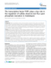
- Chen et al. Journal of Experimental & Clinical Cancer Research 2010, 29:172 Page 4 of 8 http://www.jeccr.com/content/29/1/172 Figure 1 Verification of difference of OPN and c-Myb expression in HCC cell lines. HCCLM6 cells expressed high level of OPN and c-Myb compared with SMMC-7721 cells. (A) Relative OPN and c-Myb mRNA levels in different cell lines by RT-PCR analysis. (B) Real-time quantitative PCR analysis confirmed the difference of c-Myb mRNA expression in different cell lines. Graph depicted relative expression of OPN mRNA normalized to that of GAPDH. The mRNA expression of c-Myb in HCCLM6 was used as control. Data expressed as means ± SD (*P < 0.05, SMMC-7721 vs. HCCLM6). (C)Western blot analysis of OPN and c-Myb protein expression in HCC cell line SMMC-7721 and HCCLM6. Blot was representative of three experiments. Table 2 Differential activity of transcription factorsin two HCC cell lines (SMMC-7721, HCCLM6) with different OPN expression levels (> 2 fold or
- Chen et al. Journal of Experimental & Clinical Cancer Research 2010, 29:172 Page 5 of 8 http://www.jeccr.com/content/29/1/172 containing c-Myb site, a specific retarded complex was M HCC97-L cells, and at a much lower level in observed. In contrast, incubation with the oligonucleotides SMMC-7721 cells, and barely detectable in normal cell containing mutant c-Myb site significantly abrogated bind- line L02 cells. Corresponding to different OPN expres- ing (Figure 2A). In addition, the oligonucleotides contain- sion level (HCCLM6 > MHCC-97-L> SMMC-7721), ing the c-Myb site incubated with nuclear extracts from the expression level of c-Myb increased sharply in SMMC-7721 cells formed a weakly specific retarded com- HCCLM6 cells (Figure 1A). Similar results were plex (Figure 2A). These data demonstrate that the c-Myb obtained in real-time PCR analysis. When normalized site in the OPN promoter can be specifically bound by to the internal standard control, mRNA expression of transcription factor c-Myb in HCCLM6 cells. c-Myb in HCCLM6 cells was significantly higher than To further determine whether the c-Myb site in the OPN SMMC-7721 cells (Figure 1B). Similar to the result of promoter was required for transcription activation, mRNA expression, the difference of c-Myb protein HCCLM6 cells were transfected with an OPN promoter expression between HCCLM6 and SMMC-7721 cells reporter plasmid. To assess whether down-regulation of was also significant. (Figure 1C) c-Myb could suppress the transcription activity of the OPN promoter, HCCLM6 cells were co-transfected with 3.2 Transcription factor c-Myb contributing to the OPN promoter reporter and siRNA targeting c-Myb. transcription activation of the OPN promoter in HCCLM6 Inhibition of c-Myb expression by siRNA significantly cells decreased OPN promoter activity in HCCLM6 cells. In Having shown that c-Myb was over-expressed in HCCLM6 contrast, co-transfection of the OPN promoter reporter cells, we next sought to establish whether it has a function- and a scramble siRNA had no effect on the activity of the ally important role in the regulation of OPN expression. OPN promoter (Figure 2B). These data demonstrate that To establish if functional c-Myb is present in HCCLM6 c-Myb is essential for transcription activity of OPN in cells, nuclear extracts were incubated with the oligonucleo- HCCLM6 cells. tides containing c-Myb-binding site and the formation of specific complexes was determined by EMSA. A double- stranded biotin-labeled oligonucleotides encompassing 3.3 OPN expression was down-regulated after c-Myb was the c-Myb site or a mutant form of the c-Myb site in the inhibited in HCCLM6 cells OPN promoter were used. When nuclear extracts from To further validate c-Myb regulating OPN expression in HCCLM6 cells was incubated with the oligonucleotides HCCLM6 cells, we examined the level of OPN expression Figure 2 Electrophoretic mobility shift sssays (EMSA) of c-Myb binding to OPN promoter and transient transfection analysis of OPN promoter activity. (A). EMSA were performed using nuclear extract prepared from SMMC-7721 and HCCLM6 cells. Assays utilized a labeled probe of 25-nt fragment containing the area of c-Myb binding site in the OPN promoter or a mutant form of the c-Myb binding site (c-Myb- binding site TAACGG was mutated to TATCGG). The blot was representative of three experiments. (B) To confirm the role of c-Myb in the increased OPN protein expression in HCCLM6 cells, Human OPN promoter (-1488 to +185 nt) was cloned into the pGL3-basic luciferase reporter vector. The OPN promoter reporter constructs were transfected into HCCLM6 cells. In certain instances, c-Myb siRNA or scramble siRNA was co- transfected. Luciferase activity was normalized to that of b-galactosidase activity. Data are presented as means ± SD of three experiments. (*P < 0.05, c-Mb siRNA-treated group vs. scramble siRNA group).
- Chen et al. Journal of Experimental & Clinical Cancer Research 2010, 29:172 Page 6 of 8 http://www.jeccr.com/content/29/1/172 Figure 3 The effect of c-Myb on OPN expression of HCCLM6 cells. (A) OPN mRNA expression in HCCLM6 cells transfected with c-Myb siRNA was significantly decreased. (*P < 0.05, vs control). The mRNA expression of OPN in cells transfect with scramble siRNA was used as control. (B) OPN protein expression in HCCLM6 cells transfected with c-Myb siRNA was significantly reduced compared with cells transfected with sramble siRNA. Blot was representative of three experiments. activity. Inhibition of c-Myb by siRNA decreased the i n HCCLM6 cells transfected with siRNA targeting transcription activity of the OPN promoter, reduced the c-Myb. The results showed that inhibition of c-Myb expression of OPN, and compromised the ability for expression by siRNA significantly decreased OPN mRNA migration and invasion of HCC cells. Therefore, our and protein expression (Figure 3A, B), suggesting that results demonstrate that c-Myb plays an important role c-Myb contributes to the regulation of OPN expression in regulating OPN expression in HCC cells, suggesting in HCCLM6 cells. c-Myb might be a novel target for therapeutic intervention. OPN is known to mediate correlates of metastatic 3.4 Migration and invasion of HCCLM6 cells in vitro were biology in a variety of cancers including HCC. Thus, inhibited by c-Myb siRNA modulating OPN expression might be a novel approach As migratory and invasive behaviors are the indicators of suppressing tumor metastasis [17-19]. Transcription of the metastatic potential, we examined migration and invasion of HCCLM6 cells in vitro using the transwell factors are located at endpoints of signaling pathways and integrate various stimuli to determine which genes assay after c-Myb expression was inhibited by c-Myb are expressed or suppressed [10]. To search for the siRNA. The average numbers of HCCLM6 cells trans- determinant transcription factors regulating OPN in fected with c-Myb siRNA migrating toward the condi- HCC, we used transcription factor microassays to com- tioned medium or invading through the Matrigel were pare differential activities of transcription factors significantly fewer than those transfected with scramble siRNA (Migration assay: 17.60 ± 4.04 vs 33.60 ± 4.67, P between two HCC lines with different OPN expression < 0.05; Invasion assay: 8.00 ± 2.55 vs 18.8 ± 4.15, P < 0.05, Figure 4), This result showed that the capabil- ity of migration and invasion in HCCLM6 cells was sig- nificantly decreased after inhibition of c-Myb, suggesting that c-Myb is an important contributor to the migration and invasion of HCC cells. Discussion Metastasis remains one of the major challenges for HCC patients undergoing various therapies including liver resection, local ablation and chemoembolization [2,3]. Previous work at our institute has shown that OPN gene is over-expressed in the metastatic HCC [6]. In this Figure 4 Migration and invasion of HCCLM6 cells in response study, we searched for transcription factors that were to transfection of c-Myb siRNA. The c-Myb siRNA could correlated with OPN expression in HCC cells and significantly inhibit the migration and invasion of HCCLM6 cells compared with cells treated with scramble siRNA (*P < 0.05). The revealed that transcription factor c-Myb was positively migration and invasion assays were assessed by transwell chambers. associated with OPN expression in HCC cells, which can Data were expressed as means ± SD of three experiments. bind the OPN promoter and increase its transcription
- Chen et al. Journal of Experimental & Clinical Cancer Research 2010, 29:172 Page 7 of 8 http://www.jeccr.com/content/29/1/172 Considering transcription factors including AP-1, Sp- level. Through microarray analysis, we found that eleven 1, v-Src, Runx and Tcf-4 participating in the transcrip- transcription factors were highly expressed meanwhile tion regulation of OPN in other types of cancers [20,29], twelve were down-regulated in metastatic HCC cells. and transcription factor along with co-activators or co- Transcription factor c-Myb was selected for further repressors strategically binding to specific sites of target investigation. The reasons are the following: (1) after gene promoters [30], it is possible that c-Myb interacts predicting the potential transcription factors in the OPN with other transcription factors to modulate the promoter in the TRANSFAC database http://www.gene- OPN expression in HCC cells. This requires further regulation.com and searching the reported transcription validation. factor which can bind to the OPN promoter in the Apart from demonstrating the function of c-Myb in literature [20], we have found that among the eleven the regulating OPN expression in HCC cells, we also up-regulated transcription factors, c-Myb and IRF-1 showed that down-regulation of c-Myb by siRNA have the definitive binding sites in the OPN promoter. decreased OPN expression and also inhibited the migra- Although the rests of transcription factors were up- tion and invasion of HCCLM6 cell in vitro, indicating regulated in gene-chip analysis, they lacked the reported that modulating OPN expression by targeting c-Myb binding site in the OPN promoter and may act by the might be a new approach for intervening HCC invasion way of combining with co-activators or other transcrip- and metastasis. Antisense oligodeoxynucleotides target- tion factors, and then together binding to specific sites ing c-Myb, a dominant negative c-Myb or c-Myb of the OPN promoter. (2) Interestingly, Schultz J and vaccine has shown an effective approach for therapy of colleagues [21] have reported that differential capability c-Myb dependent haematopoietic and epithelial malig- of c-Myb binding to -443T/C osteopontin promoter nancies [31-33]. influences osteopontin gene expression in melanoma In summary, our data demonstrate that transcription cells, suggesting the importance of c-Myb regulating factor c-Myb is over-expressed in the metastatic HCC OPN expression in tumor progression. In this study, cells and has a functionally important role in the regula- c-Myb expression increased corresponding to OPN tion of OPN expression, suggesting that c-Myb might be levels in different HCC cell lines, suggesting that c-Myb a new target for therapeutic intervention in the HCC is associated with OPN expression. The differences of invasion and metastasis by modulating OPN expression. OPN expression might reflect the differential activities of c-Myb among HCC cell lines. EMAS and luciferase assays further demonstrated that c-Myb is essential for Acknowledgements transcription activity of OPN in HCC cells. This work was sponsored by grants from China State Key Basic Research The transcription factor c-Myb has a key role in regu- Program Grant (No. 2004CB518708), National Natural Science Foundation of China (No. 81000909), and Shanghai Natural Science Foundation lating the exquisite balance among cell division, differ- (09ZR1406400). entiation and survival and has now been identified as an oncogene involved in some human leukemia and solid Author details 1 Liver Cancer Institute and Zhongshan Hospital, Fudan University, Shanghai, cancers [22-24]. Recently, it is reported that oncogene China. 2Key Laboratory of Carcinogenesis and Cancer Invasion (Fudan c-Myb participates in the process of hepatitis B virus- University), the Chinese Ministry of Education, Shanghai, 200032, China. induced liver carcinogenesis [21]. When inappropriately Authors’ contributions expressed, c-Myb appears to activate important gene CRX and SLY designed the study. CRX, YHX and TCX performed experiments. targets to promote cancer progression and metastasis. CRX drafted the manuscript. All authors read and approved the final These genes include cyclooxygenase-2 (COX-2) [25], manuscript. Bcl-2, BclX(L) [26] and c-Myc [27], which influence Competing interests diverse processes such as angiogenesis, proliferation and The authors declare that they have no competing interests. apoptosis. As for HCC, Yang et al [28] has documented Received: 7 November 2010 Accepted: 30 December 2010 that increased expression of c-Myb and Sp1 binding to Published: 30 December 2010 the methionine adenosyltransferase 2A (MAT2A) pro- moter contribute to the up-regulation of MAT2A References expression. MAT2A can catalyze the formation of S- 1. Parkin DM, Bray F, Ferlay J, Pisani P: Global cancer statistics, 2002. CA Cancer J Clin 2005, 55:74-108. adenosylmethionine to facilitate HCC growth. In the 2. Llovet JM, Burroughs A, Bruix J: Hepatocellular carcinoma. Lancet 2003, present study, we first demonstrate that c-Myb is a new 362:1907-1917. transcription factor of regulating OPN expression in 3. Tang ZY, Ye SL, Liu YK, Qin LX, Sun HC, Ye QH, Wang L, Zhou J, Qiu SJ, Li Y, et al: A decade’s studies on metastasis of hepatocellular carcinoma. J HCC cells, providing at least one mechanism for up- Cancer Res Clin Oncol 2004, 130:187-196. regulation of OPN expression in HCC invasion and 4. Coppola D, Szabo M, Boulware D, Muraca P, Alsarraj M, Chambers AF, metastasis. Yeatman TJ: Correlation of osteopontin protein expression and
- Chen et al. Journal of Experimental & Clinical Cancer Research 2010, 29:172 Page 8 of 8 http://www.jeccr.com/content/29/1/172 pathological stage across a wide variety of tumor histologies. Clin Cancer 27. Greco C, Alvino S, Buglioni S, Assisi D, Lapenta R, Grassi A, Stigliano V, Res 2004, 10:184-190. Mottolese M, Casale V: Activation of c-MYC and c-MYB proto-oncogenes 5. Rangaswami H, Bulbule A, Kundu GC: Osteopontin: role in cell signaling is associated with decreased apoptosis in tumor colon progression. and cancer progression. Trends Cell Biol 2006, 16:79-87. Anticancer Res 2001, 21:3185-3192. 6. Ye QH, Qin LX, Forgues M, He P, Kim JW, Peng AC, Simon R, Li Y, Robles AI, 28. Yang H, Huang ZZ, Wang J, Lu SC: The role of c-Myb and Sp1 in the up- Chen Y, et al: Predicting hepatitis B virus-positive metastatic regulation of methionine adenosyltransferase 2A gene expression in hepatocellular carcinomas using gene expression profiling and human hepatocellular carcinoma. FASEB J 2001, 15:1507-1516. supervised machine learning. Nat Med 2003, 9:416-423. 29. Chakraborty G, Jain S, Behera R, Ahmed M, Sharma P, Kumar V, Kundu GC: Donati V, Boldrini L, Dell’Omodarme M, Prati MC, Faviana P, Camacci T, 7. The multifaceted roles of osteopontin in cell signaling, tumor Lucchi M, Mussi A, Santoro M, Basolo F, Fontanini G: Osteopontin progression and angiogenesis. Curr Mol Med 2006, 6:819-830. expression and prognostic significance in non-small cell lung cancer. 30. Ali SA, Zaidi SK, Dacwag CS, Salma N, Young DW, Shakoori AR, Clin Cancer Res 2005, 11:6459-6465. Montecino MA, Lian JB, van Wijnen AJ, Imbalzano AN, et al: Phenotypic 8. Macri A, Versaci A, Lupo G, Trimarchi G, Tomasello C, Loddo S, Sfuncia G, transcription factors epigenetically mediate cell growth control. Proc Natl Caminiti R, Teti D, Famulari C: Role of osteopontin in breast cancer Acad Sci USA 2008, 105:6632-6637. patients. Tumori 2009, 95:48-52. 31. Abaza MS, Al-Attiyah RJ, Al-Saffar AM, Al-Sawan SM, Moussa NM: Antisense 9. Yeatman TJ, Chambers AF: Osteopontin and colon cancer progression. oligodeoxynucleotide directed against c-myb has anticancer activity and Clin Exp Metastasis 2003, 20:85-90. potentiates the antiproliferative effect of conventional anticancer drugs 10. Stein GS, Stein JL, Van Wijnen AJ, Lian JB, Montecino M, Croce CM, Choi JY, acting by different mechanisms in human colorectal cancer cells. Ali SA, Pande S, Hassan MQ, et al: Transcription factor-mediated Tumour Biol 2003, 24:241-257. epigenetic regulation of cell growth and phenotype for biological 32. Ramsay RG, Barton AL, Gonda TJ: Targeting c-Myb expression in human control and cancer. Adv Enzyme Regul 50:160-167. disease. Expert Opin Ther Targets 2003, 7:235-248. 11. Kajanne R, Miettinen P, Tenhunen M, Leppa S: Transcription factor AP-1 33. Funato T, Satou J, Kozawa K, Fujimaki S, Miura T, Kaku M: Use of c-myb promotes growth and radioresistance in prostate cancer cells. Int J Oncol antisense oligonucleotides to increase the sensitivity of human colon 2009, 35:1175-1182. cancer cells to cisplatin. Oncol Rep 2001, 8:807-810. 12. Song Y, Wu J, Oyesanya RA, Lee Z, Mukherjee A, Fang X: Sp-1 and c-Myc doi:10.1186/1756-9966-29-172 mediate lysophosphatidic acid-induced expression of vascular Cite this article as: Chen et al.: Transcription factor c-Myb promotes the endothelial growth factor in ovarian cancer cells via a hypoxia-inducible invasion of hepatocellular carcinoma cells via increasing osteopontin factor-1-independent mechanism. Clin Cancer Res 2009, 15:492-501. expression. Journal of Experimental & Clinical Cancer Research 2010 29:172. 13. Blyth K, Cameron ER, Neil JC: The RUNX genes: gain or loss of function in cancer. Nat Rev Cancer 2005, 5:376-387. 14. Li Y, Tian B, Yang J, Zhao L, Wu X, Ye SL, Liu YK, Tang ZY: Stepwise metastatic human hepatocellular carcinoma cell model system with multiple metastatic potentials established through consecutive in vivo selection and studies on metastatic characteristics. J Cancer Res Clin Oncol 2004, 130:460-468. 15. Deregibus MC, Cantaluppi V, Doublier S, Brizzi MF, Deambrosis I, Albini A, Camussi G: HIV-1-Tat protein activates phosphatidylinositol 3-kinase/AKT- dependent survival pathways in Kaposi’s sarcoma cells. J Biol Chem 2002, 277:25195-25202. 16. Hijiya N, Setoguchi M, Matsuura K, Higuchi Y, Akizuki S, Yamamoto S: Cloning and characterization of the human osteopontin gene and its promoter. Biochem J 1994, 303(Pt 1):255-262. 17. Shevde LA, Das S, Clark DW, Samant RS: Osteopontin: An Effector and an Effect of Tumor Metastasis. Curr Mol Med 2010, 10(1):71-81. Johnston NI, Gunasekharan VK, Ravindranath A, O’Connell C, Johnston PG, 18. El-Tanani MK: Osteopontin as a target for cancer therapy. Front Biosci 2008, 13:4361-4372. 19. Jain S, Chakraborty G, Bulbule A, Kaur R, Kundu GC: Osteopontin: an emerging therapeutic target for anticancer therapy. Expert Opin Ther Targets 2007, 11:81-90. 20. Wai PY, Kuo PC: Osteopontin: regulation in tumor metastasis. Cancer Metastasis Rev 2008, 27:103-118. 21. Schultz J, Lorenz P, Ibrahim SM, Kundt G, Gross G, Kunz M: The functional -443T/C osteopontin promoter polymorphism influences osteopontin gene expression in melanoma cells via binding of c-Myb transcription factor. Mol Carcinog 2009, 48:14-23. 22. Ramsay RG, Gonda TJ: MYB function in normal and cancer cells. Nat Rev Cancer 2008, 8:523-534. Submit your next manuscript to BioMed Central 23. Ramsay RG: c-Myb a stem-progenitor cell regulator in multiple tissue compartments. Growth Factors 2005, 23:253-261. and take full advantage of: 24. Fang F, Rycyzyn MA, Clevenger CV: Role of c-Myb during prolactin- induced signal transducer and activator of transcription 5a signaling in • Convenient online submission breast cancer cells. Endocrinology 2009, 150:1597-1606. 25. Ramsay RG, Friend A, Vizantios Y, Freeman R, Sicurella C, Hammett F, • Thorough peer review Armes J, Venter D: Cyclooxygenase-2, a colorectal cancer nonsteroidal • No space constraints or color figure charges anti-inflammatory drug target, is regulated by c-MYB. Cancer Res 2000, • Immediate publication on acceptance 60:1805-1809. Biroccio A, Benassi B, D’Agnano I, D’Angelo C, Buglioni S, Mottolese M, 26. • Inclusion in PubMed, CAS, Scopus and Google Scholar Ricciotti A, Citro G, Cosimelli M, Ramsay RG, et al: c-Myb and Bcl-x • Research which is freely available for redistribution overexpression predicts poor prognosis in colorectal cancer: clinical and experimental findings. Am J Pathol 2001, 158:1289-1299. Submit your manuscript at www.biomedcentral.com/submit
CÓ THỂ BẠN MUỐN DOWNLOAD

-

báo cáo khoa học: " Uncharacterized conserved motifs outside the HD-Zip domain in HD-Zip subfamily I transcription factors; a potential source of functional diversity"
 19 p |
  47
|
47
|  5
5
-

báo cáo khoa học: " Roles of arabidopsis WRKY18, WRKY40 and WRKY60 transcription factors in plant responses to abscisic acid and abiotic stress"
15 p | 58
| 5
-

Báo cáo y học: "Transcriptional regulation of collagenase (MMP-1, MMP-13) genes in arthritis: integration of complex signaling pathways for the recruitment of gene-specific transcription factor"
8 p | 58
| 4
-

báo cáo khoa học: " Arabidopsis WRKY2 transcription factor mediates seed germination and postgermination arrest of development by abscisic acid"
14 p | 53
| 4
-

báo cáo khoa học: " The HaDREB2 transcription factor enhances basal thermotolerance and longevity of seeds through functional interaction with HaHSFA9"
12 p | 50
| 4
-

báo cáo khoa học: " SoyDB: a knowledge database of soybean transcription factors"
12 p | 59
| 4
-

báo cáo khoa học: "SND2, a NAC transcription factor gene, regulates genes involved in secondary cell wall development in Arabidopsis fibres and increases fibre cell area in Eucalyptus"
51 p | 57
| 4
-

báo cáo khoa học: "The grapevine guard cell-related VvMYB60 transcription factor is involved in the regulation of stomatal activity and is differentially expressed in response to ABA and osmotic stress"
15 p | 59
| 4
-
báo cáo khoa học: "A single amino acid change within the R2 domain of the VvMYB5b transcription factor modulates affinity for protein partners and target promoters selectivity"
14 p | 42
| 4
-

báo cáo khoa học: " WRKY Transcription Factors Involved in Activation of SA Biosynthesis Genes"
12 p | 49
| 4
-

báo cáo khoa học: "Loss-of-function mutations affecting a specific Glycine max R2R3 MYB transcription factor result in brown hilum and brown seed coats"
12 p | 40
| 3
-

báo cáo khoa học: " DNA polymorphisms and haplotype patterns of transcription factors involved in barley endosperm development are associated with key agronomic traits"
11 p | 53
| 3
-

báo cáo khoa học: "Induction of endogenous γ-globin gene expression with decoy oligonucleotide targeting Oct-1 transcription factor consensus sequence"
11 p | 46
| 3
-

báo cáo khoa học: " Identification and expression analysis of WRKY transcription factor genes in canola (Brassica napus L.) in response to fungal pathogens and hormone treatments"
19 p | 60
| 3
-

báo cáo khoa học: " Transcriptional profiling of Medicago truncatula under salt stress identified a novel CBF transcription factor MtCBF4 that plays an important role in abiotic stress responses"
19 p | 47
| 3
-
báo cáo khoa học: " The transcription factor PHR1 plays a key role in the regulation of sulfate shoot-to-root flux upon phosphate starvation in Arabidopsis"
10 p | 82
| 3
-

báo cáo khoa học: " Identification of a GCC transcription factor responding to fruit colour change events in citrus through the transcriptomic analyses of two mutants"
14 p | 72
| 3


Chịu trách nhiệm nội dung:
Nguyễn Công Hà - Giám đốc Công ty TNHH TÀI LIỆU TRỰC TUYẾN VI NA
LIÊN HỆ
Địa chỉ: P402, 54A Nơ Trang Long, Phường 14, Q.Bình Thạnh, TP.HCM
Hotline: 093 303 0098
Email: support@tailieu.vn
Giấy phép Mạng Xã Hội số: 670/GP-BTTTT cấp ngày 30/11/2015 Copyright © 2022-2032 TaiLieu.VN. All rights reserved.

