


Báo cáo sinh học: "Characterization and targeting of phosphatidylinositol-3 kinase (PI3K) and mammalian target of rapamycin (mTOR) in renal cell cancer"
lượt xem 4
download
 Download
Vui lòng tải xuống để xem tài liệu đầy đủ
Download
Vui lòng tải xuống để xem tài liệu đầy đủ
Tuyển tập báo cáo các nghiên cứu khoa học quốc tế ngành hóa học dành cho các bạn yêu hóa học tham khảo đề tài: Characterization and targeting of phosphatidylinositol-3 kinase (PI3K) and mammalian target of rapamycin (mTOR) in renal cell cancer
Bình luận(0) Đăng nhập để gửi bình luận!
Nội dung Text: Báo cáo sinh học: "Characterization and targeting of phosphatidylinositol-3 kinase (PI3K) and mammalian target of rapamycin (mTOR) in renal cell cancer"
- Elfiky et al. Journal of Translational Medicine 2011, 9:133 http://www.translational-medicine.com/content/9/1/133 RESEARCH Open Access Characterization and targeting of phosphatidylinositol-3 kinase (PI3K) and mammalian target of rapamycin (mTOR) in renal cell cancer Aymen A Elfiky1, Saadia A Aziz2, Patricia J Conrad2, Summar Siddiqui3, Wolfgang Hackl4, Michel Maira4, Camp L Robert3 and Harriet M Kluger2* Abstract Background: PI3K and mTOR are key components of signal transduction pathways critical for cell survival. Numerous PI3K inhibitors have entered clinical trials, while mTOR is the target of approved drugs for metastatic renal cell carcinoma (RCC). We characterized expression of p85 and p110a PI3K subunits and mTOR in RCC specimens and assessed pharmacologic co-targeting of these molecules in vitro. Methods: We employed tissue microarrays containing 330 nephrectomy cases using a novel immunofluorescence- based method of Automated Quantitative Analysis (AQUA) of in situ protein expression. In RCC cell lines we assessed synergism between PI3K and mTOR inhibitors and activity of NVP-BEZ235, which co-targets PI3K and mTOR. Results: p85 expression was associated with high stage and grade (P < 0.0001 for both). High p85 and high mTOR expression were strongly associated with decreased survival, and high p85 was independently prognostic on multi- variable analysis. Strong co-expression of both PI3K subunits and mTOR was found in the human specimens. The PI3K inhibitor LY294002 and rapamycin were highly synergistic in all six RCC cell lines studied. Similar synergism was seen with all rapamycin concentrations used. NVP-BEZ235 inhibited RCC cell growth in vitro with IC50s in the low hM range and resultant PARP cleavage. Conclusions: High PI3K and mTOR expression in RCC defines populations with decreased survival, suggesting that they are good drug targets in RCC. These targets tend to be co-expressed, and co-targeting these molecules is synergistic. NVP-BEZ235 is active in RCC cells in vitro; suggesting that concurrent PI3K and mTOR targeting in RCC warrants further investigation. Background metastases [5]. The immunogenicity of RCC has been the basis for use of cytokines such as interleukin-2 and Renal cell carcinoma (RCC) is among the ten leading interferon for metastatic RCC, which benefit about 15% causes of cancer-related deaths, and the incidence has of patients [6,7]. Alternative drugs are needed for been increasing by approximately 2% per year [1-4]. patients who are not responsive and/or are intolerant to RCC is typically resistant to chemotherapy and radiation these therapies. therapy. The five-year survival rate is 90.8% for localized A growing understanding of the pathogenesis of RCC RCC (confined to primary site), 63.3% for cases with has enabled us to identify factors pertinent to develop- regional disease, and 11.1% in patients with distant ment of RCC-targeting therapies. The discovery of VHL tumor-suppressor gene inactivation and consequent * Correspondence: Harriet.Kluger@yale.edu hypoxia-induced factor (HIF) activation of genes and 2 Section of Medical Oncology, Yale Cancer Center, Yale University, 333 Cedar downstream pathways important to tumor progression, St, New Haven, CT 06520, United States of America Full list of author information is available at the end of the article © 2011 Elfiky et al; licensee BioMed Central Ltd. This is an Open Access article distributed under the terms of the Creative Commons Attribution License (http://creativecommons.org/licenses/by/2.0), which permits unrestricted use, distribution, and reproduction in any medium, provided the original work is properly cited.
- Elfiky et al. Journal of Translational Medicine 2011, 9:133 Page 2 of 10 http://www.translational-medicine.com/content/9/1/133 specific [21]. Activation of mTOR can also up-regulate h ave provided the impetus for development of new HIF gene expression, which, in patients with VHL muta- agents that target angiogenesis and proliferation path- tions, can magnify HIF accumulation and expression of ways. Specifically, therapies that have demonstrated ben- HIF-inducible genes. efit in metastatic RCC include the small molecule In RCC, data from moderate sized studies support tyrosine kinase inhibitors sunitinib, sorafenib and pazo- activation of the mTOR signaling pathway. Immunos- panib [8-10], the anti-VEGF antibody bevacizumab [11], tained tissue microarray sections of 150 RCCs showed temsirolimus and everolimus, inhibitors of mTOR, significantly higher expression of phosphorylated which has been implicated in HIF transcription [12]. p70S6K (p-p70S6K), phosphorylated-mTOR (p-mTOR) Although these new agents improve progression free and phosphorylated Akt (p-Akt) compared to normal survival, none have shown a statistically significant kidney, p < 0.05 [22]. Additionally, Robb et. al found improvement in overall survival. In effect none are cura- strong co-expression of phosphorylated-S6 (p-S6) and p- tive, and duration of response is often limited. mTOR in 14 of 29 clear cell carcinomas [23]. Signifi- The PI3K pathway is activated and/or up-regulated in cantly decreased mean disease-free survival was cancers, and plays a critical role in tumor progression observed when caveolin was co-expressed p-AKT, p- [13,14]. There are three classes of PI3Ks; each has its mTOR, p-S6 and phosphorylated-4EBP1 [24]. Therefore, own substrate specificity [15,16]. Class I A PI3Ks, the inhibition of mTOR has the potential to inhibit tumor most widely implicated in cancer, primarily phosphory- progression at multiple levels, and along with PI3K inhi- late phosphatidylinositol-4,5-bisphosphate to generate bition is particularly attractive for development for RCC the second messenger phosphatidylinositol-3,4,5-trispho- treatment. sphate. This enzyme is a heterodimer consisting of a Despite the literature demonstrating the importance of p85 regulatory and a p110 catalytic subunit. Class I A PI3K and mTOR in RCC pathogenesis, there is limited PI3K is activated by receptor tyrosine kinase (RTK) sig- information on total protein expression and co-expres- naling [17,18]. Binding of p85 to activated RTKs serves sion in large cohort RCC tumor studies in the context both to recruit the p85-p110 heterodimer to the plasma of patient survival. A previous meta-analysis of mRNA membrane, where its substrate phosphatidylinositol-4,5- expression microarrays revealed signature alternations in bisphosphate resides, and to relieve basal inhibition of p110a by p85. Downstream mediators, including Akt the PI3K/AKT pathway that are associated with tumor versus benign renal tissue [25]. Merseberger et. al deter- and PDK1, directly bind to phosphatidylinositol-3,4,5- mined expression patterns of PI3K, PTEN, p-Akt for trisphosphate. Akt phosphorylates several cellular pro- possible prognostic value in 176 RCC cases, and found teins, including GSK3, GSK3ß, FOXO transcription fac- that activation of the PI3K pathway is associated with tors, MDM2, and BAD, to facilitate cell survival and cell adverse clinical outcome [26]. In a more recent study, cycle entry [15]. Akt phosphorylation also results in acti- metastatic RCC samples from 132 patients and a subset vation of the mTOR/raptor complex, which regulates of 25 matched primary RCC specimens were stained for protein synthesis, cell growth, and proliferation [19]. PI3K, PTEN, p-Akt, p-mTOR, and p70S6. p-mTOR was There are two distinct functional mTOR complexes, associated with decreased survival [27]. mTORC1 and mTORC2. mTORC1 (rapamycin sensi- The relevance of the PI3K/Akt/mTOR signaling path- tive) consists of mTOR and Raptor, and its activation way in RCC is the focus of ongoing research. Single- results in phosphorylation of p70S6 and 4E-BP1. agent mTOR inhibitors have some efficacy in RCC, and mTORC2 consists of mTOR and the rapamycin-insensi- co-targeting additional PI3K pathway members along tive companion of mTOR (Rictor), and causes Akt phos- with mTOR might be a valuable strategy for overcoming phorylation. Akt promotes protein synthesis and cell the escape mechanisms that can limit activity of mTOR growth by alleviating TSC1/2 suppression of mTOR, inhibitors. Seeing that PI3K inhibitors are currently in allowing the latter to act as part of the mTOR-raptor clinical development, our purpose was to assess co- complex on 4EBP1 and S6 kinases. expression of PI3K subunits, p110 a and p85, and Activation of the PI3K pathway in cancers has been mTOR in RCC tumors in a quantitative fashion and demonstrated in numerous studies. The two most com- study pharmacological co-inhibition of these targets in mon mutations are of p110a (PIK3CA) and loss of the vitro. To thoroughly assess co-expression of mTOR and tumor suppressor PTEN. Amplification of PIK3CA and PI3K subunits in a quantitative fashion, we employed a Akt are occasionally observed in epithelial cancers [15]. Recently, high expression of the PI3K/p110 g isoform new method of automated, quantitative analysis (AQUA) of in situ protein expression, which has been was implicated in pancreatic adenocarcinoma progres- validated and used in a number of previous studies sion [20]. There is specific evidence of PI3K pathway [28,29]. Expression of mTOR and PI3K, p85 and p110a activation in RCC; it is constitutively activated in RCC subunits was assessed in a large cohort of human cells regardless of VHL status, and activation is tumor
- Elfiky et al. Journal of Translational Medicine 2011, 9:133 Page 3 of 10 http://www.translational-medicine.com/content/9/1/133 rabbit) HRP-decorated polymer backbone (Envision, specimens and we determined associations with stan- Dako North America, Carpinteria, CA) was used as a dard clinical/pathological variables. We further studied secondary reagent. To create a tumor mask, slides were co-targeting these molecules in RCC cell lines, and simultaneously incubated with rabbit (for p85) or mouse assessed the effects on cell growth and apoptosis using a (for p110a and mTOR) anti-cytokeratin (Dako) at 1:100, clinical quality compound, NVP-BEZ235. and visualized with an appropriate secondary antibody Methods conjugated to Alexa 488 (Molecular Probes, Inc., Eugene, OR). The target antibody was visualized with Tissue Microarray (TMA) Construction Cy5-tyramide (Perkin-Elmer, Boston, MA, and mounted Briefly, representative regions were selected for coring with ProLong Gold antifade reagent with 4, 6-diami- by pathologists based on the corresponding H&E- dine-2-phenylindole (DAPI) (Invitrogen, Carlsbad, CA). stained full sections. The tissue microarray was con- To verify that there was no background staining from structed with single 0.6 mm-diameter cores of each case the Alexa 488, slides were stained with and without Cy5 spaced 0.8 mm apart in a grid format using a Tissue tyramide. Microarrayer (Beecher Instruments, Sun Prairie, WI). The tissue microarray block was then cut into 5 μm sec- tions with a microtome, adhered to the slide by an Automated Image Acquisition and Analysis (AQUA) adhesive tape-transfer method (Instrumedics, Inc., Hack- Images were acquired and analyzed using extensively ensack, NJ) and UV crosslinked. TMAs were con- described algorithms [30]. Briefly, monochromatic, high- structed using RCC cores from 330 patients. Tumors resolution (1024 × 1024 pixel) images were obtained of were represented by two cores from different areas of each histospot. Tumor was distinguished from stroma the specimen. Specimens and clinical information were by cytokeratin/streptavidin signal. Cell surface coales- collected with approval of a Yale University Institutional cence of cytokeratin was used to localize membranes Review Board. Histological subtypes included clear cell and DAPI to identify nuclei. The target signal (p85, p110a or mTOR) from the pixels within the cytoplasm (71%), papillary (14%), chromophobe (2%), mixed histol- ogy (4%), oncocytomas (6%), and sarcomatoid tumors was normalized to area of tumor mask and scored on a (3%). Oncocytomas were excluded from survival ana- scale of 0-255 (the AQUA score). Histospots were lyses given that they have low metastatic potential and excluded if the tumor mask represented < 3% of the his- are curable by nephrectomy. Eight percent had stage II tospot area. and III disease, 56% had stage I and 28% had stage IV disease. 12% were Fuhrman nuclear grade I, 52% grade Statistical Analysis II, 27% grade III and 9% grade IV. Specimens were Statview and JMP 5.0 software were used (SAS Institute, resected between 1987 and 1999; follow-up time was 2- Cary, NC). AQUA scores for replicate tumor cores were 240 months (median-89.7). Age at diagnosis was 25-87 averaged. Prognostic significance of parameters was years (median-63). No patients were treated with suniti- assessed using the Cox proportional hazards model with nib, sorafenib, pazopanib, bevacizumab, everolimus or RCC-specific survival as an endpoint. Associations temsirolimus, although a few were previously treated between continuous AQUA scores of the target and with interferon or interleukin-2 in the metastatic setting. clinical and pathological parameters were assessed using Performance status, LDH, hemoglobin and calcium ANOVA. For demonstrating survival analyses, continu- levels were unavailable. ous target AQUA scores were divided into quartiles and survival curves were generated using the Kaplan-Meier method, with significance evaluated using the Mantel- Immunofluorescence Cox log-rank test. One set of two slides (each containing a core from dif- ferent areas of tumor for each patient) was stained indi- vidually for the three target markers, p85 and p110 a Human Cell Lines PI3K subunits, and mTOR. Antibody validation was A498, ACHN, Caki-1, Caki-2, 769-P, and 786-0 cells conducted by immunoblots to verify presence of a single were obtained from American Type Culture Collection and maintained per the supplier’s instructions (Mana- band of the appropriate size (not shown). AQUA stain- ing was performed as described [30]. Slides were incu- ssas, VA). bated with mouse monoclonal anti-human PI3K p85, (BD transduction Laboratories, Franklin Lakes, NJ) at Viability and Synergism Studies 1:50, rabbit anti-human PI3K p110a, clone C73F8 (Cell At a density of 103, cells were plated in triplicate in 96 Signaling Technology, Danvers, MA) at 1:200 or rabbit well plates with growth medium and allowed to monoclonal anti-human mTOR, clone 7C10 at 1:40, adhere overnight. The PI3K inhibitor, LY294002 (LC (Cell Signaling Technology). Goat anti-mouse (or anti- Laboratories, Woburn MA), was used alone and in
- Elfiky et al. Journal of Translational Medicine 2011, 9:133 Page 4 of 10 http://www.translational-medicine.com/content/9/1/133 We found a moderate correlation between expression c ombination with the mTORC1 inhibitor, Rapamycin of the two PI3K subunits ( r = 0.129, P = 0.046) and (LC Laboratories), at 5-25 μmol/L and 0.02-0.5 μmol/ stronger correlations between mTOR and the two PI3K L, respectively for 48 hours. NVP-BEZ235 was studied subunits; r = 0.251 for p85 and r = 0.385 for p110a (P alone at concentrations of 10-500 h mol/L for 48 < 0.0001 for both). Expression of both PI3K sub-units hours. The relative number of viable cells was and mTOR was significantly higher in sarcomatoid assessed by the luminometric Cell-Titer Glo assay tumors (P = 0.002, P = 0.04 and P = 0.02, respectively), (Promega), and luminescent quantification was mea- and expression of p110 a and mTOR was also signifi- sured using a Viktor plate reader (Perkin Elmer). cantly higher in oncocytomas. Expression of mTOR was Using CalcuSyn software (Biosoft, Ferguson, MO), also somewhat higher in papillary carcinomas (P = 0.02) results were analyzed for synergistic, additive, or (Figure 2A). We found significant differences in p85 antagonistic effects. Synergism is indicated by a Com- expression between early and late stage disease, and bination Index (CI) of < 0.9, additivity by CI values of expression of mTOR was higher in high grade tumors), 0.9-1.1, and antagonism by CI > 1.1 [31]. To deter- (Figure 2B), ( P < 0.0001 for all). p85 expression was mine the IC 50 for NVP-BEZ235, we used XLfit soft- higher in cases with high Fuhrman grade (P < 0.0001) ware (IDBS, Surrey, UK). (Figure 2B). No association was found between expres- sion of p110a and stage or grade (Figure 2B). Immunoblots After treatment with NVP-BEZ235 at 100 hM for 1, 6 AQUA provides continuous output scores rather than divisions into “high” and “low” categories. We therefore and 24 hours, cells were lysed using standard methods. arbitrarily divided the continuous AQUA scores for the Primary rabbit anti-human antibodies were used: phos- porylated AKT Ser-473, phosphorylated p70S6K Thr389 three markers into quartiles. For p85 and mTOR, survi- and phosphorylated pS6 Ser235/236 at 1:1000 (Cell Signal- val of patients with AQUA scores in the top quartile was significantly lower (Figure 2C). Using Cox univariate ing Technologies). To assess apoptosis, cells were trea- ted with 100 hM, 500hM and 1000 hM NVP-BEZ235 analysis of continuous AQUA scores, high p85 PI3K expression was strongly associated with decreased survi- for 72 hours. Levels of cleaved PARP (rabbit polyclonal val (P < 0.0001). No association was found between con- antibody, Cell Signaling) and cleaved caspase-2 (mouse tinuous p110a scores and survival (P = 0.8287), while monoclonal antibody, BD Biosciences) were measured at 1:1000 for both. Mouse or rabbit anti-b-actin antibodies continuous mTOR AQUA scores were associated with decreased survival (P = 0.0099). (Sigma Aldrich) were used to visualize protein gel Using the Cox Proportional Hazards Model, we per- loading. formed multivariable analyses. Expression of p85 Results retained its independent prognostic value, as did stage and Fuhrman grade (Table 1). AQUA analyses To assess intra-tumor heterogeneity, two separate slides, each containing a core from a different area of the Synergism between PI3K and mTOR inhibition Using 5, 25 and 50 μM of LY294002, we studied syner- tumor for each patient, were used for each marker (p85, p110a and mTOR). None of the markers had nuclear gism with a range of concentrations of rapamycin (20, 100 and 500hM). Synergism was seen in all six cell lines staining, and only membranous/cytoplasmic compart- at 5 μM LY294002 with all three concentrations of rapa- ments were analyzed. By log-regression analysis, scores for matching histospots were highly correlated (R = 0.7 mycin (Table 2). We note that the degree of viability for p85, R = 0.8 for p110 a and R = 0.7 for mTOR). inhibition with all concentrations of rapamycin was Scores from the automated analysis are continuous from almost identical, as shown in Figure 3, using A498 and 0 to 255. The range of AQUA scores was 3.6-91.4 (med- Caki-2 cells as examples (p > 0.5 for comparison ian-32.3) for p85, 1.8-46.5 (median-7.9) for p110a and between combinations of rapamycin and LY294002). 4.1-75.5 (median-25.38) for mTOR. Examples of strong Viability of cells treated with LY294002, rapamycin or AQUA staining for p85, p110a and mTOR are shown the combination is calculated as a percent of the viabi- in Figure 1A-C. lity of the untreated (control) cells. Scores from the two slides were combined for a single dataset. Spots were deemed uninterpretable if they had Activity of the dual PI3K-mTOR inhibitor NVP-BEZ235 in insufficient tumor, loss of tissue or abundant necrosis. A RCC cell lines composite score was formed by averaging the scores. Given the synergism seen between the LY294002 and rapamycin in RCC cell lines, we studied the in vitro Patients with only one core were excluded from the ana- lysis. The combined dataset had 264 cases for p85, 237 activity of NVP-BEZ235, which has been given to solid for p110a and 267 for mTOR. tumor patients in phase I clinical trials. In all 6 RCC
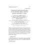
- Elfiky et al. Journal of Translational Medicine 2011, 9:133 Page 5 of 10 http://www.translational-medicine.com/content/9/1/133 Figure 1 Automated, Quantitative Analysis (AQUA) of expression of p85, p110a and mTOR in renal cell carcinoma. AQUA uses cytokeratin to create a tumor mask (two upper left quadrants at × 10). Cytokeratin staining was cytoplasmic and the mask is made by filling in holes (lower left quadrants on left). 4’, 6-diamidino-2-phenylindole (DAPI) defines the nuclear compartment within the tumor mask, which is then subtracted from the tumor mask to create a cytoplasmic compartment within the tumor mask. Target expression (A - p85, B - p110a and C - mTOR) expression is measured within the cytoplasmic compartments, within the tumor mask (lower right quadrants), and each clinical case is assigned a score based on pixel intensity per unit area within the tumor mask. Squares on right show × 40 magnification. in the tissue microarray. The correlation with p110a was particularly strong.
- Elfiky et al. Journal of Translational Medicine 2011, 9:133 Page 6 of 10 http://www.translational-medicine.com/content/9/1/133 Figure 2 Associations between marker expression and clinical/pathological variables. (A) Associations between target expression (p85, p110a and mTOR) and histologic subtype. Expression of all three targets was higher in sarcomatoid tumors. (B) Associations between target expression and tumor stage and Fuhrman grade. p85 and mTOR were associated with high grade and p85 with high stage. p110a levels were not associated with stage or grade. (C) Kaplan-Meier survival curves of quartiles of AQUA scores for the three targets. High levels of both p85 and mTOR were associated with decreased survival. hours. b-actin is shown as a loading control. p-P70S6K cell lines the IC 50 s of this compound were in the hM levels are undetectable at all concentrations and time range (Table 3). points studied, whereas levels of p-Akt and p-S6 decrease after 4 hours of drug exposure in a dose- NVP-BEZ235 target inhibition and induction of apoptosis dependent fashion (Figure 4). Exposure of RCC cells to Targets of NVP-BEZ235, p-P70S6K, p-Akt and p-S6 were decreased in Caki-1, 769-P, A498 and 786-0 cells with exposure to the drug. Cells were exposed to 0.1 Table 2 Combination indexes assessing synergism/ and 1.0 μ M NVP-BEZ235, or DMSO for 4 and 24 additivity/antagonism in rapamycin and LY294002 LY294002 Rapamycin A498 ACHN CAKI- CAKI- 769- 786- Table 1 PI3K, p85 and p110 and mTOR Multivariate 1 2 P O Analysis* (nM) (nM) (CI) (CI) (CI) (CI) (CI) (CI) P-value Marker Relative 95% Confidence 5000 500 0.476 0.371 0.593 0.201 0.430 0.242 Risk Interval 5000 100 0.461 0.193 0.492 0.212 0.444 0.262 PI3K - p85 1.026 [1.004 - 1.048] 0.02 5000 20 0.441 0.173 0.472 0.184 0.385 0.281 PI3K - p110a 0.946 [0.872 - 1.025] 0.1762 25000 500 1.388 0.678 1.409 1.270 1.356 0.830 mTOR 1.017 [0.992 - 1.042] 0.178 25000 100 1.420 0.586 1.193 1.074 1.430 0.847 Stage 4.087 [2.120 - 5.469] < 0.0001 25000 20 1.393 0.484 1.227 1.020 1.530 0.910 Nuclear Grade 2.898 [1.536 - 5.469] 0.001 Synergism is indicated by a CI of < 0.9, additivity by CI values between 0.9 *Statistically significant markers are bolded and 1.1, and antagonism by CI of > 1.1.
- Elfiky et al. Journal of Translational Medicine 2011, 9:133 Page 7 of 10 http://www.translational-medicine.com/content/9/1/133 Figure 3 Synergism between PI3K and mTOR inhibition. Cell viability assays in A498 (A) and Caki-2 cells (B): Cells were treated with LY294002 alone, rapamycin alone (at three concentrations) or the combination of LY294002 and rapamycin. The combinations were highly synergistic, with similar viability seen for all three concentrations of rapamycin used. There was no significant difference between the different combination therapies (p > 0.5 for all). grade or survival. mTOR was associated with survival ascending concentrations of NVP-BEZ235 at 72 hours on uni-variable analysis; however on multi-variable ana- resulted in PARP cleavage and cleavage of caspase-2 lysis it lost its independence as a prognostic marker. (Additional file 1, Figure 1). Caspase-2 was selected as it The association between PI3K and mTOR and disease has been shown in other publications to be activated in progression suggests that they might be valuable drug response to treatment with NVP-BEZ235 [32,33]. targets. The p85 subunit has both a regulatory and a sti- Discussion mulatory role in activity of the PI3K pathway. The p110a subunit is thought to be stimulatory only. The We studied expression patterns of PI3K pathway mem- functional roles of the subunits, in conjunction with our bers critical for cell survival and proliferation in a large findings of stronger co-expression of the p110a subunit cohort of RCC specimens. We used a novel method of and mTOR, suggest that pharmacological co-targeting quantitative immunofluorescence, AQUA. This method of p110 a and mTOR might be a useful strategy for is void of the pathologist-based bias associated with treating RCC. DAB staining. The p85 subunit was associated with high Activation of the PI3K-Akt pathway and its role in grade, high stage and decreased survival, and remained RCC progression was previously evaluated in a small an independent prognostic marker on multi-variable analysis. p110 a was not associated with high stage, study of 48 patients with RCC by immunohistochemis- try using an antibody to p-Akt, showing that p-Akt was associated with high tumor grade and metastatic disease. Table 3 IC50 values of RCC cell lines treated with NVP- In addition, high p-Akt immunostaining was signifi- BEZ235 cantly associated with decreased cancer-specific survival IC50 NVP-BEZ235 (hM) Cell Line [34]. Activation of the PI3K-Akt signaling pathway was A498 25 also examined in RCC cell lines treated with PI3K inhi- ACHN 30 bitors, wortmannin and LY294002 in previous studies Caki-1 26 [21]. This study demonstrated that the PI3K-Akt signal- Caki-2 164 ing pathway is constitutively activated in RCC cells, 769-P 67 regardless of VHL status, and that activation of this 786-0 87 pathway is tumor specific relative to corresponding nor- mal renal tissue [21]. The same group conducted in vivo In all 6 RCC cell lines the IC50s of NVP-BEZ235 were in the hM range.
- Elfiky et al. Journal of Translational Medicine 2011, 9:133 Page 8 of 10 http://www.translational-medicine.com/content/9/1/133 Figure 4 Effects of NVP-BEZ235 on renal cell carcinoma cells in vitro. Targets of NVP-BEZ235, p-P70S6K, p-Akt and p-S6 were decreased in Caki-1, 769-P, A498 and 786-0 cells with exposure to the drug. Cells were exposed to 0.1 and 1.0 μM NVP-BEZ235, or DMSO (vehicle) for 4 and 24 hours. b-actin is shown as a loading control. II trials [43]. GDC-0941 (Genentech/Piramed/Roche) is s tudies of nude mice bearing human RCC xenografts a Pan-class I PI3K inhibitor in Phase I trials. The Exe- treated with LY294002. LY294002 inhibited tumor lexis compounds XL-147 and XL-765 are also in Phase I growth, and p-Akt was reduced in these tumors [21]. trials. The recognition that the PI3K pathway has gained as a In our models, activity of LY294002 alone in RCC cell putative target in cancer therapy is reflected by the lines was limited, with IC50s in the micromolar range. recent increase in literature regarding novel PI3K inhibi- tors [4,15,35-37]. Preliminary data from a phase I study While this compound is also a weak inhibitor of mTOR, of the oral PI3K/mTOR inhibitor, NVP-BEZ235 was there are a number of potential mechanisms of resis- conducted in patients with histologically confirmed, tance to PI3K inhibitors when administered alone. For advanced, unresectable solid tumors[38]. The findings in example, Akt can be activated by PI3K-independent the breast and colorectal patients which were reported mechanisms such as mTORC2 activation [44]. Members showed that NVP-BEZ235 was well tolerated with a of the MAPK pathway have been shown to activate Akt favorable safety profile. as well: ERK and RSK inhibit TSC2, which can result in There is also emerging evidence that mTOR activation mTOR activation despite effective PI3K inhibition, as may play a role in promoting cell survival through the reviewed [45]. Others have shown that inhibition of activation of antiapoptotic proteins that contribute to PI3K results in down-regulation of S6K, a negative regu- tumor progression. Given that the mTOR pathway is lator of PI3K through phosphorylation and inhibition of deregulated in a number of cancers, it was anticipated insulin receptor substrate 1, causing a negative feedback that mTOR inhibitors would have broad therapeutic loop, as reviewed by Chalhoub and Baker [46]. One application across many tumor types. Two mTOR inhi- potential method to overcome this resistance to pure bitors have been approved for use in metastatic RCC. PI3K inhibition is co-inhibition of the down-stream Both have clinical activity in this disease, however pri- mediator, mTOR. mary and acquired resistance limit their use, and our We found that the combination of LY294002 and studies suggest that the addition of a PI3K inhibitor rapamycin was highly synergistic in all six RCC cell might result in improved outcome. While both wort- lines studied. We used concentrations of rapamycin that ranged from 20hM to 500hM. Similar inhibition of via- mannin and LY294002 have provided tools to study PI3K inhibition in pre-clinical models, the clinical use of bility was seen with all rapamycin concentrations used. these compounds is limited due to their chemical prop- This is most important when designing novel therapies erties, lack of specificity and poor tolerability [39-41]. and novel drug combinations, particularly as toxicity Given the diversity of activity of PI3K family members, associated with higher doses of mTOR inhibitors can be isoform-selective inhibitors could potentially be better quite remarkable [12]. Grade 3 adverse events occur in tolerated [42]. Compounds that inhibit the p110a and a subset of patients treated with temsirolimus mono- p85 subunits with a high degree of selectivity are in therapy and include hematologic toxicities, hyperlipide- development. Examples include the semi-synthetic viri- mia, hyperglycemia, asthenia and dyspnea. Similar din and wortmannin derivative PX-866 (Oncothyreon/ toxicities were seen in patients treated with everolimus ProIX Pharmaceuticals) which has entered Phase I trials, [47]. Moreover, combinations of mTOR inhibitors and the LY294002 RGDS-conjugated pro-drug SF-1126 other targeted therapies have sometimes been surpris- (Semafore Pharmaceuticals) which has entered Phase I/ ingly toxic [48].
- Elfiky et al. Journal of Translational Medicine 2011, 9:133 Page 9 of 10 http://www.translational-medicine.com/content/9/1/133 D ue to the poor pharmacologic properties of Oncology, Yale Cancer Center, Yale University, 333 Cedar St, New Haven, CT 06520, United States of America. 3Department of Pathology, Yale University, LY294002, we further investigated the co-targeting of 333 Cedar St, New Haven, CT 06520, United States of America. 4Novartis PI3K and mTOR using a clinical-grade dual inhibitor, Institutes for Biomedical Research, AG CH-4002, Basel, Switzerland. NVP-BEZ235. Previously, significant toxicity in preclini- Authors’ contributions cal models has been an issue in combined PI3K and AAE, SAA and PJC performed experiments. AAE and HMK designed mTOR inhibitor studies. NVP-BEZ235 has an advanta- experiments. AAE, HMK and SAA wrote the manuscript. RLC performed the geous pharmacologic profile and in vivo administration statistical analysis. HMK supervised the project. All authors read and approved the final manuscripts. results in high and sustained exposure in tumor tissue [49]. It inhibits both mTORC1 and mTORC2, resulting Competing interests in enhanced inhibition of p-Akt compared to either RLC is a co-founder, stockholder and consultant for a company called HistoRx that has licensed the technology for automated tissue analysis used LY294002 or rapamycin, or the combination of in this study. WH and MM are employees and stock-holders of Novartis LY294002 and rapamycin, as shown in other malignan- Pharmaceuticals. AE, SAA, PJC, SS and HMK have no competing interests. cies [50]. We found that this compound was highly active Received: 10 February 2011 Accepted: 11 August 2011 in vitro, inhibiting RCC cell growth with IC50s in the low Published: 11 August 2011 hM range. Our studies further support results published by Cho et. al demonstrating growth arrest in RCC cell References lines in vitro and in vivo using NVP-BEZ235 [51]. 1. Motzer RJ, Bander NH, Nanus DM: Renal-cell carcinoma. N Engl J Med 1996, 335:865-875. 2. Vogelzang NJ, Stadler WM: Kidney cancer. Lancet 1998, 352:1691-1696. Conclusion 3. Pantuck AJ, Zisman A, Belldegrun AS: The changing natural history of Expression of PI3K and mTOR is upregulated in aggres- renal cell carcinoma. J Urol 2001, 166:1611-1623. 4. Ruckle T, Schwarz MK, Rommel C: PI3Kgamma inhibition: towards an sive RCC tumor cells, suggesting that these are valuable ‘aspirin of the 21st century’? Nat Rev Drug Discov 2006, 5:903-918. drug targets. Co-expression of the p110 a subunit and 5. SEER Cancer Statistics Review, 1975-2008. . mTOR further indicate that co-targeting these molecules 6. Wolchok JD, Motzer RJ: Management of renal cell carcinoma. Oncology (Williston Park) 2000, 14:29-34, discussion 34-26, 39. in RCC might be a useful therapeutic approach. We 7. McDermott DF, Regan MM, Clark JI, Flaherty LE, Weiss GR, Logan TF, found that concurrent use of PI3K and mTOR targeting Kirkwood JM, Gordon MS, Sosman JA, Ernstoff MS, et al: Randomized drugs in RCC cell lines was synergistic in all cell lines phase III trial of high-dose interleukin-2 versus subcutaneous interleukin- 2 and interferon in patients with metastatic renal cell carcinoma. J Clin studied. The dual PI3K/mTOR inhibitor NVP-BEZ235 Oncol 2005, 23:133-141. that is currently in clinical development is highly active 8. Motzer RJ, Hutson TE, Tomczak P, Michaelson MD, Bukowski RM, Rixe O, in RCC models, and further evaluation of this com- Oudard S, Negrier S, Szczylik C, Kim ST, et al: Sunitinib versus interferon alfa in metastatic renal-cell carcinoma. N Engl J Med 2007, 356:115-124. pound in RCC is warranted. 9. Escudier B, Eisen T, Stadler WM, Szczylik C, Oudard S, Siebels M, Negrier S, Chevreau C, Solska E, Desai AA, et al: Sorafenib in advanced clear-cell Funding renal-cell carcinoma. N Engl J Med 2007, 356:125-134. 10. Hutson TE, Figlin RA: Novel therapeutics for metastatic renal cell AAE is supported by a Young Investigator Award from carcinoma. Cancer 2009, 115:2361-2367. the American Society of Clinical Oncology. RLC is sup- 11. Yang JC, Haworth L, Sherry RM, Hwu P, Schwartzentruber DJ, Topalian SL, ported by NIH Grant R21 CA116265. HMK is sup- Steinberg SM, Chen HX, Rosenberg SA: A randomized trial of bevacizumab, an anti-vascular endothelial growth factor antibody, for ported by NIH grants RO-1 R0-1 CA158167 (to H. metastatic renal cancer. N Engl J Med 2003, 349:427-434. Kluger) R0-1 CA129034 (to F. Waldman) and by Ameri- 12. Hudes G, Carducci M, Tomczak P, Dutcher J, Figlin R, Kapoor A, can Cancer Society Award M130572 (to H. Kluger). Staroslawska E, Sosman J, McDermott D, Bodrogi I, et al: Temsirolimus, interferon alfa, or both for advanced renal-cell carcinoma. N Engl J Med 2007, 356:2271-2281. Additional material 13. Nicholson KM, Anderson NG: The protein kinase B/Akt signalling pathway in human malignancy. Cell Signal 2002, 14:381-395. 14. Hanada M, Feng J, Hemmings BA: Structure, regulation and function of Additional file 1: Induction of apoptosis by NVP-BEZ-235. Western PKB/AKT–a major therapeutic target. Biochim Biophys Acta 2004, blots demonstrating caspase-2 induction and PARP cleavage in RCC cells 1697:3-16. exposed to NVP-BEZ-235 15. Engelman JA, Luo J, Cantley LC: The evolution of phosphatidylinositol 3- kinases as regulators of growth and metabolism. Nat Rev Genet 2006, 7:606-619. 16. Katso R, Okkenhaug K, Ahmadi K, White S, Timms J, Waterfield MD: Cellular List of abbreviations function of phosphoinositide 3-kinases: implications for development, The following abbreviations were used: AQUA: Automated; Quantitative homeostasis, and cancer. Annu Rev Cell Dev Biol 2001, 17:615-675. Analysis; DAPI: 4; 6-diamidine-2-phenylindole; HIF: hypoxia-induced factor; 17. Kurosu H, Maehama T, Okada T, Yamamoto T, Hoshino S, Fukui Y, Ui M, mTOR: Mammalian target of Rapamycin; PI3K: phospahtidylinositol-3 kinase; Hazeki O, Katada T: Heterodimeric phosphoinositide 3-kinase consisting RCC: renal cell carcinoma; RTK: receptor tyrosine kinase; and TMA: tissue of p85 and p110beta is synergistically activated by the betagamma microarray. subunits of G proteins and phosphotyrosyl peptide. J Biol Chem 1997, 272:24252-24256. Author details 18. Roche S, Downward J, Raynal P, Courtneidge SA: A function for 1 Division of Medical Oncology, Dana-Farber Cancer Institute, 450 Brookline phosphatidylinositol 3-kinase beta (p85alpha-p110beta) in fibroblasts Ave., Boston, MA 02215, United States of America. 2Section of Medical
- Elfiky et al. Journal of Translational Medicine 2011, 9:133 Page 10 of 10 http://www.translational-medicine.com/content/9/1/133 during mitogenesis: requirement for insulin- and lysophosphatidic acid- 40. Ng SS, Tsao MS, Nicklee T, Hedley DW: Wortmannin inhibits pkb/akt mediated signal transduction. Mol Cell Biol 1998, 18:7119-7129. phosphorylation and promotes gemcitabine antitumor activity in 19. Manning BD, Cantley LC: United at last: the tuberous sclerosis complex orthotopic human pancreatic cancer xenografts in immunodeficient gene products connect the phosphoinositide 3-kinase/Akt pathway to mice. Clin Cancer Res 2001, 7:3269-3275. mammalian target of rapamycin (mTOR) signalling. Biochem Soc Trans 41. Ng SSW, Tsao MS, Chow S, Hedley DW: Inhibition of phosphatidylinositide 2003, 31:573-578. 3-kinase enhances gemcitabine-induced apoptosis in human pancreatic 20. Edling CE, Selvaggi F, Buus R, Maffucci T, Di Sebastiano P, Friess H, cancer cells. Cancer Res 2000, 60:5451-5455. Innocenti P, Kocher HM, Falasca M: Key role of phosphoinositide 3-kinase 42. Stein RC: Prospects for phosphoinositide 3-kinase inhibition as a cancer class IB in pancreatic cancer. Clin Cancer Res 2010, 16:4928-4937. treatment. Endocr Relat Cancer 2001, 8:237-248. 21. Sourbier C, Lindner V, Lang H, Agouni A, Schordan E, Danilin S, Rothhut S, 43. Garlich JR, De P, Dey N, Su JD, Peng X, Miller A, Murali R, Lu Y, Mills GB, Jacqmin D, Helwig JJ, Massfelder T: The phosphoinositide 3-kinase/Akt Kundra V, et al: A vascular targeted pan phosphoinositide 3-kinase pathway: a new target in human renal cell carcinoma therapy. Cancer inhibitor prodrug, SF1126, with antitumor and antiangiogenic activity. Res 2006, 66:5130-5142. Cancer Res 2008, 68:206-215. 22. Lin F, Zhang PL, Yang XJ, Prichard JW, Lun M, Brown RE: Morphoproteomic 44. Serra V, Markman B, Scaltriti M, Eichhorn PJ, Valero V, Guzman M, and molecular concomitants of an overexpressed and activated mTOR Botero ML, Llonch E, Atzori F, Di Cosimo S, et al: NVP-BEZ235, a dual PI3K/ pathway in renal cell carcinomas. Ann Clin Lab Sci 2006, 36:283-293. mTOR inhibitor, prevents PI3K signaling and inhibits the growth of 23. Robb VA, Karbowniczek M, Klein-Szanto AJ, Henske EP: Activation of the cancer cells with activating PI3K mutations. Cancer Res 2008, mTOR signaling pathway in renal clear cell carcinoma. J Urol 2007, 68:8022-8030. 177:346-352. 45. Carracedo A, Pandolfi PP: The PTEN-PI3K pathway: of feedbacks and 24. Campbell L, Jasani B, Edwards K, Gumbleton M, Griffiths DF: Combined cross-talks. Oncogene 2008, 27:5527-5541. expression of caveolin-1 and an activated AKT/mTOR pathway predicts 46. Chalhoub N, Baker SJ: PTEN and the PI3-kinase pathway in cancer. Annu reduced disease-free survival in clinically confined renal cell carcinoma. Rev Pathol 2009, 4:127-150. Br J Cancer 2008, 98:931-940. 47. Motzer RJ, Escudier B, Oudard S, Hutson TE, Porta C, Bracarda S, 25. Mackler NJ, Bhandari M, Redman B, Rhodes D, Chinnaiyan A: The PI3K/AKT Grunwald V, Thompson JA, Figlin RA, Hollaender N, et al: Efficacy of pathway in renal (clear cell) carcinoma via meta-analysis of expression everolimus in advanced renal cell carcinoma: a double-blind, microarrays. 2005 ASCO Annual Meeting Orlando, FL 2005. randomised, placebo-controlled phase III trial. Lancet 2008, 372:449-456. 26. Merseburger AS, Hennenlotter J, Kuehs U, Simon P, Kruck S, Koch E, 48. Sosman J, Puzanov I: Combination targeted therapy in advanced renal Stenzl A, Kuczyk MA: Activation of PI3K is associated with reduced cell carcinoma. Cancer 2009, 115:2368-2375. survival in renal cell carcinoma. Urol Int 2008, 80:372-377. 49. Maira SM, Stauffer F, Brueggen J, Furet P, Schnell C, Fritsch C, Brachmann S, 27. Abou Youssif T, Fahmy MA, Koumakpayi IH, Ayala F, Al Marzooqi S, Chen G, Chene P, De Pover A, Schoemaker K, et al: Identification and Tamboli P, Squire J, Tanguay S, Sircar K: The mammalian target of characterization of NVP-BEZ235, a new orally available dual rapamycin pathway is widely activated without PTEN deletion in renal phosphatidylinositol 3-kinase/mammalian target of rapamycin inhibitor cell carcinoma metastases. Cancer 2011, 117:290-300. with potent in vivo antitumor activity. Mol Cancer Ther 2008, 7:1851-1863. 28. Kluger HM, Siddiqui SF, Angeletti C, Sznol M, Kelly WK, Molinaro AM, 50. Roccaro AM, Sacco A, Husu EN, Pitsillides C, Vesole S, Azab AK, Azab F, Camp RL: Classification of renal cell carcinoma based on expression of Melhem M, Ngo HT, Quang P, et al: Dual targeting of the PI3K/Akt/mTOR VEGF and VEGF receptors in both tumor cells and endothelial cells. Lab pathway as an antitumor strategy in Waldenstrom macroglobulinemia. Invest 2008, 88:962-972. Blood 2010, 115:559-569. 29. Camp RL, Chung GG, Rimm DL: Automated subcellular localization and 51. Cho DC, Cohen MB, Panka DJ, Collins M, Ghebremichael M, Atkins MB, quantification of protein expression in tissue microarrays. Nat Med 2002, Signoretti S, Mier JW: The efficacy of the novel dual PI3-kinase/mTOR 8:1323-1327. inhibitor NVP-BEZ235 compared with rapamycin in renal cell carcinoma. 30. Camp RL, Chung GG, Rimm DL: Automated subcellular localization and Clin Cancer Res 2010, 16:3628-3638. quantification of protein expression in tissue microarrays. Nat Med 2002, doi:10.1186/1479-5876-9-133 8:1323-1327. Cite this article as: Elfiky et al.: Characterization and targeting of 31. Chou TC, Talalay P: Quantitative analysis of dose-effect relationships: the phosphatidylinositol-3 kinase (PI3K) and mammalian target of combined effects of multiple drugs or enzyme inhibitors. Adv Enzyme rapamycin (mTOR) in renal cell cancer. Journal of Translational Medicine Regul 1984, 22:27-55. 2011 9:133. 32. Brachmann SM, Hofmann I, Schnell C, Fritsch C, Wee S, Lane H, Wang S, Garcia-Echeverria C, Maira SM: Specific apoptosis induction by the dual PI3K/mTor inhibitor NVP-BEZ235 in HER2 amplified and PIK3CA mutant breast cancer cells. Proc Natl Acad Sci USA 2009, 106:22299-22304. 33. Aziz SA, Jilaveanu LB, Zito C, Camp RL, Rimm DL, Conrad P, Kluger HM: Vertical targeting of the phosphatidylinositol-3 kinase pathway as a strategy for treating melanoma. Clin Cancer Res 2010, 16:6029-6039. 34. Horiguchi A, Oya M, Uchida A, Marumo K, Murai M: Elevated Akt activation and its impact on clinicopathological features of renal cell carcinoma. J Urol 2003, 169:710-713. 35. Vivanco I, Sawyers CL: The phosphatidylinositol 3-Kinase AKT pathway in human cancer. Nat Rev Cancer 2002, 2:489-501. Submit your next manuscript to BioMed Central 36. Wymann MP, Marone R: Phosphoinositide 3-kinase in disease: timing, location, and scaffolding. Curr Opin Cell Biol 2005, 17:141-149. and take full advantage of: 37. Marone R, Cmiljanovic V, Giese B, Wymann MP: Targeting phosphoinositide 3-kinase: moving towards therapy. Biochim Biophys • Convenient online submission Acta 2008, 1784:159-185. 38. Burris H, Rodon J, Sharma S, Herbst S, Tabernero J, Infante JR, Silva A, • Thorough peer review Demanse D, Hackl W, Baselga J: First-in-human phase I study of the oral • No space constraints or color figure charges PI3K inhibitor BEZ235 in patients (pts) with advanced solid tumors. 2010 • Immediate publication on acceptance ASCO Annual Meeting Chicago, IL 2010. 39. Gupta AK, Cerniglia GJ, Mick R, Ahmed MS, Bakanauskas VJ, Muschel RJ, • Inclusion in PubMed, CAS, Scopus and Google Scholar McKenna WG: Radiation sensitization of human cancer cells in vivo by • Research which is freely available for redistribution inhibiting the activity of PI3K using LY294002. Int J Radiat Oncol Biol Phys 2003, 56:846-853. Submit your manuscript at www.biomedcentral.com/submit
CÓ THỂ BẠN MUỐN DOWNLOAD

-

Báo cáo sinh học: " CODEHOP-mediated PCR – A powerful technique for the identification and characterization of viral genomes"
 24 p |
  53
|
53
|  8
8
-

báo cáo hóa học:" Proteomic characterization of HIV-modulated membrane receptors, kinases and signaling proteins involved in novel angiogenic pathways"
24 p | 49
| 8
-
báo cáo hóa học:" CODEHOP-mediated PCR – A powerful technique for the identification and characterization of viral genomes"
24 p | 39
| 7
-
Báo cáo khoa học: " Using the reduced La(Co,Cu)O3 nanoperovskites as catalyst precursors for CO hydrogenation"
11 p | 75
| 7
-

Báo cáo sinh học: "Functional characterization of human Cd33+ And Cd11b+ myeloid-derived suppressor cell subsets induced from peripheral blood mononuclear cells co-cultured with a diverse set of human tumor cell lines"
20 p | 62
| 7
-

Báo cáo sinh học: "Construction and characterization of a BAC library from a gynogenetic channel catfish Ictalurus punctatus"
11 p | 56
| 7
-

Báo cáo sinh học: "Construction and characterization of an infectious clone of coxsackievirus A16"
22 p | 46
| 5
-
báo cáo hóa học:" Gene profiling, biomarkers and pathways characterizing HCV-related hepatocellular carcinoma"
14 p | 49
| 5
-

Báo cáo sinh học: " Demographic characterization, inbreeding and maintenance of genetic diversity in the endangered "
0 p | 58
| 5
-

Báo cáo sinh học: "Small-molecule modulators of Hedgehog signaling: identification and characterization of Smoothened agonists and antagonists."
19 p | 75
| 4
-
Báo cáo sinh học: "Cytogenetic and molecular characterization of eight new reciprocal translocations in the pig species. Estimation of their incidence in French populations"
18 p | 52
| 4
-

Báo cáo sinh học: "Characterization of porcine ENO3: genomic and cDNA structure, polymorphism and expression"
17 p | 57
| 4
-
báo cáo hóa học:" Gene expression profiling for molecular distinction and characterization of laser captured primary lung cancers"
17 p | 66
| 3
-
Báo cáo sinh học: " Molecular cloning and partial sequence characterization of the duplicated amylase genes from Drosophila erecta"
0 p | 54
| 3
-
Báo cáo sinh học: " A specific pattern of splicing for the horse αS1-Casein mRNA and partial genomic characterization of the relevant locus"
11 p | 41
| 3
-

Báo cáo y học: "Characterization of erythrovirus B19 genomes isolated in liver tissues from patients with fulminant hepatitis and biliary atresia who underwent liver transplantation"
5 p | 48
| 3
-
Báo cáo khoa hoc:" Construction and characterization of a bovine BAC library with four genome-equivalent coverage"
0 p | 40
| 2


Chịu trách nhiệm nội dung:
Nguyễn Công Hà - Giám đốc Công ty TNHH TÀI LIỆU TRỰC TUYẾN VI NA
LIÊN HỆ
Địa chỉ: P402, 54A Nơ Trang Long, Phường 14, Q.Bình Thạnh, TP.HCM
Hotline: 093 303 0098
Email: support@tailieu.vn
Giấy phép Mạng Xã Hội số: 670/GP-BTTTT cấp ngày 30/11/2015 Copyright © 2022-2032 TaiLieu.VN. All rights reserved.

