


Báo cáo y học: "A phase I, randomized study of combined IL-2 and therapeutic immunisation with antiretroviral therapy"
lượt xem 4
download
 Download
Vui lòng tải xuống để xem tài liệu đầy đủ
Download
Vui lòng tải xuống để xem tài liệu đầy đủ
Tuyển tập báo cáo các nghiên cứu khoa học quốc tế ngành y học dành cho các bạn tham khảo đề tài: A phase I, randomized study of combined IL-2 and therapeutic immunisation with antiretroviral therapy...
Bình luận(0) Đăng nhập để gửi bình luận!
Nội dung Text: Báo cáo y học: "A phase I, randomized study of combined IL-2 and therapeutic immunisation with antiretroviral therapy"
- Journal of Immune Based Therapies and Vaccines BioMed Central Open Access Original research A phase I, randomized study of combined IL-2 and therapeutic immunisation with antiretroviral therapy Gareth AD Hardy†1, Nesrina Imami†1, Mark R Nelson2, Ann K Sullivan2, Ron Moss3, Marlén MI Aasa-Chapman4, Brian Gazzard2 and Frances M Gotch*1 Address: 1Department of Immunology, Imperial College London, Chelsea and Westminster Hospital, 369 Fulham Road, London, SW10 9NH, UK, 2Department of HIV/GU Medicine, Imperial College London, Chelsea and Westminster Hospital, 369 Fulham Road, London, SW10 9NH, UK, 3Immune Response Corporation, Carlsbad, CA, USA and 4Wohl Virion Centre, Windeyer Institute of Medical Sciences, UCL, London, W1T 4JF, UK Email: Gareth AD Hardy - gareth.hardy@case.edu; Nesrina Imami - n.imami@imperial.ac.uk; Mark R Nelson - sandra.mead@chelwest.nhs.uk; Ann K Sullivan - ann.sullivan@chelwest.nhs.uk; Ron Moss - shotdoc92130@yahoo.com; Marlén MI Aasa-Chapman - m.aasa- chapman@ucl.ac.uk; Brian Gazzard - eileen.witney@chelwest.nhs.uk; Frances M Gotch* - f.gotch@imperial.ac.uk * Corresponding author †Equal contributors Published: 11 April 2007 Received: 29 September 2006 Accepted: 11 April 2007 Journal of Immune Based Therapies and Vaccines 2007, 5:6 doi:10.1186/1476-8518-5-6 This article is available from: http://www.jibtherapies.com/content/5/1/6 © 2007 Hardy et al; licensee BioMed Central Ltd. This is an Open Access article distributed under the terms of the Creative Commons Attribution License (http://creativecommons.org/licenses/by/2.0), which permits unrestricted use, distribution, and reproduction in any medium, provided the original work is properly cited. Abstract Background: Fully functional HIV-1-specific CD8 and CD4 effector T-cell responses are vital to the containment of viral activity and disease progression. These responses are lacking in HIV-1-infected patients with progressive disease. We attempted to augment fully functional HIV-1-specific CD8 and CD4 effector T-cell responses in patients with advanced chronic HIV-1 infection. Design: Chronically infected patients with low CD4 counts T-cell counts who commenced antiretroviral therapy (ART) were subsequently treated with combined interleukin-2 and therapeutic vaccination. Methods: Thirty six anti-retroviral naive patients were recruited and initiated on combination ART for 17 weeks before randomization to: A) ongoing ART alone; B) ART with IL-2 twice daily for 5 days every four weeks starting at week 17 for 3 cycles; C) ART with IL-2 as in group B and Remune HIV-1 vaccine administered once every 3 months, starting at week 17; and D) ART with Remune vaccine as in group C. Patients were studied for 65 weeks following commencement of ART, with an additional prior 6 week lead-in observation period. CD4 and CD8 T-cell counts, evaluations of HIV-1 RNA levels and proliferative responses to recall and HIV-1 antigens were complemented with assessment of IL-4-secretion alongside quantification of anti-HIV-1 CD8 T-cell responses and neutralizing antibody titres. Results: Neither IL-2 nor Remune™ vaccination induced sustained HIV-1-specific T-cell responses. However, we report an inverse relationship between HIV-1-specific proliferative responses and IL-4 production which continuously increased in patients receiving immunotherapy, but not patients receiving ART alone. Conclusion: Induction of HIV-1-specific cell-mediated responses is a major challenge in chronically HIV- 1-infected patients even when combining immunisation with IL-2 therapy. An antigen-specific IL-4- associated suppressive response may play a role in attenuating HIV-specific responses. Page 1 of 12 (page number not for citation purposes)
- Journal of Immune Based Therapies and Vaccines 2007, 5:6 http://www.jibtherapies.com/content/5/1/6 17 patients were randomized to receive immunotherapy Background Immune recovery subsequent to antiretroviral therapy with IL-2 and/or therapeutic immunisation with a gp120- (ART) often appears to be partial and does not comprise depleted whole inactivated HIV-1 immunogen. Sufficient the HIV-1-specific CD4 or CD8 T-cell proliferative and IL- Remune™ was donated for use in 20 patients by Immune 2-producing responses that are associated with protection Response Corporation (IRC), Carlsbad, CA, USA. Patients from disease progression [1-5]. These potentially protec- were randomized at week 17 only if their viral load was
- Journal of Immune Based Therapies and Vaccines 2007, 5:6 http://www.jibtherapies.com/content/5/1/6 tion and IL-4 data were assessed with repeated measures Lymphocyte Proliferative Assays Proliferation assays and supernatant collection for IL-4 analysis of variance using MIXED procedure in SAS statis- assessment were conducted as previously described [2]. tical software. For log-transformed antigen-specific prolif- Stimulation indices (SI) for triplicates (standard error erative stimulation indices, between and within subject 5. 95% confidence interval. Differences in overall viral load from week 18–26, between groups B and C were assessed using the Pearson Chi-squared test. Measurement of Cytokine Production IL-4 bioassays were carried out as previously described [2] using IL-4 (CT.h4S) dependent cell lines. Briefly, IL-4 was Results measured in culture supernatants of HIV-1 antigen stimu- Patient demographics lated proliferation assays. Proliferation of the IL-4- Fifty two anti-retroviral naïve patients were screened for dependent cell line CT.h4S was measured by incorpora- this study which was carried out at the Chelsea and West- tion of tritiated thymidine. Results are presented as IL-4 minster Hospital, London over a six year period. Of the driven cpm. screened patients 16 dropped out prior to randomisation at week 17. All the remaining 36 patients who were ran- domised completed the study. The mean age of patients at Delayed hypersensitivity (DTH) tests to Remune™ In vivo delayed-type hypersensitivity (DTH) skin tests to study entry was 38.75 years. There was one female patient Remune™ antigen were performed to assess HIV-1-specific (2.78%) and 100% of patients were of white European cell-mediated immune responses as described elsewhere ethnicity. [29] in all patients at weeks 17, 29 and 53. Viral loads and CD4 T-cell counts Median viral loads and CD4 T-cell counts for each group HLA-typing HLA haplotypes of patients were assessed by PCR-SSP are depicted in Figure 1. The median week 0 viral load for [30]. all patients was 88,699 copies/ml plasma (range 50– 779,254). The median absolute CD4 T-cell count was 294 IFN-γ ELIspot assays cells/μl whole blood (range 76–551). Nineteen of 36 IFN-γ ELIspots were conducted as described previously patients (53%) had CD4 T-cell counts below 300 cells/μl [31]. Briefly, 2.5 × 10⁵ PBMC from individual patients blood at pre-ART time points. who had been tissue typed were cultured with or without 10 μg/ml of appropriate HLA-restricted peptides or PHA By week 17 the median viral load in all groups was less (positive control) in 96-well anti-IFN-γ (Mabtech, Stock- than 50 copies. All patients, except 5 (one in Group A, holm, Sweden) coated PVDF-backed plates (Millipore, three in Group B, one in group D), maintained viral sup- Watford, UK). After overnight incubation IFN-γ spot- pression at the main study time points from week 17 forming cells (SFC) were detected according to the manu- onwards while receiving ongoing ART. One patient in facturer's instructions (Mabtech). Group C (patient 11) elected to discontinue ART at week 47 causing subsequent rebound in viraemia. At week 17 the median CD4 T-cell count in each complete group was Neutralisation assays as follows: Group A, 456 cells/μl (range 261–673) (Figure Immunoglobulin (Ig)G was purified from patient and 1A); Group B, 377 cells/μl (range 81–742) (Figure 1B); HIV-1 negative plasma using the MAbTrap kit (Amersham Group C, 468 cells/μl (range 306–680) (Figure 1C); Biosciences, Little Chalfont, UK), and quantified by Pro- Group D, 337 cells/μl (range 289–702) (Figure 1D). IL-2- tein Assay (Bio-Rad, Munich, Germany). Two-fold serial dilutions of purified IgG, from 1 mg/ml, were incubated induced increases in absolute CD4 T-cell counts are also with 100 focus forming units (FFU) of HIV-1SF162 for 1 hr apparent in groups B and C. By week 65 these values were 602 cells/μl (range 300–918) for Group A, 731 cells/μl at 37°C and then plated onto NP2/CD4+/CCR5+ cells. (range 253–1025) for Group B, 619 cells/μl (range 358– Infection was detected after 48 hrs by p24-immunostain- 1404) for Group C and 505 cells/μl (range 345–768) for ing, as detailed before [32]. The percentage reduction of infection was calculated: 100 × (1-(mean FFU with patient Group D. IgG)/(mean FFU with seronegative IgG at 1 mg/ml)). Lymphocyte proliferation to HIV-1 antigens HIV-1-specific proliferative responses increased tran- Statistical Analysis Changes in lymphocyte subsets and viral load between siently for many patients. Regression analysis of these week 0 and 16, weeks 0 and 65 and weeks 16 and 65, were responses revealed no significant changes in group A (Fig. assessed using the Wilcoxon Signed Rank test. Prolifera- 2A). In group-B from week 0 to 65 a significant increase in Page 3 of 12 (page number not for citation purposes)
- Journal of Immune Based Therapies and Vaccines 2007, 5:6 http://www.jibtherapies.com/content/5/1/6 plasma absolute CD4 with counts per μl of whole ranges over the entire study median HIV-1 RNA viral load per ml of Median (open squares) T-cell1st and 3rd interquartile blood (closed diamonds) andperiod for group A (A), group B (B), group C Figure group D (D) (C), and1 Median absolute CD4 T-cell counts per μl of whole blood (closed diamonds) and median HIV-1 RNA viral load per ml of plasma (open squares) with 1st and 3rd interquartile ranges over the entire study period for group A (A), group B (B), group C (C), and group D (D). A subset of patients were assessed for viral load and CD4 T-cell counts at day 5 of each IL-2 cycle in groups B and C. Results are shown for all patients. Elevations in viral loads occurred more often in viral loads of patients receiving ART and IL-2 without Remune™ (group B), than in patients receiving ART and IL-2 with Remune™ (group C). the np24 response (p = 0.005) was observed (Fig. 2B). In Lymphocyte proliferation to recall antigens group C no increase in HIV-1-specific proliferative Baseline to week 16 T-cell responses have been described responses were seen (Fig. 2C). Of note was an outstanding in detail previously [1]. Regression analysis revealed sig- response to Remune™ with an SI of 95 at week 65 for nificant increases in recall responses in all groups between patient 11 in this group (data not shown) coincident with week 0 and 65 (Fig. 2E–H), predominantly for the persist- rebound viraemia (152,536 copies/ml) following self- ent recall antigens: For group A (Figure 2E) responses to imposed ART discontinuation. In group D a positive CMV and Candida significantly increased (p = 0.027 and regression was seen in responses to Nef (p = 0.004) and p = 0.006 respectively); for group B (Figure 2F) HSV whole Remune™ antigen (p = 0.005) from week 0 to 65. responses significantly increased (p = 0.007), CMV (Fig. 2D). The only response which significantly increased responses increased p = 0.002 and Candida responses between week 17 and 65 in any group was the p24 increased (p = 0.0006); in group C (Figure 2G) CMV and response in group D (p = 0.039). Patient 8 in this group Candida responses increased (p = 0.002 and p = 0.007 demonstrated a very large response to Remune™ (SI = respectively); in group D (Figure 2H) HSV and CMV 261) and np24 (SI = 144) at week 29 following break- responses significantly increased (p = 0.002 and p = through of resistant virus at week 25 (6,315 copies/ml) 0.001). Responses to transient antigens were less apparent. (data not shown). No other substantial increases in In group A PPD and tetanus responses showed significant responses occurred between week 17 and 65 for any group positive regressions over the 65 weeks (p = 0.018 and p = or at any time point between the groups. 0.018 respectively); in group B only the PPD response regression curve was significant (p = 0.044); and in group C only the tetanus response was significant (p = 0.0003). In vivo delayed type hypersensitivity reactions to There were no significant differences in recall responses Remune™ Induration size of hypersensitivity reactions to Remune™ between treatment groups. did not become positive (>9 mm diameter) in any patient at any of the time points tested (weeks 17, 29 and 53). HIV-1-specific IL-4 production Antigen-specific IL-4 production may be associated with suppression of proliferative responses and dampening of inflammatory immune responses. Therefore we measured Page 4 of 12 (page number not for citation purposes)
- Journal of Immune Based Therapies and Vaccines 2007, 5:6 http://www.jibtherapies.com/content/5/1/6 tive and 2 Figure group (right-hand panel) proliferative (B), group C (C) and panel) D (D) and mean group A (E), lymphocyte group C (G) responses D (H)lymphocyte(A), grouptimes of IL-2 and Remune™ administration, for recall-specificgroup Badministration, showing responses Mean HIV-1-specificfor group A with the B responses (left-hand group with the times of IL-2 and Remune™ (F), prolifera- Mean HIV-1-specific lymphocyte proliferative responses (left-hand panel) with the times of IL-2 and Remune™ administration, showing responses for group A (A), group B (B), group C (C) and group D (D) and mean recall-specific lymphocyte prolifera- tive responses (right-hand panel) with the times of IL-2 and Remune™ administration, for group A (E), group B (F), group C (G) and group D (H). The scale of HIV-1-specific responses is displayed as half the magnitude of that for recall responses. Page 5 of 12 (page number not for citation purposes)
- Journal of Immune Based Therapies and Vaccines 2007, 5:6 http://www.jibtherapies.com/content/5/1/6 IL-4 production in proliferation assay culture superna- The median viral load change (from 50 copies/ml) for the tants in response stimulation with each HIV-1 antigen, as patients who had day 5 viral load measurements in group previously described [1]. Significant increases in HIV-1- B were 23.5 copies/ml, 53 copies/ml and 0 copies/ml at specific IL-4 production from weeks 17 to 65 were the end of each IL-2 cycle. In comparison the median observed in response to p24 in group-B (p = 0.023), and change in viral load for patients in group C who had day group C (p = 0.032) and in response to gp120 in group D 5 viral load measurements was 0 at each time point. (p = 0.037) (Fig. 3A). HIV-1-specific IL-4 production did Change in viral load was not significantly different when not significantly increase in group A (data not shown). comparing the sub-study patients in group B with those in group C, at any of the 3 individual IL-2 day 5 time points. We observed an apparent relationship between IL-4 pro- However when taking all three day 5 time points together duction and loss/lack of proliferation in 17 of 28 patients for each group, comparison between the two groups tested, distributed evenly between groups, evident from revealed a trend towards significance (p = 0.071, Pearson week 0 to 16 and week 17 to 65. Patient 11 (group C) is Chi-Squared). shown as an example, where HIV-1-specific proliferative responses to p24, gp120, nef and Remune™ are high at Neutralising antibody responses in discordant virologic week 29 (Fig. 3B) with absence of IL-4 (Fig. 3C), while at responders week 41 IL-4 secretion increases as proliferation declines. As Remune™ displays the transmembrane (gp41) enve- lope antigen with strong neutralization epitopes [33-35], CD8 T-cell IFN-γ responses to HLA class I restricted it may prime neutralizing antibodies to new epitopes. We tested the ability of plasma IgG from patients in Group C peptides Where possible we investigated CD8 T-cell IFN-γ produc- who did not experience day 5 viral blips to neutralize HIV- tion in response to characterised HLA class I restricted 1SF162 at weeks 0, 16 and 65. There was no effect of immu- peptides (listed in Table 1), by ELISpot assay using cryo- notherapy or ART on virus neutralization (data not preserved PBMCs. As reported previously [15] Remune™ shown). did not induce increases in CD8 T-cell responses. Although fluctuations occurred in CD8 T-cell IFN-γ Discussion responses these were not sustained and could not be In this pilot study of combined immunotherapy with the attributed to ART or immunotherapy. Summary data is therapeutic HIV-1 vaccine Remune™ and subcutaneous IL- presented from week 0, following ART alone at week 16 2 in the context of ART, neither IL-2 nor Remune™ immu- and at week 65 in Table 2. Assays in which responses were nisation confered any substantial benefits to the effect of not observed to the postive control, PHA, were considered ART with regard to HIV-1-specific CD4 or CD8 T-cell to have failed, as denoted by "F". Of note is the observa- responses, or neutralising antibodies. For the majority of tion that no patient in groups B, C or D demonstrated an these chronically HIV-1-infected patients responses to increase in responses between week 16 and week 65. HIV-1 antigens remained negligible and there was little Patient 11 is an exception, although this patient had high difference between the four immunotherapy arms. Possi- baseline responses and by week 65 had discontinued ART ble reasons for this could be: the low CD4 T-cell counts of causing considerable rebound viraemia, concomitant these patients when ART was initiated resulting in pro- with strong HIV-1-specific lymphocyte proliferative tracted immunosuppression and/or clonal dysfunction; a responses as detailed above. need for longer duration of ART before immunotherapeu- tic intervention, thus allowing greater immune recovery; the permanent depletion of HIV-1-specific CD4 T-cells by Discordant viraemia between patients receiving IL-2 with direct HIV-1 infection; and/or the timing of IL-2 therapy or without Remune™ Secondary to the main study protocol, further viral load with immunisation which may have important conse- and lymphocyte subset analysis was conducted at addi- quences for induction or suppression of induced tional time points – week 18, week 22 and week 26 – for responses. a sub-group of 15 patients receiving IL-2 in groups B (n = 9) and C (n = 6) on the last day of each IL-2 cycle. Small, Previous Remune™ studies which induced proliferative transient elevations in HIV-1 RNA load were apparent on responses comprised patients with higher pre-ART mean CD4 T-cell counts of 617 cells/μl [36] and 700 cells/μl 12 occasions for 7 of 9 (78%) patients in group-B, and 2 blood [15]. The mean in this study was 303 cells/μl. 53% of 6 patients (33%) in group-C. All these viral load eleva- of these patients had CD4 nadirs below 300 cells/μl. tions had spontaneously resolved by the next viral load test 3 weeks later at weeks 21, 25 and 29. The difference in Immune reconstitution in patients who initiate ART with occurrence of these viral load elevations remains evident low CD4 T-cell counts is impaired compared to those who when comparing the median viral loads for all the initiate ART with higher CD4 T-cell counts [37,38]. The patients in groups B and C as shown in Fig. 1B and 1C. ability to respond to immunisation is also dependant on Page 6 of 12 (page number not for citation purposes)
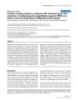
- Journal of Immune Based Therapies and Vaccines 2007, 5:6 http://www.jibtherapies.com/content/5/1/6 A) Mean of IL-4-specific Log10 CPM 3 2.5 2 1.5 1 p24 (Group B p24 (Group C) gp120 (Group D) y = 0.0199 x + 0.836 y = 0.0161x + 1.4304 y = 0.0171x + 1.2146 R = 0.5645 R = 0.8722 R= 0.5937 0.5 p = 0.0234 p = 0.0318 p = 0.0370 0 0 10 20 30 40 50 60 70 Study Week B) 60 p24 gp120 nef Remune np24 50 Lymphocyte proliferation SI 40 SI 30 20 10 0 Week 0 Week 16 Week 29 Week 41 C) 600 p24 gp120 nef Remune np24 500 400 PM IL-4 Ccpm 300 200 100 0 Week 0 Week 16 Week 29 Week 41 Figure 3 HIV-1-specific IL-4 production and lymphocyte proliferative responses HIV-1-specific IL-4 production and lymphocyte proliferative responses. Significant increases in CT.h4S delta cpm in response to HIV-1-specific IL-4 production were seen over the week 17–65 period (A) for groups B (ART + IL-2), C (ART + IL-2 + Remune™) and D (ART + Remune™), but not for group A (ART alone) (data not shown). Patient 11 is shown as an example of an inverse relationship between lymphocyte proliferative stimulation index (SI) (B) and IL-4 production as delta cpm (C) which was evident for 17 of 28 patients who were assessed for IL-4 production. Page 7 of 12 (page number not for citation purposes)
- Journal of Immune Based Therapies and Vaccines 2007, 5:6 http://www.jibtherapies.com/content/5/1/6 Table 1: HLA class I restricted peptides used for IFN-γ ELISpot assays Peptide Protein Sequence HLA-restriction Gag g1 p17 (71–79) GSEELRSLY A1 g2 p17 (77–85) SLYNTVATL A*0201 g3 p17 (20–28) RLRPGGKKK A3 g4 p24 (217–227) ACQGVGGPGHK A11 g5 p17 (84–92) TLYCVHQRI A11 g6 p17 (28–36) KYKLKHIVW A24 g7 p24 (35–43) EVIPMFSAL A26 g8 p24 (16–23) SPRTLNAW B7 g9 p17 (74–82) ELRSLYNTV B8 g10 p24 (197–205) DCKTILKAL B8 g11 p17 (93–101) EIKDTKEAL B8 g12 p17 (24–31) GGKKKKYKL B8 g13 p24 (131–140) KRWIILGLNK B27 B35A g14 p17 (36–44) WASRELERF B35A g15 p17 (124–132) NSSKVSQNY B35A g16 p24 (122–130) PPIPVGDIY g17 p24 (174–184) AEQASQDVKNW B44 g18 p24 (83–92) VHPVHAGPIA B55 B57B g19 p24 (108–117) TSTLQEQIGW B57B g20 p24 (176–184) QASQEVKNW B57B g21 p24 (15–23) ISPRTLNAW B57B g22 p24 (32–40) FSPEVIPMF g23 p17 (92–101) IEIKDTKEAL B61 Pol p1 RT (309–317) ILKEPVHGV A*0201 p2 RT (179–187) VIYQYMDDL A2 p3 RT (485–493) ALQDSGLEV A2 p4 RT (341–350) IYQEPFKLNK A11 p5 RT (158–166) AIFQSSMTK A11 p6 IN (179–188) AVFIHNFKRK A11 p7 RT (448–457) RETKLGKAGY A29 p8 RT (392–401) PIQKETWETW A32 p9 RT (18–26) GPKVKQWPL B8 B35A p10 RT (175–183) HPDIVIYQY B35A p11 RT (156–164) SPAIFQSSM B35A p12 RT (432–441) EPIVGAETFY p13 RT (203–212) EELRQHLLRW B44 p14 RT (397–406) TWETWWTEYW B44 Nef n1 nef (180–189) VLEWRFDSRL A2 n2 nef (73–82) QVPLRPMTYK A11 n3 nef (75–82) PLRPMTYK A11 n4 nef (84–92) AVDLSHFLK A11 n5 nef (128–137) TPGPGVRYPL B7 n6 nef (90–97) FLKEKGGL B8 n7 nef (13–20) WPTVRERM B8 n8 nef (135–143) YPLTFGWCY B18/B49 B35A n10 nef (186–193) DSRLAFHH B35A n11 nef (74–84) VPLRPMTY B57B n12 nef (116–125) HTQGYFPDWQ n13 nef (92–100) KEKGGLEGL B61 Peptides were identified from the NIH HIV Molecular Immunology Database website [50]. References for each peptide are also available at this website. RT (reverse transcriptase antigen), IN (integrase antigen). AHLA B35 is associated with rapid progression in HIV-1 infection. BHLA B57 is associated with slow/non progression in HIV-1 infection Page 8 of 12 (page number not for citation purposes)
- Journal of Immune Based Therapies and Vaccines 2007, 5:6 http://www.jibtherapies.com/content/5/1/6 Table 2: Patient HLA types and ELISpot results, showing the sum of delta (background subtracted) IFN-γ spot forming cells (SFC) at weeks 0, 16 and 65 for each patient for whom ELISpot analysis was possible. Sum of Δ IFN-γ SFC/106 PBMC Patient HLA type (peptides used) Week 0 Week 16 Week 65 Group A Gag Pol Nef Gag Pol Nef Gag Pol Nef 1 A1 (g1) 20 20 260 0 0 150 20 10 680 A2 (g2, p1, p2, p3, n1) B7 (g8, n2) B18 (n8) 16 A2, (g2, p1, p2, p3, n1) 50 90 110 940 850 320 740 1200 20 B44 (g17, p13, p14) 36 A2, (g2, p1, p2, p3) F F F 70 60 30 230 280 370 A23, (g13) B27, (n9) B49 (n8) 52 A11, (g4, g5, p5, n2) 670 470 295 910 405 1280 370 215 165 A9, (none identified) B35A, (g14, p10) B61 (g23, p15, n13) 54 A2, (g2, p2, n1) 80 190 30 F F F 290 240 160 A11, (g5, p5) B27, (n9) B35A (g14, p10) Group B Gag Pol Nef Gag Pol Nef Gag Pol Nef 7 A2, (g2, p2, p3, n1) 500 30 0 1310 120 20 1340 190 0 A24, (g6) B55, (g18) B57B (g19, g20, 21, g22) 35 A3, (g3) 50 110 75 130 70 25 185 135 115 A32, (p8) B7 (g8, n5) 57 A1, (g1) 490 50 130 190 30 20 450 70 120 B8 (g9, g10, g11, g12, p9, n6, n7) Group C Gag Pol Nef Gag Pol Nef Gag Pol Nef 4 A1, (g1) 220 nd 50 130 nd 40 520 nd 60 A26, (g7) B27, (n9) B57B(g19, g20, g21, g22, n12) 11 A1, (g1) 3820 3140 2990 90 50 20 2820 2760 960 A2, (g2, p1, p2, p3, n1) B35A, (g14, g15, g16, p10, p11, p12, n10, n11) B57B(g19, g20, g21, g22, n12) 14 A1, (g1) F F F 1140 130 20 100 30 70 A2, (g2, p1, p2, p3, n1) B57B,(g19, g20, g21, g22, n12) B62 (none identified) 34 A3, (g3) 320 100 0 F F F 520 80 10 A29, (p7) B35A, (g14, g15, p10, n10) B44 (g17, p13, p14) Group D Gag Pol Nef Gag Pol Nef Gag Pol Nef 13 A2, (g2, p1, p2, n1) 50 20 70 730 1890 1190 390 640 240 A11, (g4, p4, n2) B8, (g9, p9, n6) B62 (none identified) 22 A1, (g1) 270 80 120 F F F 60 40 260 A2, (g2, p2, n1) B7, (g8, n5) B35A (g14, g15, p10, n11) F denotes assay failure. IFN-γ (interferon-gamma), PBMC (peripheral blood mononuclear cells), SFC (spot forming cells). AHLA B35 is associated with rapid progression in HIV-1 infection. BHLA B57 is associated with slow/non progression in HIV-1 infection. Page 9 of 12 (page number not for citation purposes)
- Journal of Immune Based Therapies and Vaccines 2007, 5:6 http://www.jibtherapies.com/content/5/1/6 higher CD4 T-cell count nadirs and earlier initiation of ART tion of responses [43] utilized a co-administered model. [39]. Future trials will address such important issues. The fate of missing HIV-1-specific CD4 T-cell responses is We observed low-levels of transient self-limiting viraemia unclear. HIV-1 preferentially infects and deletes many resulting from IL-2 therapy as previously reported [44]. HIV-1-specific CD4 T-cells [7], while some remain detect- Despite the lack of detectable cell-mediated responses able, expressing IFN-γ, though unable to proliferate or induced by immunotherapy in these patients, the tran- express IL-2 [40,41]. The significant increase in HIV-1-spe- sient viral load blips we have reported appear to occur less cific IL-4 production from weeks 17 to 65 in group B in patients receiving IL-2 when administered with (p24), C (p24) and D (gp120) could indicate clonal dys- Remune™ in group C, with a trend towards significance (p function in these patients. Only group A (ART alone) had = 0.071). Larger group sizes may have revealed a stronger no increase in HIV-1-specific IL-4 production. Thus relationship between Remune™ and protection from IL-2 immunotherapy in these patients appears to be associated induced transient viraemia, but this pilot study was not with increasing levels of HIV-1-specific IL-4 production. powered for such observations. This possible effect of IL-4, an anti-inflammatory type-2 cytokine, has a suppres- Remune™ was not due to induction of neutralising anti- sive effect on lymphocyte proliferation [26,42]. The obser- bodies, by the display of potentially sensitive epitopes on vation in a number of patients that HIV-1-specific IL-4 gp41 resulting from the removal of gp120 from Remune™ increases when proliferative responses diminish may indi- [45]. We have separately reported the induction of anti- cate a mechanism by which clonal proliferation remains body responses against HLA-B62 and HLA-DR4 in some suppressed, although this needs further investigation. We of these patients who received Remune™ immunisation previously found a balanced IL-4/IL-2 phenotype in HIV- [46], as Remune™ contains these antigens derived from 1-specific CD4 T-cell responses in patients who remain the HUT-78 cell line in which it is grown. Group sizes are disease free [27]. While IL-4 expression by HIV-1-specific too small in this study to determine whether such CD4 T-cells may be thought of as a pathological anti-pro- responses may have played a role in potential protection liferative effect, it must be considered that this phenotype from viral blips in group C. could be protective, by dampening immune activation and quashing viral replication. This relationship requires Conclusion further investigation. Combined Remune™ and IL-2 with ART in advanced HIV- 1 infection conferred no immunological benefits to ART. Despite the lack of induced HIV-1-specific T-cell responses Taking together the absence of induced HIV-1-specific lymphocyte proliferative responses, CD8 T-cell IFN-γ in these patients we report significant increases in recall antigen proliferative responses, particularly for persistent responses and in vivo DTH responses these results imply antigens. These increases were largely evident between that induction of renewed HIV-1-specific cell-mediated week 0 and 65 with no differences between groups, sug- responses by therapeutic immunisation, even when sup- gesting immunotherapy was ineffective in this respect. plemented with IL-2, is extremely problematic in patients While some significant increases in proliferative responses who initiate ART with lower CD4 T-cell counts. Although to HIV-1 antigens were apparent from week 0 to 65 (group a recently reported clinical trial of the vCP1433 canary B and D), the same cannot be said regarding the immuno- pox-based therapeutic vaccine elicited p24-specific therapy period between weeks 17 and 65. Nor were there responses which were significantly associated with time any significant differences in responses between groups. off therapy in a subsequent treatment discontinuation These results suggest that any improvements were singu- protocol, these responses also remained transient, dimin- larly attributable to duration of ART. ishing at study end [47]. This underscores the difficulties in inducing protective immune responses by therapeutic Of note is one patient in group B, vaccinated with tetanus immunisation of chronically infected patients. Further- 4 weeks before receiving IL-2. High tetanus-specific prolif- more immunological protection in chronically infected eration was enhanced and sustained by subsequent IL-2, individuals may be best conferred by autologous virus, as as reported separately [25]. In contrast a second patient opposed to vaccine derived antigens as found with the received tetanus vaccination after IL-2 and did not vCP1452 canary pox vaccine [48] to which T-cell respond. These responses may be dependent on adminis- responses may in fact be associated with more rapid viral tration of IL-2 subsequent to antigen priming, during T- rebound following treatment interruptions [49]. In our cell contraction. Animal models demonstrate IL-2 admin- study we saw the greatest proliferative responses to HIV-1 istration during T-cell contraction enhances and prolongs antigens in patients who experienced virological responses, unlike co-administration of IL-2 with antigen rebounds, demonstrating that autologous virus induces when the survival of responding T cells is abrogated [24]. greater responses, albeit transiently, than immunisation Our protocol and others that also achieved limited induc- or IL-2 therapy. Page 10 of 12 (page number not for citation purposes)
- Journal of Immune Based Therapies and Vaccines 2007, 5:6 http://www.jibtherapies.com/content/5/1/6 In these chronically infected treated patients, we found Acknowledgements that immunotherapy was associated with increasing HIV- The authors would like to extend their gratitude to The Welcome Trust (Grant numbers 050020 and 058700) and the St Stephen's AIDS Trust/Cru- 1-specific IL-4 production, which appears to negatively said for funding. NI and FG were also funded by EU (Grant number LSHP- impact proliferative responses. HIV-1-specific IL-4 pro- CT-2004-503487) and MRC (Grant number G0501957). Clade B HIV-1 duction may result from a general dysfunction of HIV-1- recombinant antigens were provided by the EU Programme EVA/MRC specific CD4 T-cells with pathological implications for Centralised Facility for AIDS Reagents, NIBSC, UK (Grants QLK-CT 1999- induction of HIV-1-specific responses. We suggest that 00609 and G9828102). Remune™ vaccine, Remune™ and native p24 anti- these results underscore the importance of early initiation gens were provided by Ron Moss, at the time of writing with Immune of ART. As Remune™ may have positive effects in less Response Corporation, Carlsbad, CA, USA. We would also like to thank advanced patients [17], we suggest further investigations, Aine McKnight for contributions to the study and design and implementa- tion of neutralization experiments, Sundhiya Mandalia and Benigno Rod- with or without cytokine adjuvants, be conducted in riguez for help with statistical analysis, the routine laboratory staff at the patients for whom extensive immunological damage has Department of Immunology, the nursing staff, and most importantly the been prevented with earlier initiation of ART. patients at the St Stephen's Centre, Chelsea and Westminster Hospital, London, UK Abbreviations ART (antiretroviral therapy), CMV (Cytomegalovirus), References cpm (counts per minute), DNA (Deoxyribonucleic Acid), 1. Hardy GA, Imami N, Sullivan AK, Pires A, Burton CT, Nelson MR, et HIV-1 (Human Immunodeficiency Virus type-1), HLA al.: Reconstitution of CD4+ T cell responses in HIV-1 infected individuals initiating highly active antiretroviral therapy (Human Leukocyte Antigen), HSV (Herpes Simplex (HAART) is associated with renewed interleukin-2 produc- Virus), IgG (Immunoglobulin G), IFN (Interferon), IL tion and responsiveness. Clin Exp Immunol 2003, 134:98-106. 2. Imami N, Hardy G, Gotch F: Development of immunotherapeu- (Interleukin), I/M (Intramuscular), PBMC (Peripheral tic strategies for HIV-1. Expert Opin Biol Ther 2001, 1:803-816. Blood Mononuclear Cells), PCR (Polymerase Chain Reac- 3. Jansen CA, Piriou E, De Cuyper IM, van Dort K, Lange JM, Miedema tion), RNA (Ribonucleic Acid), S/C (subcutaneous), SI F, van Baarle D: Long-term highly active antiretroviral therapy in chronic HIV-1 infection: evidence for reconstitution of (Stimulation Index). antiviral immunity. Antivir Ther 2006, 11:105-116. 4. Pontesilli O, Kerkhof-Garde S, Notermans DW, Foudraine NA, Roos MT, Klein MR, et al.: Functional T cell reconstitution and Competing interests human immunodeficiency virus-1-specific cell-mediated The author(s) declare that they have no competing inter- immunity during highly active antiretroviral therapy. J Infect ests. Dis 1999, 180:76-86. 5. Kalams SA, Goulder PJ, Shea AK, Jones NG, Trocha AK, Ogg GS, Walker BD: Levels of human immunodeficiency virus type 1- Authors' contributions specific cytotoxic T-lymphocyte effector and memory All authors have read and approved the final version of responses decline after suppression of viremia with highly active antiretroviral therapy. J Virol 1999, 73:6721-6728. this manuscript. GH, FG, NI and BG conceived the study 6. Rosenberg ES, Walker BD: HIV type 1-specific helper T cells: a and co-coordinated the design of the study. GH and BG critical host defense. AIDS Res Hum Retroviruses 1998, 14(Suppl 2):S143-147. participated in the application for government and ethical 7. Douek DC, Brenchley JM, Betts MR, Ambrozak DR, Hill BJ, Okamoto approval, and in co-ordination of the study; he conducted Y, et al.: HIV preferentially infects HIV-specific CD4+ T cells. laboratory work and cellular immunological assays, con- Nature 2002, 417:95-98. 8. Valdez H, Carlson NL, Post AB, Asaad R, Heeger PS, Lederman MM, ducted data analysis and Interpretation, and preparation et al.: HIV long-term non-progressors maintain brisk CD8 T and completion of the manuscript. NI had responsibility cell responses to other viral antigens. Aids 2002, 16:1113-1118. 9. Jin X, Bauer DE, Tuttleton SE, Lewin S, Gettie A, Blanchard J, et al.: for overall management and coordination of the study, Dramatic rise in plasma viremia after CD8(+) T cell deple- she conducted laboratory work and cellular immunologi- tion in simian immunodeficiency virus-infected macaques. J cal assays, coordinated data analysis and interpretation, Exp Med 1999, 189:991-998. 10. Garcia F, Lejeune M, Climent N, Gil C, Alcami J, Morente V, et al.: and participated in preparation of the manuscript. FG and Therapeutic immunization with dendritic cells loaded with NI secured funding for the study. FG participated in data heat-inactivated autologous HIV-1 in patients with chronic analysis and interpretation, and in preparation of the HIV-1 infection. J Infect Dis 2005, 191:1680-1685. 11. Kinloch-de Loes S, Hoen B, Smith DE, Autran B, Lampe FC, Phillips manuscript. RM provided Remune™ vaccine and AN, et al.: Impact of therapeutic immunization on HIV-1 Remune™ antigen and its native p24 antigen and partici- viremia after discontinuation of antiretroviral therapy initi- ated during acute infection. J Infect Dis 2005, 192:607-617. pated in study design. MAA conducted virus neutralisa- 12. Levy Y, Durier C, Lascaux AS, Meiffredy V, Gahery-Segard H, Goujard tion assays. BG participated in the design of the study, co- C, et al.: Sustained control of viremia following therapeutic coordinated application for government and ethical immunization in chronically HIV-1-infected individuals. Aids 2006, 20:405-413. approval, and coordinated patient management. AS 13. Salk J: Prospects for the control of AIDS by immunizing sero- undertook patient care and management and participated positive individuals. Nature 1987, 327:473-476. in study co-ordination, and data interpretation. MN par- 14. Kahn JO, Cherng DW, Mayer K, Murray H, Lagakos S: Evaluation of HIV-1 immunogen, an immunologic modifier, administered ticipated in patient care and management and data inter- to patients infected with HIV having 300 to 549 × 10(6)/L pretation. CD4 cell counts: A randomized controlled trial. Jama 2000, 284:2193-2202. Page 11 of 12 (page number not for citation purposes)
- Journal of Immune Based Therapies and Vaccines 2007, 5:6 http://www.jibtherapies.com/content/5/1/6 15. Robbins GK, Addo MM, Troung H, Rathod A, Habeeb K, Davis B, et anti-gp41 human monoclonal antibody. Proc Natl Acad Sci USA al.: Augmentation of HIV-1-specific T helper cell responses in 1994, 91:3348-3352. chronic HIV-1 infection by therapeutic immunization. Aids 34. Trkola A, Pomales AB, Yuan H, Korber B, Maddon PJ, Allaway GP, et 2003, 17:1121-1126. al.: Cross-clade neutralization of primary isolates of human 16. Turner JL, Kostman JR, Aquino A, Wright D, Szabo S, Bidwell R, et al.: immunodeficiency virus type 1 by human monoclonal anti- The effects of an HIV-1 immunogen (Remune) on viral load, bodies and tetrameric CD4-IgG. J Virol 1995, 69:6609-6617. CD4 cell counts and HIV-specific immunity in a double-blind, 35. Zwick MB, Labrijn AF, Wang M, Spenlehauer C, Saphire EO, Binley randomized, adjuvant-controlled subset study in HIV JM, et al.: Broadly neutralizing antibodies targeted to the infected subjects regardless of concomitant antiviral drugs. membrane-proximal external region of human immunodefi- HIV Med 2001, 2:68-77. ciency virus type 1 glycoprotein gp41. J Virol 2001, 17. Chantratita W, Sukeepaisarncharoen W, Chandeying V, Kulpradist S, 75:10892-10905. Israngkura Na Ayudhtaya B, Rugpao S, et al.: Delayed progression 36. Valentine FT, Paolino A, Saito A, Holzman RS: Lymphocyte-prolif- to AIDS in volunteers treated with long-term HIV-1 Immu- erative responses to HIV antigens as a potential measure of nogen (REMUNE) therapy in Thailand. HIV Med 2004, immunological reconstitution in HIV disease. AIDS Res Hum 5:317-325. Retroviruses 1998, 14(Suppl 2):S161-166. 18. Davey RT Jr, Chaitt DG, Albert JM, Piscitelli SC, Kovacs JA, Walker 37. Lange CG, Valdez H, Medvik K, Asaad R, Lederman MM: CD4+ T- RE, et al.: A randomized trial of high- versus low-dose subcu- lymphocyte nadir and the effect of highly active antiretrovi- taneous interleukin-2 outpatient therapy for early human ral therapy on phenotypic and functional immune restora- immunodeficiency virus type 1 infection. J Infect Dis 1999, tion in HIV-1 infection. Clin Immunol 2002, 102:154-161. 179:849-858. 38. Lederman HM, Williams PL, Wu JW, Evans TG, Cohn SE, McCutchan 19. Davey RT Jr, Chaitt DG, Piscitelli SC, Wells M, Kovacs JA, Walker RE, JA, et al.: Incomplete immune reconstitution after initiation of et al.: Subcutaneous administration of interleukin-2 in human highly active antiretroviral therapy in human immunodefi- immunodeficiency virus type 1-infected persons. J Infect Dis ciency virus-infected patients with severe CD4+ cell deple- 1997, 175:781-789. tion. J Infect Dis 2003, 188:1794-1803. 20. Hengge UR, Goos M, Esser S, Exner V, Dotterer H, Wiehler H, et al.: 39. Lange CG, Lederman MM, Medvik K, Asaad R, Wild M, Kalayjian R, Randomized, controlled phase II trial of subcutaneous inter- Valdez H: Nadir CD4+ T-cell count and numbers of CD28+ leukin-2 in combination with highly active antiretroviral CD4+ T-cells predict functional responses to immunizations therapy (HAART) in HIV patients. Aids 1998, 12:F225-234. in chronic HIV-1 infection. Aids 2003, 17:2015-2023. 21. Kovacs JA, Baseler M, Dewar RJ, Vogel S, Davey RT Jr, Falloon J, et al.: 40. Wilson JD, Imami N, Watkins A, Gill J, Hay P, Gazzard B, et al.: Loss Increases in CD4 T lymphocytes with intermittent courses of CD4+ T cell proliferative ability but not loss of human of interleukin-2 in patients with human immunodeficiency immunodeficiency virus type 1 specificity equates with pro- virus infection. A preliminary study. N Engl J Med 1995, gression to disease. J Infect Dis 2000, 182:792-798. 332:567-575. 41. Younes SA, Yassine-Diab B, Dumont AR, Boulassel MR, Grossman Z, 22. Davey RT Jr, Murphy RL, Graziano FM, Boswell SL, Pavia AT, Cancio Routy JP, Sekaly RP: HIV-1 viremia prevents the establishment M, et al.: Immunologic and virologic effects of subcutaneous of interleukin 2-producing HIV-specific memory CD4+ T interleukin 2 in combination with antiretroviral therapy: A cells endowed with proliferative capacity. J Exp Med 2003, randomized controlled trial. Jama 2000, 284:183-189. 198:1909-1922. 23. Levy Y, Durier C, Krzysiek R, Rabian C, Capitant C, Lascaux AS, et al.: 42. Imami N, Brookes PA, Lombardi G, Hakooz B, Johns M, Goldman JM, Effects of interleukin-2 therapy combined with highly active et al.: Association between interleukin-4-producing T lym- antiretroviral therapy on immune restoration in HIV-1 infec- phocyte frequencies and reduced risk of graft-versus-host tion: a randomized controlled trial. Aids 2003, 17:343-351. disease. Transplantation 1998, 65:979-988. 24. Blattman JN, Grayson JM, Wherry EJ, Kaech SM, Smith KA, Ahmed R: 43. Kuekrek H, Schlingmann T, Valdez H, Boehm BO, Pollard RB, Mitsu- Therapeutic use of IL-2 to enhance antiviral T-cell responses yasu R, et al.: Differential effect of interleukin-2 treatment on in vivo. Nat Med 2003, 9:540-547. primary and secondary immunizations in HIV infected indi- 25. Hardy GA, Imami N, Sullivan AK, Nelson MR, Gazzard B, Gotch FM: viduals. Aids 2005, 19:1967-1974. Tetanus vaccination with IL-2 during highly active antiretro- 44. Sullivan AK, Hardy GA, Nelson MR, Gotch F, Gazzard BG, Imami N: viral therapy induces sustained and pronounced specific CD4 Interleukin-2-associated viral breakthroughs induce HIV-1- T-cell responses. Aids 2004, 18:2199-2202. specific CD4 T cell responses in patients on fully suppressive 26. Lombardi G, Hargreaves R, Sidhu S, Imami N, Lightstone L, Fuller- highly active antiretroviral therapy. Aids 2003, 17:628-629. Espie S, et al.: Antigen presentation by T cells inhibits IL-2 pro- 45. Burton DR, Desrosiers RC, Doms RW, Koff WC, Kwong PD, Moore duction and induces IL-4 release due to altered cognate sig- JP, et al.: HIV vaccine design and the neutralizing antibody nals. J Immunol 1996, 156:2769-2775. problem. Nat Immunol 2004, 5:233-236. 27. Imami N, Pires A, Hardy G, Wilson J, Gazzard B, Gotch F: A bal- 46. Page M, Ojugo A, Imami N, Hardy G, Gotch F, Almond N: Specifi- anced type 1/type 2 response is associated with long-term city of anti-human leukocyte antigen antibody responses nonprogressive human immunodeficiency virus type 1 infec- after immunization with Remune, an inactivated HIV-1 vac- tion. J Virol 2002, 76:9011-9023. cine. Aids 2007, 21:375-377. 28. Moss RB, Wallace MR, Giermakowska WK, Webb E, Savary J, Cham- 47. Tubiana R, Carcelain G, Vray M, Gourlain K, Dalban C, Chermak A, berlin-Brandt C, et al.: Phenotypic analysis of human immuno- et al.: Therapeutic immunization with a human immunodefi- deficiency virus (HIV) type 1 cell-mediated immune ciency virus (HIV) type 1-recombinant canarypox vaccine in responses after treatment with an HIV-1 immunogen. J Infect chronically HIV-infected patients: The Vacciter Study Dis 1999, 180:641-648. (ANRS 094). Vaccine 2005, 23:4292-4301. 29. Turner JL, Trauger RJ, Daigle AE, Carlo DJ: HIV-1 immunogen 48. Jacobson JM, Pat Bucy R, Spritzler J, Saag MS, Eron JJ Jr, Coombs RW, induction of HIV-1-specific delayed-type hypersensitivity: et al.: Evidence That Intermittent Structured Treatment results of a double-blind, adjuvant-controlled, dose-ranging Interruption, but Not Immunization with ALVAC-HIV trial. Aids 1994, 8:1429-1435. vCP1452, Promotes Host Control of HIV Replication: The 30. Bidwell J: Advances in DNA-based HLA-typing methods. Results of AIDS Clinical Trials Group 5068. J Infect Dis 2006, Immunol Today 1994, 15:303-307. 194:623-632. 31. Lalvani A, Brookes R, Hambleton S, Britton WJ, Hill AV, McMichael 49. Autran B: Time to ART Resumption following Treatment AJ: Rapid effector function in CD8+ memory T cells. J Exp Med Interruption Is Shorter for Individuals Immunized with HIV- 1997, 186:859-865. Recombinant Canarypox Vaccine (Vcp1452) Compared to 32. Aasa-Chapman MM, Hayman A, Newton P, Cornforth D, Williams I, Placebo: The Manon-02 Trial. Conference on Retroviruses and Borrow P, et al.: Development of the antibody response in Opportunistic Infections 2007. Abstract 126LB acute HIV-1 infection. Aids 2004, 18:371-381. 50. NIH HIV Molecular Immunology Database [http:// 33. Conley AJ, Kessler JA 2nd, Boots LJ, Tung JS, Arnold BA, Keller PM, www.hiv.lanl.gov/content/immunology/ctl_search] et al.: Neutralization of divergent human immunodeficiency virus type 1 variants and primary isolates by IAM-41-2F5, an Page 12 of 12 (page number not for citation purposes)
CÓ THỂ BẠN MUỐN DOWNLOAD

-

Báo cáo y học: "A 12 Week, Open Label, Phase I/IIa Study Using Apatone® for the Treatment of Prostate Cancer Patients Who Have Failed Standard Therapy"
 6 p |
6 p |  55
|
55
|  7
7
-

Báo cáo y học: "A multicenter, double-blind, randomized, controlled phase III clinical trial of chicken type II collagen in rheumatoid arthritis"
10 p | 60
| 6
-

Báo cáo y học: "IMP321 (sLAG-3), an immunopotentiator for T cell responses against a HBsAg antigen in healthy adults: a single blind randomised controlled phase I study"
15 p | 56
| 5
-

Báo cáo y học: " Masitinib in the treatment of active rheumatoid arthritis: results of a multicentre, open-label, dose-ranging, phase 2a study"
12 p | 54
| 5
-
Báo cáo y học: "Cardiovascular risk factors and acute-phase response in idiopathic ascending aortitis: a case control study"
6 p | 45
| 5
-

Báo cáo y học: "Circadian phase-shifting effects of a laboratory environment: a clinical trial with bright and dim light"
9 p | 62
| 4
-

Báo cáo y học: " Failure of a non-authorized copy product to maintain response achieved with imatinib in a patient with chronic phase chronic myeloid leukemia: a case report"
3 p | 58
| 4
-

Báo cáo y học: "Biologic activity and safety of belimumab, a neutralizing anti-B-lymphocyte stimulator (BLyS) monoclonal antibody: a phase I trial in patients with systemic lupus erythematosus"
15 p | 52
| 3
-
Báo cáo y học: " A phase 1 trial of nebulised heparin in acute lung injury"
8 p | 58
| 3
-

Báo cáo y học: " Assessment of pulmonary antibodies with induced sputum and bronchoalveolar lavage induced by nasal vaccination against Pseudomonas aeruginosa: a clinical phase I/II study"
10 p | 41
| 3
-
Báo cáo khoa học: "A phase II randomized trial comparing radiotherapy with concurrent weekly cisplatin or weekly paclitaxel in patients with advanced cervical cancer"
8 p | 49
| 3
-
Báo cáo khoa học: "A phase I radiation dose-escalation study to determine the maximal dose of radiotherapy in combination with weekly gemcitabine in patients with locally advanced pancreatic adenocarcinoma"
7 p | 70
| 3
-
Báo cáo y học: "Control of hyperuricemia in subjects with refractory gout, and induction of antibody against poly(ethylene glycol) (PEG), in a phase I trial of subcutaneous PEGylated urate oxidase"
10 p | 68
| 2
-

Báo cáo khoa học: " Prospective phase II study of preoperative short-course radiotherapy for rectal cancer with twice daily fractions of 2.9 Gy to a total dose of 29 Gy - Long-term results"
9 p | 44
| 2
-

Báo cáo y học: "Acute phase reactants add little to composite disease activity indices for rheumatoid arthritis: validation of a clinical activity score"
11 p | 38
| 2
-

Báo cáo y học: " WT1 PEPTIDE VACCINATION IN COMBINATION WITH IMATINIB THERAPY FOR A PATIENT WITH CML IN THE CHRONIC PHASE"
10 p | 51
| 2
-

Báo cáo khoa học: " A phase III trial comparing an anionic phospholipid-based cream and aloe vera-based gel in the prevention of radiation dermatitis in pediatric patients"
8 p | 48
| 2


Chịu trách nhiệm nội dung:
Nguyễn Công Hà - Giám đốc Công ty TNHH TÀI LIỆU TRỰC TUYẾN VI NA
LIÊN HỆ
Địa chỉ: P402, 54A Nơ Trang Long, Phường 14, Q.Bình Thạnh, TP.HCM
Hotline: 093 303 0098
Email: support@tailieu.vn
Giấy phép Mạng Xã Hội số: 670/GP-BTTTT cấp ngày 30/11/2015 Copyright © 2022-2032 TaiLieu.VN. All rights reserved.

