


Báo cáo y học: "Demonstration of the histopathological and immunohistochemical effects of a novel hemostatic agent, ankaferd blood stopper, on vascular tissue in a rat aortic bleeding mode"
lượt xem 4
download
 Download
Vui lòng tải xuống để xem tài liệu đầy đủ
Download
Vui lòng tải xuống để xem tài liệu đầy đủ
Tuyển tập các báo cáo nghiên cứu về y học được đăng trên tạp chí y học Wertheim cung cấp cho các bạn kiến thức về ngành y đề tài: Demonstration of the histopathological and immunohistochemical effects of a novel hemostatic agent, ankaferd blood stopper, on vascular tissue in a rat aortic bleeding model...
Bình luận(0) Đăng nhập để gửi bình luận!
Nội dung Text: Báo cáo y học: "Demonstration of the histopathological and immunohistochemical effects of a novel hemostatic agent, ankaferd blood stopper, on vascular tissue in a rat aortic bleeding mode"
- Kandemir et al. Journal of Cardiothoracic Surgery 2010, 5:110 http://www.cardiothoracicsurgery.org/content/5/1/110 RESEARCH ARTICLE Open Access Demonstration of the histopathological and immunohistochemical effects of a novel hemostatic agent, ankaferd blood stopper, on vascular tissue in a rat aortic bleeding model Ozer Kandemir1*, Mustafa Buyukates1, Nilufer Onak Kandemir2, Erol Aktunc3, Aylin Ege Gul4, Sanser Gul5, S Akin Turan1 Abstract Background: Ankaferd Blood Stopper® (ABS) is a folkloric medicinal plant extract used as a hemostatic agent in traditional Turkish medicine. This experimental study investigated the histopathological and immunohistochemical effects of ABS on vascular tissue in a rat model of aortic bleeding. Methods: Four groups of 11 Wistar albino rats were used. The abdominal aortas of the rats were wounded; an ABS-soaked tampon was applied to rats in Groups 1 and 3, and a plain gauze tampon was applied to rats in Groups 2 and 4 until the bleeding stopped. The bleeding time was recorded. Immediately following sacrificing, the arteriotomy sites from Groups 1 and 2 were removed. The abdominal incisions in Groups 3 and 4 were closed following hemostasis. On Day 7 of the study, Group 3 and 4 rats were sacrificed and the abdominal aorta arteriotomy sites were removed for histopathological and immunohistochemical evaluation. Results: The mean bleeding time in 15 animals in Groups 2 and 4 was 4.9 ± 0.6 s, and in 22 animals in Groups 1 and 3 was 3.1 ± 0.6 s. Distal aortic occlusion was not observed on either Day 1 or 7 in any group. Significantly more widespread and dense endothelial nitric oxide synthase (eNOS) staining was observed in Group 1 animals than Group 2. On Days 1 and 7 after application of ABS, histopathological changes, consisting of necrosis, inflammation, and endothelial cell loss, in the rat abdominal aortas did not differ between Groups 1 and 2. The basophilic discoloration in the ABS group on the operation day was a result of a foreign body reaction and hemosiderin-loaded histiocyte accumulation, which occurred on Day 7. Conclusions: In this study, hemostasis was successfully achieved with ABS in rat abdominal aortas. No histopathological change was found in the rat abdominal aortas between the ABS and control groups on Days 1 and 7. Further studies on the long-term effects of foreign body reactions and hemosiderin-loaded histiocyte accumulation are required. Background bleeding from anastomosis si tes is usually controlled Impaired tissue integrity and uncontrollable hemorrhage with pressure or additional suturing techniques. Occa- are important causes of morbidity and mortality, espe- sionally, these techniques may be insufficient, requiring cially in the presence of coagulopathies [1]. Various tissue adhesives as supportive agents [4,5]. Additionally, hemostatic agents have been developed to achieve suffi- blind suturing for blood oozing from sutured vascular cient hemostasis [2,3]. In cardiovascular surgery, segments may impair the quality of anastomosis. To preserve the quality of anastomosis, adjuvant topical hemostatic agents are favored in cardiac and vascular sur- * Correspondence: ozerkandemir@isnet.net.tr gery. However, topical hemostatic agents may have disad- 1 Department of Cardiovascular Surgery, Zonguldak Karaelmas University, vantages, such as limited e fficacy, limited availability, Zonguldak, Turkey Full list of author information is available at the end of the article © 2010 Kandemir et al; licensee BioMed Central Ltd. This is an Open Access article distributed under the terms of the Creative Commons Attribution License (http://creativecommons.org/licenses/by/2.0), which permits unrestricted use, distribution, and reproduction in any medium, provided the original work is properly cited.
- Kandemir et al. Journal of Cardiothoracic Surgery 2010, 5:110 Page 2 of 7 http://www.cardiothoracicsurgery.org/content/5/1/110 limited vascular biological compatibility, expensiveness, the vascular wound, and the bleeding time was and risk of infection as a result of the requirement for recorded. In case of insufficient hemostasis using either human blood for commercial production of collagen, of the tampons, an 8/0 Prolene suture (Prodek, Sutures thrombin, and prothrombin [6]. Surgeons should also be Ltd, UK) was used to provide hemostasis. Aortic sam- trained in the use of hemostatic agents, such as fibrin pling was performed in all rats to search for immediate glues. and Day-7 postoperative histopathological changes in Ankaferd Blood Stopper® (ABS) is a folkloric medicinal vascular tissues as a result of ABS. plant extract used as a hemostatic agent in traditional Turkish medicine [7]. The use of this product Bleeding assay was approved by the Ministry of Health, Turkey, on The duration of bleeding was measured using a chron- October 26, 2007. ometer and defined as the time from wounding until In a recent literature search, we found no study on the the time bleeding stopped. histopathological and immunohistochemical effects of ABS on vascular tissue. In this experimental study, we Animal groups investigated the effects of ABS on vascular tissue in a The abdominal aortas of the animals were wounded. rat model of aortic bleeding. ABS-soaked tampons were applied in Group 1 (n = 11), and plain gauze tampons were applied in Group 2 (n = Methods 11) until the bleeding stopped. All of these animals were Wistar albino (WA) rats were used to demonstrate the sacrificed by cervical dislocation on the operation day. vascular histopathological and immunohistochemical The abdominal aortas of Groups 3 (n = 11) and 4 (n = changes following the application of ABS (Trend Tekno- 11) were wounded, and hemostasis was provided with loji Ilac AS, Istanbul, Turkey) on the abdominal aorta. ABS-soaked tampons in Group 3 and plain gauze tam- The experimental procedure was approved by the pons in Group 4. The abdominal incisions in these two Committee for Animal Research at Zonguldak Karaelmas groups were closed following hemostasis. They were University School of Medicine. All animal studies con- kept alive for 7 days and fed ad libitum. On Day 7 after formed with the animal experiment guidelines of the the operation, all Group 3 and 4 animals were sacrificed Committee for Humane Care. All animals received care by cervical dislocation. Immediately following sacrifice, in compliance with the “Principles of Laboratory Animal the arteriotomy sites from all 44 animals were removed Care ” formulated by the National Society for Medical en bloc with a safety margin of 1 mm of untouched aor- Reseacrh and “Guide for the Care and the Use of Labora- tic vascular tissue both distal and proximal to the tory Animlas” prepared by the US Natinoal Academy of wound site. Sciences and published by the US Natinoal Institute of Health (NIH Publications, No:80-23) Histopathological procedure All specimens were fixed in 10% phosphate-buffered for- maldehyde solution for 24 h at room temperature. Each Animals Male adult WA rats (Zonguldak Karaelmas University specimen was cut into three sections: the proximal, Laboratories, Zonguldak, Turkey), weighing 250-300 g, intact part of the aorta, the wounded part of the aorta, were maintained on a 12/12-h light/dark cycle and fed ad and the distal, intact part of the aorta. libitum. All animals were housed in individual cages in a Following the dehydration process using graded etha- temperature-controlled environment (20 ± 2°C). The rats nols, specimens were embedded in paraffin blocks and cut into 5- μ m-thick sections to be mounted on glass were randomly assigned into ABS and control groups. slides. Sections were then deparaffinized with xylene and counterstained with hematoxylin and eosin (H&E), Surgical procedure All animals were anesthetized with intramuscularly iron blue, and Elastic van Gieson (EVG). EVG staining administered ketamine hydrochloride (75 mg/kg). Post- was performed to identify the external and internal elas- operative analgesia was provided by 1-2 mg/mL parace- tic lamina. Iron blue staining was performed to identify tamol added to the drinking water. The abdominal aorta hemosiderin. All of the sections were examined in 10 was accessed surgically by a midline abdominal incision random fields at ×40 magnification using a light micro- using sterile technique. The retroperitoneum was scope. Blinded light microscopic examinations were per- explored and the aorta was exposed. The abdominal formed by two of the coauthors (NOK, AEG). aorta was wounded just proximal to the iliac bifurcation using an iris blade. ABS solution (1 mL) in a glass vial Histopathological grading of the specimens was poured on a gauze tampon through a syringe. Either Light microscopic findings were graded semi-quantita- an ABS-soaked or plain gauze tampon was applied to tively from 0 (no histopathological change) to +3 (severe
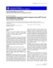
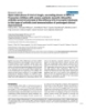
- Kandemir et al. Journal of Cardiothoracic Surgery 2010, 5:110 Page 3 of 7 http://www.cardiothoracicsurgery.org/content/5/1/110 histopathological change). This histopathological grading covering nearly the whole area of the specimen was clas- was performed for vascular (endothelial cell loss, inflam- sified as 3. Grade 2 was intermediate between 1 and 3. matory reaction, medial necrosis, fibrin plug formation, and erythrocyte aggregation) and perivascular (inflam- Statistical analysis matory reaction, hemosiderin-loaded histiocytes, and Statistical analyses were carried out using the SPSS soft- granulation tissue formation) connective tissue reactions ware (v. 11.0 for Windows; SPSS Inc.; Chicago, IL). All in the specimens. values are expressed as means ± SD. P-values less than 0.05 were deemed to be statistically significant. Group comparisons were made by one-way analysis of variance Immunohistochemical procedure In immunohistochemical surveys, anti-CD31 was used (Kruskall-Wallis) followed, in cases of significance, by to monitor vascular endothelial cells, and anti-eNOS the Mann-Whitney U test. antibodies were used to determine eNOS expression of Results endothelial cells. For immunohistochemical studies, immunostaining was performed according to the avidin- Bleeding did not stop in four of the Group 2 animals biotin-peroxidase (BSA-DAB) complex technique. Paraf- and in three of the Group 4 animals, for a total of seven fin sections were collected on slides, deparaffinized, and in the plain gauze tampon groups. The ABS-soaked dehydrated. Endogenous peroxidase activity was blocked gauze tampon stopped bleeding in all Group-1 and -3 using a 3% hydrogen peroxide solution for 10 min. To animals. enhance staining, heat-induced epitope retrieval was The mean bleeding time in 15 animals with the gauze performed. Primary antibodies against CD31 (rabbit tampon in Groups 2 and 4 was 4.9 ± 0.6 s, and in 22 monoclonal, JC70, Dako, Copenhagen, Denmark) and animals in the ABS-soaked tampon Groups 1 and 3 was endothelial nitric oxide synthase (eNOS; rabbit polyclo- 3.1 ± 0.6 s. The mean bleeding time in the ABS-applied nal, RR-1711-R7, Neomarkers; Lab Vision, Fremont, CA, groups was 36.7% shorter than that of the plain gauze USA) were used. The sections were incubated with pri- tampon groups, producing a significantly shorter dura- mary antisera (including CD31 or eNOS) for 1 h at tion of bleeding in the ABS groups (p = 0.0001). room temperature. After washing in phosphate-buffered Distal aortic occlusion was not observed on Days 1 saline, the tissues were incubated with biotin-conjugated or 7 after the operation in any group. secondary antibody and then a streptavidin-biotin sys- Comparisons of the histopathological changes in tem for 30 min at room temperature. The reactions Group 1 and 2 animals are depicted in Table 1. Necrosis were visualized using diaminobenzidine tetrahydrochlor- was absent, and the intensity of the inflammatory reac- ide. The sections were counterstained using hematoxy- tion together with endothelial cell loss did not differ sig- lin, then cleared and mounted. nificantly between the groups. Fibrin plug formation and erythrocyte aggregation at the arteriotomy site were more prominent in Group 1 Controls and grading of the immunostaining Appropriate positive (placenta, capillary endothelium for than in Group 2. In the ABS tampon groups, a micro- CD31 and eNOS) and negative (omitted primary anti- scopically evident basophilic discoloration in the perivas- body) controls were evaluated simultaneously in all cular tissue was observed (Figure 1). cases. All cytoplasmic staining was recorded as positive Significantly more widespread and dense eNOS stain- for eNOS and CD31. The extent and intensity of eNOS ing was observed in Group 1 animals than Group 2 reactions were semi-quantitatively evaluated using a (Figure 2). The immunostaining of the unaffected vascu- four-level grading system. Grade 0 was no apparent lar segments in ABS tampon and plain gauze tampon reaction product. Focal and minimal staining intensity groups did not differ significantly for eNOS expression was graded 1, and the most prominent staining reaction (Table 1). Comparisons of the histopathological changes Table 1 Light microscopical and immunohistochemical findings in the rats abdominal aorta on the operation day Group I (ABS) Group II (Plain gauze) P Histopathological changes Inflammatory reaction 1.0 ± 0.5 1.0 ± 0.6 0.7 Necrosis none none N/A Endothelial cell loss 1.0 ± 0.3 1.1 ± 0.4 0.5 Fibrin plug formation/Erythrocyte aggregation 1.5 ± 0.6 0.18 ± 0.4 0.0001 Immunohistochemical changes e-NOS staining 1.9 ± 0.7 1.1 ± 0.4 0.0001
- Kandemir et al. Journal of Cardiothoracic Surgery 2010, 5:110 Page 4 of 7 http://www.cardiothoracicsurgery.org/content/5/1/110 Figure 1 Fibrin plug formation and erythrocyte aggregation at the arteriotomy site were more prominent in Group 1 (A) than in Group 2 (B). In the ABS tampon groups, microscopically evident basophilic discoloration in perivascular tissue was observed (A) (H&E, ×400). o n Day 7 of the operation are depicted in Table 2. glabra 0.18 mg, Alpina officinarum 0.14 mg, and Urtica There was no necrosis adjacent to the intimal and dioica 0.12 mg). Each of these plants has vascular endothelial regeneration in either group. actions and some effect on the hematological system. T. The microscopically evident basophilic discoloration in vulgaris has anti-oxidative effects, such as prevention of the ABS group on the operation day was a result of a lipid peroxidation [8]. V. vivifera has anti-atherosclerotic foreign body reaction (Figure 3A-B) and hemosiderin- effects [9]. G. glabra decreases vascular endothelial loaded histiocyte accumulation on Day 7 after the growth factor production and cytokine-induced neovas- operation (Figure 4A-B). Immunostaining with CD-31 cularization [10]. A. officinarum inhibits nitric oxide showed an intact endothelial cell lining, and eNOS production [11]. staining did not differ among groups on Day 7 after the The hemostatic mechanism of ABS is effected by fibri- operation. nogen-erythrocyte agglutination, resulting in the forma- tion of an encapsulated protein network that stimulates Discussion erythrocyte aggregation. This encapsulated protein net- Hemorrhage from anastomosis sites can usually be man- work occurs very rapidly, in less than 1 s [12]. The ABS aged by additional sutures or light pressure. If adequate network might cover the entire physiological hemostatic hemostasis cannot be achieved, various hemostatic process without affecting any individual clotting factor. agents may be used. The ideal hemostatic agent should Göker at al. demonstrated that coagulation factors II, V, be easy to use, require minimal training, show an effect VII, VIII, IX, X, XI, and XIII were not affected, and that within minutes, be effective in both arterial and venous plasma fibrinogen activity as well as total protein, albu- bleeding, be non-toxic, and be anaphylactic [7]. Cur- min, and globulin levels were decreased by the addition rently, no hemostatic agent possesses all of these of ABS to plasma [6,7]. These results showed that nor- characteristics. mal hemostatic elements were spared during the forma- ABS is a novel topical hemostatic agent that consists tion of the protein network. Thus, ABS might be useful of various folkloric medicinal plant extracts ( Thymus in patients with antithrombotic drug-induced primary or vulgaris 0.1 mg, Vitis vivifera 0.16 mg, Glycyrrhiza secondary hemostatic abnormalities [13,14]. Cipil et al. Figure 2 A significantly more widespread and dense eNOS staining was observed in Group 1 (A) animals compared with Group 2 (B) (Immunohistochemistry, eNOS, ×400).
- Kandemir et al. Journal of Cardiothoracic Surgery 2010, 5:110 Page 5 of 7 http://www.cardiothoracicsurgery.org/content/5/1/110 Table 2 Light microscopical and immunohistochemical findings in the rats abdominal aorta on the 7th day of the operation Group III (ABS) Group IV (Plain gauze) P Histopathological changes Necrosis none none N/A Endothelial cell egeneration all all N/A Foreign body reaction 2.0 ± 0.7 1.1 ± 0.4 0.006 Hemosiderin loaded hystiocyte 1.8 ± 0.6 0.8 ± 0.6 0.001 Immunohistochemical changes e-NOS staining 0.8 ± 0.4 0.7 ± 0.4 0.6 demonstrated that ABS also had hemostatic effects in Necrosis in vascular tissues, inflammatory reaction, animals pretreated with warfarin. The bleeding time was and endothelial cell loss are important, particularly in reduced to 44% with ABS treatment [13]. terms of graft patency. Intimal hyperplasia can cause Karakaya et al. demonstrated that ABS significantly aneurysm and thrombus formation [1]. In our study, on reduced blood loss and death in experimental rat liver Days 1 and 7 post-ABS application, histopathological laceration [15]. Also, Dogan et al. used ABS for coron- changes in the rat abdominal aorta did not differ ary artery bypass surgery patients. They sprayed 4-8 mL between Group 1 and 2 with regard to necrosis, inflam- of ABS solution to bypass suture lines and the bleeding matory reaction, or endothelial cell loss. area. They indicated that patients who had used ABS After application of ABS, brown-colored changes required no revisions [16]. Our study revealed that fibrin occurred around the tissue [21]. We believe that the plug formation and erythrocyte aggregation at the arter- encapsulated protein network caused these changes. In iotomy site were more prominent in Group 1 than in the ABS tampon groups, a microscopically evident baso- Group 2, and that bleeding time was 4.9 ± 0.6 s versus philic discoloration in the perivascular tissue was 3.1 ± 0.6 s in the ABS and control groups. Thus, ABS observed on the operation day and was caused by for- reduced bleeding time by 36.7% compared with the con- eign body reaction and hemosiderin-loaded histiocyte trol group. In clinical experiments, ABS has been suc- accumulation. This status could be explained by the for- cessfully used to control upper gastrointestinal bleeding mation of the encapsulated protein network, causing [17,18], acute anterior epistaxis [19], and bleeding due delayed degradation of erythrocytes. The long-term clin- to solitary rectal ulcers [20]. ical outcomes of this reaction must be clarified in pro- Although studies regarding the hemostatic effects spective experimental studies. and mechanism of ABS are available, there is no U. dioica , one of medicinal plant extracts in ABS, reported study regarding histopathological effects on causes vasodilatation by inducing nitric oxide produc- vascular tissue. Negative effects of tissue topical agents tion by the endothelium [22]. Significantly more wide- used in anastomoses in cardiovascular surgery can spread and dense eNOS staining was observed in Group influence the patency of grafts in both the short- and 1 animals compared with Group 2. An increased eNOS long-term. level around arteriotomy areas in the early stages Figure 3 Foreign body reaction on Day 7 after the operation in Groups 1 (A) and 2 (B) (H&E, ×400).
- Kandemir et al. Journal of Cardiothoracic Surgery 2010, 5:110 Page 6 of 7 http://www.cardiothoracicsurgery.org/content/5/1/110 Figure 4 In the ABS tampon groups, prominent hemosiderin-loaded histiocyte accumulation in perivascular tissue was observed (A-B) (A; H&E, B; iron blue, ×400). c onsistently stopped the bleeding in vitro without Competing interests The authors declare that they have no competing interests. impairing tissue oxygenation or microcirculation of ABS. The advantages of ABS when compared with other Received: 28 August 2010 Accepted: 14 November 2010 products that are readily available include effectiveness, Published: 14 November 2010 ease of application, and no requirement for technical References skills. However, as the product is relatively new, a lim- 1. Kaplan M, Bozkurt S, Kut MS, Kullu S, Demirtas MM: Histopathological ited amount of data is available related to long-term effects of ethyl 2-cyanoacrylate tissue adhesive following surgical side effects and toxicity [1,21]. application: an experimental study. Eur J Cardiothorac Surg 2004, 25(2):167-72. A limitation of this study is that only acute and early- 2. Björses K, Holst J: Various local hemostatic agents with different modes stage effects of ABS were evaluated. Long-term anasto- of action; an in vivo comparative randomized vascular surgical mosis patency effects must be evaluated in further stu- experimental study. Eur J Vasc Endovasc Surg 2007, 33(3):363-70, Epub 2006 Nov 28. dies. Additional studies are required regarding possible 3. Unlü Y, Vural U, Koçak H, Ceviz M, Becit N, Akbulut O: Comparison of the effects of ABS on vascular tissues over a period longer topical haemostatic agents for the prevention of suture hole bleeding. than 7 days. An experimental study. Eur J Vasc Endovasc Surg 2002, 23(5):441-4. 4. Kheirabadi BS, Pearson R, Rudnicka K, Somwaru L, MacPhee M, Drohan W, Tuthill D: Development of an animal model for assessment of the Conclusions hemostatic efficacy of fibrin sealant in vascular surgery. J Surg Res 2001, This is the first reported study evaluating the histo- 100(1):84-92. 5. Werker PM, Kon M: Review of facilitated approaches to vascular pathological and immunohistochemical effects of ABS anastomosis surgery. Ann Thorac Surg 1997, 63(6 Suppl):S122-7. on vascular structure. In this study, hemostasis was suc- 6. Bilgili H, Kosar A, Kurt M, Onal IK, Goker H, Captug O, Shorbagi A, Turgut M, cessfully achieved using ABS on rat abdominal aortas. Kekilli M, Kurt OK, Kirazli S, Aksu S, Haznedaroglu IC: Hemostatic efficacy of Ankaferd Blood Stopper in a swine bleeding model. Med Princ Pract 2009, No histopathological change in rat abdominal aortas 18(3):165-9, Epub 2009 Apr 6. between ABS and control groups on Days 1 and 7 was 7. Göker H, Haznedaroglu IC, Ercetin S, Kirazli S, Akman U, Ozturk Y, Firat HC: found. Further prospective studies are also required Haemostatic actions of the folkloric medicinal plant extract Ankaferd Blood Stopper®. J Int Med Res 2008, 36:163-170. regarding long-term effects of foreign body reaction and 8. Lee SJ, Umano K, Shibamato T, Lee KG: Identification of volatile hemosiderin-loaded histiocyte accumulation. components in basil (Ocimum basilicum L.) and thyme leaves (Thymus vulgaris L.) and their antioxidant properties. Food Chem 2007, 91:131-137. 9. Yamakoshi J, Kataoka S, Koga T, Ariga T: Proanthocyanidin-rich extracts form grape seede attenutaee the development of aortic atherosclerosis Author details 1 in cholesterol-fed rabbits. Atherosclerosis 1999, 142:139-149. Department of Cardiovascular Surgery, Zonguldak Karaelmas University, Zonguldak, Turkey. 2Department of Pathology, Zonguldak Karaelmas 10. Ramakrishna MK, Salimath BP: Angiogenic and proliferative effects if the University, Zonguldak, Turkey. 3Department of Family Medicine, Zonguldak cytokine VEGF in Ehrlich ascites tumor cells is inhibited by Glycyrrhiza Karaelmas University, Zonguldak, Turkey. 4Department of Pathology, Dr.Lutfu glabra. Int Immunopharmacol 2006, 6:494-498. Kirdar Research and Training Hospital, Istanbul, Turkey. 5Department of 11. Matsuda H, Ando S, Kato T, Morikawa T, Yoshikawa M: Inhibitors from the rhizomes of Alpina officinarum on production of nitric oxide in Neurosurgery, Zonguldak Karaelmas University, Zonguldak, Turkey. lipopolysaccharide-activated macrophages and the structural Authors’ contributions requirements of diarrylheptanoids for the activity. Bioorg med Chem 2006, 14:138-142. OK: Acquisition, analysis and interpretation of data, surgical procedure, 12. Haznedaroglu BZ, Haznedaroglu IC, Walker SL, Bilgili H, Goker H, Kosar A, drafting of manuscript. MB, SAT: study design. NOK, AEG: performed Aktas A, Captug O, Kurt M, Ozdemir O, Kirazli S, Firat HC: Ultrastructural microscopic and immunohistochemical evaluation and drafted the and Morphological Analyses of the In Vitro and In Vivo Hemostatic manuscript. EA: drafting of manuscript, design of the study. SG: Effects of Ankaferd Blood Stopper. Clin Appl Thromb Hemost 2010, interpretation of data, surgical procedure. 6(4):446-453. All authors have read and approved the final manuscript.
- Kandemir et al. Journal of Cardiothoracic Surgery 2010, 5:110 Page 7 of 7 http://www.cardiothoracicsurgery.org/content/5/1/110 13. Cipil HS, Kosar A, Kaya A, Uz B, Haznedaroglu IC, Goker H, Ozdemir O, Koroglu M, Kirazli S, Firat HC: In vivo hemostatic effect of the medicinal plant extract Ankaferd Blood Stopper in rats pretreated with warfarin. Clin Appl Thromb hemost 2009, 15:270-276. 14. Kosar A, Cipil HS, Kaya A, Uz B, Haznedaroglu IC, Goker H, Ozdemir O, Ercetin S, Kirazli S, Firat HC: The efficacy of Ankaferd Blood Stopper in antithrombotic drug-induced primary and secondary hemostatic abnormalities of a rat-bleeding model. Blood Coagul Fibrinolysis 2009, 20(3):185-90. 15. Karakaya K, Ucan HB, Tascilar O, Emre AU, Cakmak GK, Irkorucu O, Ankarali H, Comert M: Evaluation of a new hemostatic agent Ankaferd Blood Stopper in experimental liver laceration. J Invest Surg 2009, 22:201-206. 16. Dogan OF, Ozyurda U, Uymaz OK, Ercetin S, Haznedaroglu I: New anticoagulant for CABG surgery. 4th Clinical Vascular Biology Congress, 2008, Antalya, Turkey . 17. Kurt M, Onal I, Akdogan M, Kekilli M, Arhan M, Sayilir A, Oztas E, Haznedaroglu I: Ankaferd Blood Stopper for controlling gastrointestinal bleeding due to distinct benign lesions refractory to conventional antihemorrhagic measures. Can J Gastroenterol 2010, 24(6):380-4. 18. Kurt M, Disibeyaz S, Akdogan M, Sasmaz N, Aksu S, Haznedaroglu IC: Endoscopic application of ankaferd blood stopper as a novel experimental treatment modality for upper gastrointestinal bleeding: a case report. Am J Gastroenterol 2008, 103(8):2156-8. 19. Teker AM, Korkut AY, Kahya V, Gedikli O: Prospective, randomized, controlled clinical trial of Ankaferd Blood Stopper in patients with acute anterior epistaxis. Eur Arch Otorhinolaryngol 2010, 267(9):1377-1381. 20. Ibis M, Kurt M, Onal IK, Haznedaroglu IC: Successful management of bleeding due to solitary rectal ulcer via topical application of Ankaferd blood stopper. J Altern Complement Med 2008, 14(9):1073-4. 21. Tokgöz H, Karakaya K, Hanci V, Abdusoglu M, Erol B, Turksoy O, Akduman B, Mungan NA: Protective value of a folkloric medicinal plant extract against mortality and hemorrhage in a life-threatining renal trauma model. Urology 2010, 75:1515.e9-1515.e14. 22. Testai L, Chericoni S, Calderone V, Nencioni G, Nieri P, Morelli I, Martinotti E: Cardiovascular effects of Urtica dioica L. (Urticaceae) roots extracts: in vitro and in vivo pharmacological studies. J Ethnopharmacol 2002, 81(1):105-9. doi:10.1186/1749-8090-5-110 Cite this article as: Kandemir et al.: Demonstration of the histopathological and immunohistochemical effects of a novel hemostatic agent, ankaferd blood stopper, on vascular tissue in a rat aortic bleeding model. Journal of Cardiothoracic Surgery 2010 5:110. Submit your next manuscript to BioMed Central and take full advantage of: • Convenient online submission • Thorough peer review • No space constraints or color figure charges • Immediate publication on acceptance • Inclusion in PubMed, CAS, Scopus and Google Scholar • Research which is freely available for redistribution Submit your manuscript at www.biomedcentral.com/submit
CÓ THỂ BẠN MUỐN DOWNLOAD

-

báo cáo khoa học: " A critical review of the research literature on Six Sigma, Lean and StuderGroup's Hardwiring Excellence in the United States: the need to demonstrate and communicate the effectiveness of transformation strategies in healthcare"
 9 p |
9 p |  72
|
72
|  8
8
-

Báo cáo y học: "A critical review of the research literature on Six Sigma, Lean and StuderGroup's Hardwiring Excellence in the United States: the need to demonstrate and communicate the effectiveness of transformation strategies in healthcare"
9 p | 85
| 6
-
Báo cáo y học: "Evaluation of Applied Kinesiology meridian techniques by means of surface electromyography (sEMG): demonstration of the regulatory influence of antique acupuncture points"
9 p | 71
| 5
-

Báo cáo y học: " CD45 immunoaffinity depletion of vesicles from Jurkat T cells demonstrates that exosomes contain CD45: no evidence for a distinct exosome/HIV-1 budding pathway"
5 p | 53
| 4
-

Báo cáo Y học: Phosphorylation of initiation factor-2a is required for activation of internal translation initiation during cell differentiation
10 p | 36
| 4
-
báo cáo khoa học: "Learning to perform a new movement with robotic assistance: comparison of haptic guidance and visual demonstration"
10 p | 40
| 4
-

Báo cáo y học: " Phylogenetic history demonstrates two different lineages of dengue type 1 virus in Colombia"
12 p | 38
| 3
-

báo cáo khoa học: " Functional analysis of B and C class floral organ genes in spinach demonstrates their role in sexual dimorphism"
14 p | 53
| 3
-
Báo cáo y học: "Early NAC and DTT promote TGF-β1 monomer formation: demonstration of competitive binding"
9 p | 58
| 3
-

Báo cáo y học: "Demonstration of a novel technique to quantitatively assess inflammatory mediators and cells in rat knee joints"
8 p | 44
| 3
-
Báo cáo y học: "Portal hypertensive enteropathy diagnosed by capsule endoscopy and demonstration of the ileal changes after transjugular intrahepatic portosystemic shunt placement: a case report"
4 p | 44
| 3
-
Báo cáo y học: "Demonstrating the benefit of medical emergency teams (MET) proves more difficult than anticipated"
2 p | 47
| 3
-

Báo cáo khoa học: "Mycoplasma alkalescens demonstrated in bronchoalveolar lavage of cattle in Denmark"
3 p | 52
| 3
-

Báo cáo khoa học: "FDG PET-CT demonstration of metastatic neuroendocrine tumor of prostate"
4 p | 57
| 3
-
Báo cáo y học: "Open label phase II trial of single, ascending doses of MRA in Caucasian children with severe systemic juvenile idiopathic arthritis: proof of principle of the efficacy of IL-6 receptor blockade in this type of arthritis and demonstration of prolonged clinical improvement"
8 p | 43
| 3
-

Báo cáo y học: "Expression of the inflammatory chemokines CCL5, CCL3 and CXCL10 in juvenile idiopathic arthritis, and demonstration of CCL5 production by an atypical subset of CD8+ T cells"
11 p | 74
| 2
-

Báo cáo khoa học: "A histologic demonstration of siliceous materials in simian lung mite infected lung tissues by microincineration"
7 p | 59
| 2


Chịu trách nhiệm nội dung:
Nguyễn Công Hà - Giám đốc Công ty TNHH TÀI LIỆU TRỰC TUYẾN VI NA
LIÊN HỆ
Địa chỉ: P402, 54A Nơ Trang Long, Phường 14, Q.Bình Thạnh, TP.HCM
Hotline: 093 303 0098
Email: support@tailieu.vn
Giấy phép Mạng Xã Hội số: 670/GP-BTTTT cấp ngày 30/11/2015 Copyright © 2022-2032 TaiLieu.VN. All rights reserved.

