


Báo cáo y học: "Dendritic cells: In the forefront of immunopathogenesis and vaccine development – A review."
lượt xem 4
download
 Download
Vui lòng tải xuống để xem tài liệu đầy đủ
Download
Vui lòng tải xuống để xem tài liệu đầy đủ
Tuyển tập báo cáo các nghiên cứu khoa học quốc tế ngành y học dành cho các bạn tham khảo đề tài: Dendritic cells: In the forefront of immunopathogenesis and vaccine development – A review...
Bình luận(0) Đăng nhập để gửi bình luận!
Nội dung Text: Báo cáo y học: "Dendritic cells: In the forefront of immunopathogenesis and vaccine development – A review."
- Journal of Immune Based Therapies and Vaccines BioMed Central Open Access Review Dendritic cells: In the forefront of immunopathogenesis and vaccine development – A review Mansour Mohamadzadeh*1 and Ronald Luftig2 Address: 1Department of Medicine, Tulane University Health Science Center, New Orleans, USA and 2Department of Microbiology, Immunology and Parasitology, Louisiana State University, New Orleans, USA Email: Mansour Mohamadzadeh* - mzadeh@tulane.edu; Ronald Luftig - rlufti@lsuhsc.edu * Corresponding author Published: 13 January 2004 Received: 03 December 2003 Accepted: 13 January 2004 Journal of Immune Based Therapies and Vaccines 2004, 2:1 This article is available from: http://www.jibtherapies.com/content/2/1/1 © 2004 Mohamadzadeh and Luftig; licensee BioMed Central Ltd. This is an Open Access article: verbatim copying and redistribution of this article are per- mitted in all media for any purpose, provided this notice is preserved along with the article's original URL. dendritic cellsimmunopathogenesisvaccine developmentTh1/Th2 cellsCD4+ /CD8+ T cellsvaccines Abstract Dendritic cellls (DCs) comprise an essential component of the immune system. These cells, as antigen presenting cells (APCs) to naïve T cells, are crucial in the initiation of antigen specific immune responses. In the past years, several DC subsets have been identified in different organs which exert different effects in order to elicit adaptive immune responses. Thus, identification of such DC subsets has led to a better understanding of their distribution and function in the body. In this review, several key properties of the immunobiology, immunopathogenesis and vaccine strategies using DCs will be discussed. immune responses is due to their ability to deliver specific Review Dendritic cellls (DCs) are a complex, heterogeneous costimulatory signals which are essential for T cell activa- group of multifunctional APCs. DCs are leukocytes, dis- tion from the resting or naive state into distinct classes of tributed throughout lymphoid and non-lymphoid tissues, effector cells. These immunogen-specific immune in peripheral blood and afferent lymph vessels [1]. It has responses are critical for example, to tumor resistance, been shown that DCs after activation with different stim- prevention of metastasis, and blocking infections. DCs uli achieve maturation, where they express high levels of also can alter the function of regulatory T cells that control several molecules on the cell surface such as MHC class I activated T cells through their suppressive signals. In addi- and II, accessory molecules CD40, CD80, CD86 and early tion, DCs play an important role in innate immunity by activation markers such as CD83. These cells do not pro- secreting cytokines, e.g. IL-12 and Interferon classes I and liferate and after a certain time course they undergo apop- II, involved in host defense. Moreover, DCs activate Natu- tosis and will be replaced by a new pool of cells [1]. ral killer cells (NK) and NKT cells that rapidly eradicate Functionally, DCs exert various effects on other immune select targets [1]. Such diverse functions of DCs has begun cells, particularly in secondary lymphoid organs; DCs to shed light on their pre-eminent role in immunological present non-self peptide-MHC complexes to naïve and events. In this review we highlight several critical aspects memory T lymphocytes to mobilize specific immunity [1- of DCs in order to better understand host-pathogen 4]. By contrast, in order to induce T cell-tolerance in the interactions. thymus, DCs present self peptide-MHC complexes to thy- mocytes [5]. The capacity of DCs to initiate primary Page 1 of 11 (page number not for citation purposes)
- Journal of Immune Based Therapies and Vaccines 2004, 2 http://www.jibtherapies.com/content/2/1/1 phages, and while the phenotypic distinction between Origin and developmental processes of dendritic DCs and macrophages is not always clear; studies with cells DCs originate from hematopoietic stem cells in the bone respect to their immunostimulatory functions (e.g., pri- marrow. Recently, there have been great insights into the mary Mixed Lymphocyte Reaction) provide clear evidence origins of DC subsets [6,7] and their modulation by dis- between these two types of antigen presenting cells. In tinct cytokines of neighboring cells [8,9]. Progenitors of addition, DCs also express surface molecules which are DCs in bone marrow migrate via the blood stream and specifically expressed on T cell subsets (e.g., CD4), and a home to peripheral tissues where they encounter several DC subset residing in murine lymphoid organs express CD8α marker [7]. essential growth factors such as GM-CSF, IL-4, IL-15, TNF- α, TGF-β, and IL-3 secreted by various cell types including endothelial cells, Mast cells, keratinocytes and fibroblasts Functions of dendritic cells in the microenvironment (Figure 1). Such growth factors The role of DCs has been repeatedly highlighted in cancer and infectious diseases [1]. Human CD14+ progenitor determine the fate of the progenitors to differentiate into immature Langerhans DCs, interstitial DCs or plasmacy- DCs cultured in GM-CSF+IL-4 are equivalent to interstitial toid DCs (Figure 1). DCs (e.g., dermal DCs) and express CD1a, CD64 and Fac- tor III a [16]. By contrast, monocytes cultured with M-CSF One of the hallmarks of DC progenitors is their capacity convert to a monocyte/macrophage phenotype [18]. to migrate [10]. Cutaneous and nonlymphoid DC popu- These myeloid DCs home within lymphoid follicles, lations migrate to T-cell areas (Figure 2). For example, where they reside as germinal center DCs [19]. In this cutaneous interstitial DCs enter mesenteric lymph nodes area, germinal center DCs establish the contact between T- [11]. Liver OX62+ DCs, which reside in the portal triads and B-cells, which may lead to the stimulation of an active [12] and along the sinusoids [13], migrate into hepatic immune response [20]. DCs present processed antigenic peptides on MHC class II molecules to CD4+ T cells [21], lymph and subsequently to the celiac lymph nodes [14]. Experimentally it has been shown that isolated DCs from which will be activated in conjunction with co-stimula- several organs that were reinfused into animals, within 24 tory signals (e.g., CD40, CD86) delivered from DCs in hrs, home to the T cell rich area of the draining lymph lymphoid organs. Several receptors and their ligands are nodes. Homed DCs sample and select very rare antigen involved in the T cell/DC dialogue, e.g., CD40/CD40L specific primary T cells from the recirculating stream [15]. [22]. For instance, up-regulation of CD40L on T cells facil- itates DC maturation [23]. Activated DCs then release In addition, DC subsets are ready to confront invading cytokines such as IL-12, which modulate and stimulate the production of IFN-γ from T cells [24]. Activated DCs pathogens [1]. In such environments DCs ingest antigens can either prime naive CD8+ T cells, or they undergo apop- via several mechanisms including phagocytosis [15] and receptor-mediated endocytosis [16]. For example Langer- tosis in situ [25]. Activated T cells migrate to the area of the hans DCs phagocytose, process, and present immuno- B-cell follicles via activated adhesion molecules [26-28]. genic peptides to T cells [1,16,17]. There they interact with naïve antigen-specific B cells [29]. T- and B-cell interaction results in the clonal expansion of Antigenic infectious agents including vaccines induce pro- B cells, which takes place in the plasma foci of the T cell inflammatory cytokines (e.g., TNF-α). These cytokines rich area [30] and in the germinal centers [31]. T- and B- promote Langerhans DC maturation in lymphoid organs cell dialogue in the germinal center might be influenced where they home to the T cell rich area [18]. Langerhans by germinal center DCs [20] and follicular DCs [30] (Fig- DCs undergo phenotypic and functional changes during ure 3). their maturation and migration. These cells, which are now loaded with antigenic peptides on MHC class II, Dendritic cells and T-cell tolerance down-regulate CD1a, CCR6, and E-cadherin, and lose the T cells before they encounter immunogenic antigens must capacity to capture foreign antigens [9,18]. Mature DCs undergo a step where the T cell repertoire is tolerized to are an end stage of differentiation, and they can not be self-antigen. This process when it occurs in the thymus is converted into either macrophages or lymphocytes. called central tolerance. It occurs by deletion of develop- ing T lymphocytes; in the lymphoid organs, it is called DCs in general present marked heterogeneity in pheno- peripheral tolerance by probably eliminating or anergiz- type and function, which relate to their precise localiza- ing committed mature T cells. In both situations, as dis- tions within different tissues in the body. However, DCs cussed before, DCs not only induce primary antigen do not express phenotypic markers of T lymphocytes (e.g. specific T cell immune responses but these cells also CD3, CD16, CD19, CD28), B cells (Ig and CD19, CD20), appear to induce tolerance of T cells to self-antigens. DCs or NK cells (CD 16, CD56, CD57). In some instances, present self-antigen via MHC class molecules in the DCs express molecules that are also expressed on macro- thymic medulla. Experimentally it has been shown that if Page 2 of 11 (page number not for citation purposes)
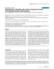
- Journal of Immune Based Therapies and Vaccines 2004, 2 http://www.jibtherapies.com/content/2/1/1 Subsets of Human Dendritic Cells ? CD14+/CD11C+ Monocytes CD14-/CD11C+ CD14-/CD11C- GM-CSF/ IL-3 TGF-β TNF/IL-4 Plasmacytoid DC Langerhans DC Interstitial DC Figure 1 Human DC subsets Human DC subsets. DC progenitors migrate from the bone marrow in the periphery and several different tissues. There they encounter various growth factors which determine the fate of these cells to differentiate into immature DC subsets. Page 3 of 11 (page number not for citation purposes)
- Journal of Immune Based Therapies and Vaccines 2004, 2 http://www.jibtherapies.com/content/2/1/1 Antigen Skin Lymph node Figure 2 Migration of immature DCs into lymphatic organs Migration of immature DCs into lymphatic organs. Skin surrounded by various immunogen antigens that can penetrate the epidermis. These antigens can be captured by immature Langerhans DCs, and processed. Cutaneous DCs will then be acti- vated, migrate, and home to the lymph nodes. Matured DCs present processed antigen to antigen specific T cells inducing spe- cific immunity. antigen-loaded DCs are administrated to developing or erance by presenting self-antigen to T cells residing in fetal thymus reactive T lymphocytes, they will be deleted specialized tissues such as the pancreas [32,33]. Presenta- indicating that DCs play a critical role in this process. tion of processed self-antigen as peptides by DCs ensures T cell tolerance probably through T cell deletion or anergy Moreover, in the cortical area of the thymus, although [32-34]. macrophages phagocytize dying T cells which did not undergo positive selection these cells seem not to be The role of dendritic cells in clinical diseases involved in deleting auto-reactive T cells. Studies show Recent studies shed light on the role of DC involvement that if MHC class II molecules are solely expressed by the in various diseases such as autoimmunity, allergy, trans- cortical epithelium and not by DCs residing in the plantation, infection and cancer. For example, studies medulla, there is a higher probability towards an autoim- showed that DCs differentiated in vitro express very mune disease. These results highlight the critical role of important co-stimulatory molecules, e.g. CD40, which DCs in educative processes of thymic T cells to self-anti- allow these cells to approach T cells and deliver signals to gens. In addition, DCs play a critical role in peripheral tol- them [22,23]. With respect to that phenomenon, Page 4 of 11 (page number not for citation purposes)
- Journal of Immune Based Therapies and Vaccines 2004, 2 http://www.jibtherapies.com/content/2/1/1 DC progenitor DC DC progenitor Macrophage Macrophage Ag Ag-capture of Immature DC Eosinophils T T NK Mature DC Ag-presentation T T B Inflamed vessels T B T T B cell Follicle B Figure 3 Induction of primary immune responses by DCs Induction of primary immune responses by DCs. The DC lineage comprises cells at different stages of differentiation and development in different tissues. The currently accepted scheme suggests that DCs from bone marrow move via the blood into non-lymphoid tissues. In these organs they undergo different changes with respect to shape, functions. In these organs DCs induce primary T cell immune responses. cytokines (e.g., GM-CSF, TNF-α) produced by keratinoc- maturation in vivo. Furthermore, it seems that the ligation ytes affect DC differentiation dramatically [35]. Moreover, of CD40 with DCs can enhance the antigen presenting DCs alone produce essential cytokines (e.g. IL-1β, TNF-α, capacity of these cells [22]. It has recently been reported IL-6), and chemokines MIP-1α, MIP-1γ, IL-15 and IL-8 that rheumatoid arthritis synovial T lymphocytes express [9,36-39]. Some of these cytokines contribute directly to CD40L at a low level. These molecules can be dramatically the DCs ability to attract and recruit T cells in sites of upregulated when T cells are activated. In this context, inflammation. A number of autoimmune diseases (rheu- stimulation of self-reactive T lymphocytes in the syno- sium will be induced through GM-CSF and TNF-α along matoid arthritis) or skin psoriasis demonstrates the accu- with CD80+ C086+ DCs [41]. mulation of DCs in diseased tissues [40]. This evidence suggests that DC enrichment within the cytokine-rich syn- osium or epidermis undergo phenotypic and functional Page 5 of 11 (page number not for citation purposes)
- Journal of Immune Based Therapies and Vaccines 2004, 2 http://www.jibtherapies.com/content/2/1/1 As mentioned above, cytokines can control the develop- example, Boczkowski et al. [50] conducted several elegant ment and differentiation of DCs. For example, the experiments to demonstrate that DCs pulsed with synthe- combination of GM-CSF and TNF-α can promote sized chicken ovalbumin (OVA) RNA were more effective differentiation of CD34+ blood stem cells into DCs in than OVA peptide-pulsed DCs in activating primary OVA humans [6]. Phenotypically these cells are CD4+CD11C+ specific-CTL responses in vitro. This finding shows that the since Langerhans DCs and other DC family numbers amplification of antigens from a small number of tumor express CD4 molecules that can bind to the HIV surface cells is feasible, thus increasing the possibility of utilizing envelope protein gp 120 [42]. This makes a possibility RNA-pulsed DC based vaccines for patients bearing very stronger that DCs may contribute to HIV pathology. On small tumors [50]. one hand, in vivo and in vitro experiments indicate that the replication of HIV-1 virus occurs during cognate CD4+ Studies demonstrate that when DCs are pulsed with T cell activation through DCs. On the other hand, there is tumor antigens in ex vivo, and these cells subsequently evidence that the features of HIV pathology are an accu- readministrated, specific immunity is established [51]. In addition, several studies showed that tumor-specific CD8+ mulation of HIV virus in the germinal centers, which is T cell rich and where a novel DC population has recently cytotoxic T lymphocytes (CTL) constitute an important been identified [20]. Both the APC function of DCs and effector arm of the anti-tumor immune response [52,53]. their close interaction with CD4+ T cells suggests that ger- In this context to elicit specific immunity against tumor minal centers of lymph nodes may provide an additional cells, DCs were pulsed with protein or peptide in the pres- site for HIV viral replication [42-44]. ence of lipid [54] or transfected with DNA [55] were capa- ble of eliciting primary CTL responses in vitro. Moreover, DCs in transplanted organs are involved and they represent potent "passenger leukocytes" that sensi- Although prior investigations have established that target- tize host graft antigens and trigger rejection [45]. Studies ing immune cells to tumors may improve immunity [47- have shown that the depletion of DCs from mouse islets 55], in the case of DCs, however, it has been shown [56- or thyroid tissue prolonged survival in allogeneic recipi- 62] that the tumor microenvironment is detrimental to ents [45]. Other studies on the function of DCs after trans- DC function, and in fact may condition DCs to induce a T plantation of skin and heart tissues to allogeneic cell response that anergizes or suppresses tumor-specific recipients have shown that soon after grafting, DCs enter immunity [56]. Thus, targeting DCs directly to tumors, as the recipient's lymphoid tissues [46]. Thus, there appears demonstrated by several studies, may be inefficient. to be a sensitization of host T cells which occurs primarily Therefore, methods should be developed in order to target in these tissues when they encounter the graft-derived, all- DCs by immunogenic TAAs outside the tumor microenvi- ogeneic DCs. Austyn et al. showed recently that host DCs ronment to improve immunity. can also present graft antigens to host T cells [46]. In this process it seems that host DCs bearing graft molecules Vaccine design by targeting dendritic cells would migrate into the secondary lymphoid organs to Given the central role of DCs in controlling immunity, sensitize and activate T lymphocytes and induce graft has brought a scientific focus to the critical role of DCs as rejection. an efficient vector in vaccine technology. Several approaches to target DCs efficiently have been designed. It is clear now, that cancer cells can express tumor associ- ated antigens, which are recognized by host T cells. These There is a large body of literature involving experimental T cells may not be able to reject tumor cells. These mole- animal models and for tumors and infection in which DC cules, then, are not immunogenic. In order to become subsets pulsed with TAAs or subunits of the pathogens immunogenic they must be processed and presented by such as HCV or HIV are to induce protective immunity professional antigen presenting cells (APC). Since DCs against tumors. However, it is even more important to cre- possess relevant features, e.g. a) internalizing of immuno- ate novel strategies by targeting immunogenic antigens or genic antigen through endocytosis, b) phagocytosis for immune regulatory agents specifically to DCs without subsequent processing and presentation of several anti- impairing the functional properties of DC subsets and in gens to T cells, and c) migration capability, they could this way modulate the immune responses in vivo. acquire tumor antigen. These novel strategies must not be too costly, not immu- In the past few years the role of DCs in cancer has been nopathogenic, but specific in order to overcome anergy suggested. There is evidence that DCs can induce immu- established through negative signals which may be pro- nity to tumors if they are administrated to animals or vided by immune component cells including DCs to the exposed to tumor associated antigen (TAA) before or microenvironment. when the tumor is inoculated into animals [47-49]. For Page 6 of 11 (page number not for citation purposes)
- Journal of Immune Based Therapies and Vaccines 2004, 2 http://www.jibtherapies.com/content/2/1/1 Figure Delivery4of immunogenic antigen to DCs by probiotic microorganisms Delivery of immunogenic antigen to DCs by probiotic microorganisms. DNA encoding sequences of DC-binding peptides and immunogenic subunit of any pathogen will be expressed in Gram positive bacteria including Lactobacillus. Lactoba- cillus will be orally administrated. These bacteria colonize the gut and express and release the immunogen in the intestine. DCs in the mucosal site will then capture the immunogen via DC-binding peptide motifs. They internalize the immunogen, process and present it to T cells inducing specific responses against released immunogen. One possible strategy is to target novel molecules The Peyer's patch is the primary mucosal site for antigen expressed on the cell surface of DCs. In this, we and others processing in the intestine. Recent in vivo studies provide utilized phage display peptide library to generate small evidence that DC network in the subepithelial dome of peptides which solely bind to DC subsets and not other Peyer's patches is a critical component in the uptake and cells. DNA sequences encoding DC-peptides can then be processing of luminal antigens. Such uptake may occur by fused genetically with TAA coding regions or with the sub- endocytosis or by phagocytosis after passage of antigen unit of the pathogen of interest. Immunogenic fusion pro- through M cells. The DCs then present the processed anti- gen to CD4+ or CD8+ T cells in the subepithelial dome, or teins can be then expressed by probiotic microorganisms such as Lactobacilli or attenuated strains of Salmonella in after maturation and migration, to the interfollicular regions where antigen is presented to CD4+/CD8+ T cells vivo (Figure 4). Such novel vaccine strategies should take advantage of mucosal sites in the body, as well as the skin [63]. In this regard, immunohistologic analysis of DC in order to be delivered specifically to DC subsets in vivo subsets including LCs in Peyer's patch has revealed that (Figure 5). the unique microanatomical localization of DC subsets Page 7 of 11 (page number not for citation purposes)
- Journal of Immune Based Therapies and Vaccines 2004, 2 http://www.jibtherapies.com/content/2/1/1 Antigen DC-pep Skin Lymph Node Figure 5 Transdermal delivery of immunogenic fusion protein by cutaneous DCs Transdermal delivery of immunogenic fusion protein by cutaneous DCs. Genetically engineered immunogenic fusion protein can be transdermaly administrated into the skin whereby cutaneous DC subsets can capture it via DC-peptide motifs fused to immunogen subunits. Loaded cutaneous DC subsets can be activated, leave the skin and enter the lymph nodes where they can present processed antigen as immunogenic peptides to T cells eliciting specific T cell immune responses. enables them to regulate specific T- and B-cell responses in facilitate rapid internalization of the immunogen into vivo [63-65]. Other, studies also clearly demonstrated that DCs. DCs will then process and present it to T cells resid- Gram-positive bacteria such as Lactobacilli can successfully ing in the gut. These cells will be activated and will circu- be used in order to deliver vaccine peptides to immune late through the body in order to elicit specific T cell component cells [66-68]. immune responses against the pathogen of interest. More specifically, in order to target any vaccine to DCs, A transdermal delivery system also offers an interesting recently, a novel strategy was proposed. Mohamadzadeh route to approach DC subsets in order to enhance immu- et al. fused a subunit of hepatitis C virus with a DC-bind- nity against cancer or pathogens. Accordingly, the ing peptide. Studies are ongoing to express such immuno- immune system of the skin harbors two very potent anti- genic fusion proteins by a strain of Lactobacilli [69]. Such gen-presenting DC subsets which induce primary antigen a Lactobacillus strain will express and secretes the immuno- specific T cell immune responses [70]. Furthermore, care- genic protein in the intestinal region. DCs will be able to ful experimentation of various vaccine delivery routes has capture such immunogen via the motifs of DC-binding shed light on the skin and its immune mechanisms. It has peptides. Such binding of an immunogen to DCs will previously been shown that cutaneous DC subsets can be Page 8 of 11 (page number not for citation purposes)
- Journal of Immune Based Therapies and Vaccines 2004, 2 http://www.jibtherapies.com/content/2/1/1 targeted and activated in situ in order to achieve specific T Acknowledgements cell mediated immune responses [70-74]. Thus, the feasi- Dr. Mohamadzadeh acknowledges support from National Institute on Drug Abuse (NIDA). Dr. Luftig acknowledges LSUHSC Institutional Funds. bility of using immunogenic DC-peptide fusion proteins should be tested to determine whether administration of References such immunogenic fusion proteins will induce the activa- 1. Banchereau J, Steinman R: Dendritic cells and the control of tion of cutaneous DC subsets that in turn prime antigen- immunity. Nature 1998, 392:245-252. specific T cells in situ. 2. Steinman RM: The dendritic cell system and its role in immunogenicity. Annu Rev Immunol 1991, 9:271-296. 3. Austyn JM: Lymphoid dendritic cells. Immunol 1987, 62:161-170. DCs play a crucial role in host-pathogen interactions. A 4. Metly JP, Pure E, Steinman RM: Control of the immune response recent example [75] involves the report in human papil- at the level of antigen-presenting cells: a comparison of the function of dendritic cells and B lymphocytes. Adv Immunol loma virus 16 which is strongly associated with the devel- 1989, 47:45-116. opment of cervical cancer, that in infected cells the E6 5. Fairchild PJ, Austyn JM: Thymic dendritic cells: phenotype and function. Intern Rev Immunol 1990, 6:187-196. oncogenic protein limits the numbers of LC in infected 6. Caux C, Dezutter-Dambuyant C, Schmitt D, Banchereau J: GM-CSF epidermis. This appears to decrease the host's ability to and TNFα cooperate in the generation of dendritic Langer- mount an effective immunological response to HPV 16. han cells. Nature 1992, 360:258-261. 7. Maraskovsky E, Brasel K, Teepe M, Roux ER, Lyman SD, Shortman We anticipate that future studies will be focused on KD, McKenna HJ: Dramatic increase in the numbers of func- enhancing functional aspects of DCs to prevent such tionally mature dendritic cells in Flt3-Ligand treated mice: multiple dendritic cell sub-populations identified. J Exp Med events and establish novel vaccine strategies to efficiently 1996, 184:1953-1961. target immunogenic antigens or inhibitory agents to DCs 8. Caux C, Banchereau J: In vitro regulation of dendritic cell devel- in order to elicit or suppress specific immune responses in opment and function. In Blood Cell Biochemistry, Hemopoietic growth factors and their receptors Volume 7. Edited by: Whetton A, Gordon J. Ple- vivo. num Press, London; 1996. 9. Mohmadzadeh M, Berard F, Essert G, Chaloini C, Pulendran B, Dav- Conclusions oust J, Palucka K, Banchereau J: IL-15 skews monocytes differen- tiation into dendritic cells with features of Langerhans cells. 1. Dendritic cells play a significant role in J Exp Med 2001, 194:1013-1019. immunopathogenesis. 10. Larsen CP, Morris PJ, Austyn JM: Migration of dendritic leuko- cytes from cardiac allografts into host spleen. A novel path- way for initiation of rejection. I Exp Med 1990, 171:307-314. 2. The functions of dendritic cells involve cancer, infec- 11. Fossum S: Lymph-borne dendritic leukocytes do not recircu- tious diseases and tolerance. late, but enter the lymph node paracortex to become inter- digitating cells. Scand J Immunol 1989, 27:97-105. 12. Matsuno K, Ezaki T, Kudo S, Uehara Y: A life stage of particle- 3. Novel approaches in vaccine design can occur by target- laden rat dendritic cells in vivo: their terminal division, active phagocytosis and translocation from the liver to hepatic ing dendritic cells. lymph. J Exp Med 1996, 183:1865-1878. 13. Kudo S, Matsuno K, Ezaki T, Ogawa M: A novel migration path- Competing interests way for rat dendritic cells from the blood: Hepatic sinusoids- lymph translocation. J Exp Med 1997, 185:777-784. None declared. 14. Brenan M, Puklavec M: The MRC OX-62 antigen: A useful marker in the purification of rat veiled cells with the bio- Author's contributions chemical properties of an integrin. J Exp Med 1992, 175:1457-1465. Dr. M. Mohamadzadeh is the corresponding author and 15. Austyn JM: New insights into the mobilization and phagocytic designed the draft of the manuscript. Dr. R. Luftig contrib- activity of dendritic cells. J Exp Med 1996, 183:1287. 16. Sallusto F, Cella M, Danielli C, Lanzavecchia A: Dendritic cells use uted to the viral-related segments and overview of the macropinocytosis and mannose receptor to concentrate manuscript. Both authors read and approved the final antigen in the MHC class II compartment. Downregulation manuscript. by cytokines and bacterial products. J Exp Med 1995, 182:389-400. 17. Mohamadzadeh M, Pavlidou A, Enk A, Knop J, Ruede E, Gradehandt Abbreviations G: Freshly isolated mouse 4F7+ splenic dendritic cells process TNF: Tumor Necrosis Factors and present exogenous antigens to T cells. Eur J Immunol 1994, 24:3170. 18. Cella M, Sallusto F, Lanzavecchia A: Origin, maturation and anti- GM-CSF: Granulocyte macrophage colony stimulating gen presenting function of dendritic cells. Curr Opin Immunol 1997, 9:10-16. Factor 19. Grouard G, Rissoan M, Filguera L, Durand I, Banchereau J, Liu YJ: The enigmatic plasmacytoid cells develop into dendritic cells CD: Cluster Density with interleukin 3 and CD40-ligand. J Exp Med 1996, 185:1101-1111. 20. Grouard G, Durand I, Filgueira L, Banchereau J, Liu Y-J: Dendritic IL-1: Interleukin-1 cells capable of stimulating T cells in germinal centres. Nature 1996, 384:364-366. 21. Dubois B: Dendritic cells enhance growth and differentiation TGF: Transforming growth factor of CD40-activated B Lymphocytes. J Exp Med 1997, 185:941-951. 22. Cella MD, Scheidegger D, Lehmann K, Lane P, Lanzavecchia A, Alber G: Ligation of CD40 on dendritic cells triggers production of Page 9 of 11 (page number not for citation purposes)
- Journal of Immune Based Therapies and Vaccines 2004, 2 http://www.jibtherapies.com/content/2/1/1 high levels of interleukin 12 and enhances T cell stimulatory 48. Nestle FO, Alijagic S, Gilliet M, Sun Y, Grabbe S, Dummer R, Burg G, capacity: T-T help via APC activation. J Exp Med 1996, Schadendorf D: Vaccination of melanoma patients with pep- 184:741-747. tide- or tumor lysate-pulsed dendritic cells. Nat Med 1998, 23. Grewal IS, Flavell RA: A central role for CD40 ligand in the reg- 4:328-332. ulation of CD4+ T cell responses. Immunol Today 1996, 49. Banchereau J, Shuler-Thumer B, Paluka AK, Schuler G: Dendritic 17:410-414. cells as vectors for therapy. Cell 2001, 106:271-274. 24. Mackey MF, Gunn JR, Maliszewski CR, Kikutani H, Noeller RJ, Barth 50. Boczkowski D, Nair SK, Snyder D, Gilboa E: Dendritic cells pulsed JR: DC require maturation via CD40 to generate protective with RNA are potent antigen-presenting cells in vitro and in anti-tumor immunity. J Immunol 1998, 161:2094-2098. vivo. J Exp Med 1996, 184:465-472. 25. Schoenberger SP, Toes REM: T-cell help for cytotoxic T lym- 51. Gyure LA, Barfoot R, Denham S, Ha II JG: Immunity to a syn- phocytes is mediated by CD40-CD40L interaction. Nature geneic sarcoma induced in rats by dendritic lymph cells 1998, 393:480-483. exposed to the tumor either in vivo or in vitro. Br J Cancer 26. Weiss JM, Sleeman J, Penkl H, Dittmar C, Termeer C, Taxis S, How- 1987, 55:17-20. ellset N, Simon JC: An essential role for CD44 variant isoforms 52. Knight SC, Hunt R, Dore C, Medawar PB: Influence of dendritic in epidermal Langerhans cell and blood dendritic cell cells on tumor growth. Proc Natl Acad Sci USA 1985, 82:4495-4497. function. JC Bio 1997, 137:1137-1147. 53. Greenberg PD: Adoptive T cell therapy of tumors: Mecha- 27. Kelso A: Th1/Th2 cells: Paradigms lost. Immunol Today 1995, nisms operative in the recognition and elimination of tumor 16:374-379. cells. Adv in Immunol 1991, 49:281. 28. Kelsoe G, Zengh B: Sites of B-cell activation in vivo. Curr Opin 54. Nair S, Babu JS, Dunham RG, Kanda P, Burke RL, Rouse BT: Induc- Immunol 1993, 5:418-422. tion of primary, anti viral cytotoxic, and proliferative 29. Pulendran B, Karvelas M, Nossal GJV: A form of immunologic tol- responses with antigens administered via dendritic cells. J erance through impairment of germinal centers. Proc Natl Virol 1993, 67:4062-4069. Acad Sci USA 1994, 91:2639-2643. 55. Rouse RJ, Nair SK, Lydy SL, Bowen JC, Rouse BT: Induction in vitro 30. Liu Y-J, Grouard G, de Bouteiller O, Banchereau J: Follicular den- of primary cytotoxic T-lymphocytes responses with DNA dritic cells and germinal centers. Int Rev Cytol 1996, 166:139-179. encoding herpes simplex virus proteins. J Virol 1994, 31. Lombardi T: Langerhans cell: structure, function and role in 68:5685-5689. oral pathological conditions. J Oral Pathology Med 1993, 56. Zou W: Stromal-derived factor-1 in human tumors recruits 22:193-202. and alters the function of plasmacytoid precursor dendritic 32. Kurts C: Constitutive class I-restricted exogenous presenta- cells. Nat Med 2001, 7:1339-1346. tion of self antigen in vivo. J Exp Med 1996, 184:923-930. 57. Gabrilovich DI, Chen HL, Girgis KR, Cunningham HT, Meny GM, 33. Kurts C, Kosaka H, Carbone ER, Miller JE, Heath WR: Class-I Nadaf S, Kavanaugh D, Carbone DP: Production of vascular restricted cross-presentation of exogenous self antigens endothelial growth factor by human tumors inhibits the leads to deletion of autoreactive CD8+ T cells. J Exp Med 1997, functional maturation of dendritic cells [published erratum 186:239-245. appears in Nat Med 1996 Nov;2(11):1267]. Nat Med 1996, 34. Forster I, Lieberam I: Peripheral tolerance of CD4 T cells fol- 2:1096-1103. lowing local activation in adolescent mice. Eur J Immunol 1996, 58. Gabrilovich DI, Ciernik IF, Carbone DP: Dendritic cells in antitu- 26:3194-3202. mor immune responses. I. Defective antigen presentation in 35. Caux C, Vanbervliet B, Massacrier C, Dezutter-Dambuyant C, de tumor-bearing hosts. Cell Immunol 1996, 170:101-110. Saint-Vis B, Jacquet C, Yoneda K, Imamura S, Schmitt D, Banchereau 59. Gabrilovich DI, Corak J, Ciernik IF, Kavanaugh D, Carbone DP: J: CD34+ hematopoietic progenitors from human cord blood Decreased antigen presentation by dendritic cells in patients differentiate along two independent dendritic cell pathways with breast cancer. Clin Cancer Res 1997, 3:483-490. in response to GM-CSF + TNFα. J Exp Med 1996, 184:695. 60. Gabrilovich DI, Nadaf S, Corak J, Berzofsky JA, Carbone DP: Den- 36. Sallusto F, Lanzavecchia A: Efficient presentation of soluble anti- dritic cells in antitumor immune responses. II. Dendritic gen by cultured human dendritic cells is maintained by gran- cells grown from bone marrow precursors, but not mature ulocyte/macrophage colony-stimulating factor and DC from tumor-bearing mice, are effective antigen carriers downregulated by tumour necrosis factor a. J Exp Med 1994, in the therapy of established tumors. Cell Immunol 1996, 179:1109-1117. 170:111-119. 37. Caux G, Vanbervliet B: Regulation of dendritic cell recruitment 61. Menetrier-Caux C, Montmain G, Dieu MC, Bain C, Favrot MC, Caux by chemokines. 1994, 73:S7-11. C, Blay JY: Inhibition of the differentiation of dendritic cells 38. Mohamadzadeh M, Takashima A, Dougherty I, Knop J, Bergstresser from CD34(+) progenitors by tumor cells: role of inter- PR, Crus PD Jr: Ultraviolet B radiation up-regulates the leukin-6 and macrophage colony-stimulating factor. Blood expression of IL-15 in human skin. J Immunol 1995, 1998, 92:4778-4791. 155:4492-4496. 62. Chomarat P, Banchereau J, Davoust J, Palucka AK: IL-6 switches the 39. Mohamadzadeh M, Poltak AN, Bergstresser PR, Beutler Takashima differentiation of monocytes from dendritic cells to BA: Dendritic cells produce macrophage inflammatory pro- macrophages. Nat Immunol 2000, 1:510-514. tei 1 g, a new member of the CC chemokine family. J Immunol 63. Kelsall BL: Distinct populations of dendritic cells are present 1996, 156:3102-3106. in the subepithelial dome and T cell regions of the murine 40. Mitra RS, Judge TA, Nestle FO, Turka LA, Nikoloff BJ: J Immunol 1995, peyers patch. J Exp Med 1996, 183:237-247. 154:2669-2677. 64. Kelsall BL, Strober W: Dendritic cells of the gastrointestinal 41. Lipsky PE, Davis LS, Cuah JJ, Oppenheimer-Marks: Springer Semin. tract. Springer Semin Immunopathol 1997, 18:409. Immunopathol 1989, 11:123-162. 65. Kelsall BL, Biron ChA, Sharma O, Kaye PM: Dendritic cells at the 42. Dalgleish AG, Beverley PCL, Clapham PR, Crawford DH, Greaves host-pathogen interface. Nature immunology 2002, 3:699-702. MF, Weis RA: The CD4 (T4) antigen is an essential component 66. Erickson KL, Hubbard NE: Probiotic immunomodulation in of the receptor for AIDS retrovirus. Nature 1984, 312:763-767. health and disease. J Nutr 2000, 130:403-409S. 43. Cameron PU: Science 1992, 257:383-387. 67. Ahrne S, Nobacek S, Jeppsson B, Adlerberth I, Wold AE, Molin G: 44. Frankel SS: Science 1996, 272:115-117. The normal Lactobacillus flora of healthy human rectal and 45. Austyn JM, Steinman RM: The passenger leukocytes – a fresh oral mucosa. J Appl Microbial 1998, 85:88. look. Transplantation reviews 1988, 2:139-176. 68. Guarner F, Schaafsma GJ: Probiotics Int J Food Microbial 1998, 39:237. 46. Larsen CP, Steinman RM, Witmer-Pack M, Hankins DF, Morris PJ, 69. Curiel T, Morris C, Brumlik M, Landry S, Mohamadzadeh M: Pep- Austyn JM: Migration and maturation of Langerhans cells in tides identified through phage display direct immunogenic skin transplants and explants. J Exp Med 1990, 172:1483-1493. antigen to dendritic cells. Submitted for publication 2003. 47. Hsu FJ, Benike C, Fagnoni F, Liles TM, Czerwinski D, Taidi B, Engle- 70. Lucas A, McPherson G: Langerhans cells: immigrants or man EG, Levy R: Vaccination of patients with B-cell lymphoma residents? Nature Immunology 2003, 3:1125-1126. using autologous antigen-pulsed dendritic cells. Nat Med 1996, 71. Filgueira L, Nestle FO, Rittig M, Joller HU, Groscuth P: Human den- 2:52-58. dritic cells phagocytose and process borrelia burgdorferi. J Immunol 1996, 157:2998-3005. Page 10 of 11 (page number not for citation purposes)
- Journal of Immune Based Therapies and Vaccines 2004, 2 http://www.jibtherapies.com/content/2/1/1 72. Guebre-Xabier M: Immunostimulant patch containing heat- labile enterotoxin from Escherichia coli enhances immune responses to inject influenza virus vaccine through activation of skin dendritic cells. J virology 2003, 77:5218-5225. 73. Glenn G: Transcutaneously immunization: a human vaccine delivery strategy using patch. Nat Med 2000, 6:1403-1406. 74. Guerena-Burgueno F: Safty and immunogenicity of prototype enterotoxigenic Escherichia coli vaccine administrated transcutaneously. Infect Immun 2002, 70:1874-1880. 75. Matthews K, Leong CM, Baxter L, Inglis E, Yun K, Blackstrom B, Doorbar J, Hibma M: Depletion of Langerhans cells in Human Papilloma virus type 16-infected skin is associated with E6- mediated down regulation of E-cadhedrin. J Virol 2003, 77:8378-8385. Publish with Bio Med Central and every scientist can read your work free of charge "BioMed Central will be the most significant development for disseminating the results of biomedical researc h in our lifetime." Sir Paul Nurse, Cancer Research UK Your research papers will be: available free of charge to the entire biomedical community peer reviewed and published immediately upon acceptance cited in PubMed and archived on PubMed Central yours — you keep the copyright BioMedcentral Submit your manuscript here: http://www.biomedcentral.com/info/publishing_adv.asp Page 11 of 11 (page number not for citation purposes)
CÓ THỂ BẠN MUỐN DOWNLOAD

-

Báo cáo y học: "Interferon-induced protein IFIT4 is associated with systemic lupus erythematosus and promotes differentiation of monocytes into dendritic cell-like cells"
 12 p |
  59
|
59
|  7
7
-
Báo cáo y học: "Indoleamine 2,3-dioxygenase-expressing dendritic cells are involved in the generation of CD4+CD25+ regulatory T cells in Peyer's patches in an orally tolerized, collagen-induced arthritis mouse model"
10 p | 51
| 6
-
Báo cáo y học: " Chitosan Interferon-c Nanogene Therapy for Lung Disease: Modulation of T-Cell and Dendritic Cell Immune Responses"
11 p | 58
| 5
-
Báo cáo y học: "Analysis of proteomic profiles and functional properties of human peripheral blood myeloid dendritic cells, monocyte-derived dendritic cells and the dendritic cell-like KG-1 cells reveals distinct characteristic"
13 p | 42
| 5
-

Báo cáo y học: "DNA-like class R inhibitory oligonucleotides (INH-ODNs) preferentially block autoantigen-induced B-cell and dendritic cell activation in vitro and autoantibody production in lupus-prone MRL-Faslpr/lpr mice in vivo"
16 p | 54
| 5
-

Báo cáo y học: "Differential induction of inflammatory cytokines by dendritic cells treated with novel TLR-agonist and cytokine based cocktails: targeting dendritic cells in autoimmunity"
12 p | 64
| 5
-

Báo cáo y học: "Dendritic cell therapy for oncology roundtable conference Sandra Tuyaerts"
6 p | 53
| 4
-
Báo cáo y học: "Dendritic cell subsets dynamics and cytokine production in SIVmac239-infected Chinese rhesus macaques"
13 p | 39
| 4
-

Báo cáo y học: "TLR2 and TLR4 triggering exerts contrasting effects with regard to HIV-1 infection of human dendritic cells and subsequent virus transfer to CD4+ T cells"
16 p | 64
| 4
-

Báo cáo y học: "Immunologists getting nervous: neuropeptides, dendritic cells and T cell activation"
6 p | 44
| 4
-

Báo cáo y học: "Protein synthesis of the pro-inflammatory S100A8/A9 complex in plasmacytoid dendritic cells and cell surface S100A8/A9 on leukocyte subpopulations in systemic lupus erythematosus"
10 p | 54
| 4
-
Báo cáo y học: " Influence of the cystic fibrosis transmembrane conductance regulator on expression of lipid metabolism-related genes in dendritic cells"
15 p | 101
| 4
-
Báo cáo y học: " Transcriptome profiling of primary murine monocytes, lung macrophages and lung dendritic cells reveals a distinct expression of genes involved in cell trafficking"
16 p | 56
| 4
-
Báo cáo y học: " Myeloid dendritic cells correlate with clinical response whereas plasmacytoid dendritic cells impact autoantibody development in rheumatoid arthritis patients treated with infliximab"
10 p | 64
| 3
-

Báo cáo y học: "Targeting Toll-like receptor signaling in plasmacytoid dendritic cells and autoreactive B cells as a therapy for lupus"
11 p | 58
| 3
-
Báo cáo y học: "CTLA-4 blockade during dendritic cell based booster vaccination influences dendritic cell survival and CTL expansion"
6 p | 74
| 3
-
Báo cáo y học: "Recruitment of dendritic cells and macrophages during T cell-mediated synovial inflammation"
13 p | 54
| 2


Chịu trách nhiệm nội dung:
Nguyễn Công Hà - Giám đốc Công ty TNHH TÀI LIỆU TRỰC TUYẾN VI NA
LIÊN HỆ
Địa chỉ: P402, 54A Nơ Trang Long, Phường 14, Q.Bình Thạnh, TP.HCM
Hotline: 093 303 0098
Email: support@tailieu.vn
Giấy phép Mạng Xã Hội số: 670/GP-BTTTT cấp ngày 30/11/2015 Copyright © 2022-2032 TaiLieu.VN. All rights reserved.

