


Báo cáo y học: " Fish bone foreign body presenting with an acute fulminating retropharyngeal abscess in a resource-challenged center: a case report"
lượt xem 4
download
 Download
Vui lòng tải xuống để xem tài liệu đầy đủ
Download
Vui lòng tải xuống để xem tài liệu đầy đủ
Tuyển tập báo cáo các nghiên cứu khoa học quốc tế ngành y học dành cho các bạn tham khảo đề tài: Fish bone foreign body presenting with an acute fulminating retropharyngeal abscess in a resource-challenged center: a case report...
Bình luận(0) Đăng nhập để gửi bình luận!
Nội dung Text: Báo cáo y học: " Fish bone foreign body presenting with an acute fulminating retropharyngeal abscess in a resource-challenged center: a case report"
- Afolabi et al. Journal of Medical Case Reports 2011, 5:165 JOURNAL OF MEDICAL http://www.jmedicalcasereports.com/content/5/1/165 CASE REPORTS CASE REPORT Open Access Fish bone foreign body presenting with an acute fulminating retropharyngeal abscess in a resource-challenged center: a case report Olushola A Afolabi1*, Joseph O Fadare1, Ezekiel O Oyewole1 and Stephen A Ogah2 Abstract Introduction: A retropharyngeal abscess is a potentially life-threatening infection in the deep space of the neck, which can compromise the airway. Its management requires highly specialized care, including surgery and intensive care, to reduce mortality. This is the first case of a gas-forming abscess reported from this region, but not the first such report in the literature. Case presentation: We present a case of a 16-month-old Yoruba baby girl with a gas-forming retropharyngeal abscess secondary to fish bone foreign body with laryngeal spasm that was managed in the recovery room. We highlight specific problems encountered in the management of this case in a resource-challenged center such as ours. Conclusion: We describe an unusual presentation of a gas-forming organism causing a retropharyngeal abscess in a child. The patient’s condition was treated despite the challenges of inadequate resources for its management. We recommend early recognition through adequate evaluation of any oropharyngeal injuries or infection and early referral to the specialist with prompt surgical intervention. Introduction of the laryngeal musculature, and its symptoms include inability to help the patient ventilate with resultant A retropharyngeal abscess is an infection with abscess rapid desaturation, which requires ICU care. We empha- collection in one of the deep spaces of the neck [1-3]. size early recognition, prevention of oropharyngeal An abscess in this location is an immediate life-threa- trauma and prompt surgical intervention for life-threa- tening emergency with the potential for airway compro- tening head and neck infections, even in the face of mise and other catastrophic complications [1]. Patients challenges. with diabetes and those who are debilitated, older adults or immunocompromised patients are more likely to get Case presentation this infection [2-4]. Delay in diagnosis results in high mortality and morbidity [4,5]. Although much has been A 16-month-old Yoruba girl was referred from a periph- written about this clinical condition and its clinical indi- eral hospital to the ear, nose and throat (ENT) unit of cators, this case report is the first case of a gas-forming our hospital with a one-week history of fever, a six-day retropharyngeal abscess in a child with a foreign body (a history of cough and a five-day history of neck swelling. fish bone) seen in North-central part of Nigeria. This Her fever was high grade with bouts of cough, and she particular case was challenging as the child developed a had no history of contact with a person with chronic laryngeal spasm postoperatively but was managed in the cough, no associated weight loss and no posttussive recovery room without a stay in the intensive care unit vomiting. Her mother noticed neck swelling five days (ICU). Other challenges were inadequate laboratory before presentation which was progressive and painful, facilities. Laryngospasm is a forceful, involuntary spasm with associated limited neck movement. The patient refused to eat, expectorated a thick tenacious secretion, and had episodes of irritability and excessive crying. The * Correspondence: droaafolabi@yahoo.com 1 Kogi State Specialist Hospital, Lokoja, Kogi State, Nigeria child had a previous history of left ear discharge which Full list of author information is available at the end of the article © 2011 Afolabi et al; licensee BioMed Central Ltd. This is an Open Access article distributed under the terms of the Creative Commons Attribution License (http://creativecommons.org/licenses/by/2.0), which permits unrestricted use, distribution, and reproduction in any medium, provided the original work is properly cited.
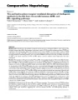
- Afolabi et al. Journal of Medical Case Reports 2011, 5:165 Page 2 of 5 http://www.jmedicalcasereports.com/content/5/1/165 h ad resolved, and there was no history of hearing Prevertebral widening impairment or nasal symptoms. About three days prior to presentation, the child was noticed to be breathless, for which she was treated at a private hospital as a case of pneumonia and was placed on an antitussive and antibiotics. The patient ’ s medical history and family and social history, as well as the review of systems, were not remarkable. An examination of the throat revealed poor oral hygiene; foul-smelling, thick, tenacious, straw- colored secretion from the oral cavity and oropharynx; and a bulging posterior pharyngeal wall. The patient’s neck showed a diffuse swelling which was tender. The ear, nose, chest and abdominal examinations were essentially normal. An assessment of retropharyngeal abscess was made to rule out parapharyngeal abscess. Investigations revealed that the packed cell volume was 41%, and the electrolyte and urea examinations showed the following concentra- tions: sodium, 142 mM/L; potassium, 3.7 mM/L; urea 6.5 mM/L; and creatinine, 101 mM/L. Figure 1 Lateral view X-ray showing the soft neck tissue and X-rays of the soft neck tissue revealed widening of the revealing widening of the prevertebral space containing areas prevertebral space containing areas of opacity and of mixed opacity and lucency extending from the base of the lucency extending from the base of the skull to the level skull to the level of the seventh cervical spine (C7), with the of the seventh cervical spine (C7), which at the level of laryngeal air column almost obliterated, anterior displacement the second cervical vertebra (C2) was about 22 mm, of the airway and straightening of the cervical spine. with the laryngeal air column almost obliterated and anterior displacement of the airway and straightening of the cervical spine (Figure 1). There was lateral displace- to extubation, residual neuromuscular block was antago- ment of the trachea to the left from the anteroposterior nized with a combination of 0.04 mg/kg neostigmine view (Figure 2). and 0.02 mg/kg atropine. The patient was extubated but The patient was resuscitated with intravenous fluid suddenly developed laryngeal spasm. Manual ventilation with a face mask was difficult as the patient’s pulse oxi- and antibiotics and was taken for examination under anesthesia and drainage of the abscess. The patient was metry was less than 80%. Anesthesia was deepened with halothane, and the patient’s trachea was resecured with placed in the anti-Trendelenburg position while under general anesthesia. Intubation was difficult but was 1 mg/kg suxamethonium. The patient was ventilated finally achieved using a size 2.5 mm endotracheal tube manually with 100% oxygen in the improvised recovery inserted by an experienced anesthetist, and light packing room on account of poor respiratory function for about with wet gauze was placed around the endotracheal 8 to 10 hours, after which she was transferred to the tube. Anesthesia was induced with halothane in oxygen, postoperative ward, where her condition was satis- and the trachea was secured with 1 mg/kg suxametho- factory. The patient was maintained on intravenous anti- nium. Anesthesia was maintained with 66% nitrous biotics, analgesics and anti-inflammatory agents. The oxide in oxygen and 0.5% to 1% halothane in oxygen, patient was discharged to home on the fifth day while muscle paralysis was induced with 0.1 mg/kg pan- postoperatively. curonium. Analgesia was ensured with 2 μg/kg fentanyl. A Boyle-Davis mouth gag was introduced gently to Discussion expose the oral cavity and oropharynx, a cruciate inci- Retropharyngeal abscess is not common nowadays with sion was made using a size 11 surgical blade and a sur- the increasing use of antibiotics in the treatment of gical probe was introduced to break down all loculi. upper respiratory tract infections. It is almost exclusively About 30 to 40 mL of foul-smelling, purulent discharge a pediatric diagnosis. Most incidents occur in children was drained with the extrusion of a fish bone remnant ages six months to six years [3-6] in whom the index from the abscess cavity (Figure 3). The culture revealed case still falls within a mean age of three to four years a growth of mixed organisms: Staphylococcus aureus , [2-4]. No racial or sex predilection has been described Klebsiella pneumoniae and anaerobic streptococci. Prior in the literature, but several studies have noted a higher
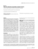
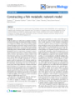
- Afolabi et al. Journal of Medical Case Reports 2011, 5:165 Page 3 of 5 http://www.jmedicalcasereports.com/content/5/1/165 Tracheal deviation Figure 2 Anteroposterior view X-ray showing lateral displacement of the trachea to the left. not uncommon in children who run and fall down after incidence of deep neck space infections in boys [3,4], they have placed an object such as a toy, stick, pencil or which is at variance with our present report of the case toothbrush into their mouths [4,7-10]. There also are of a young girl. iatrogenic causes, which include instrumentation with The retropharyngeal space is located immediately pos- laryngoscopy, endotracheal intubation, surgery, endo- terior to the nasopharynx, oropharynx, hypopharynx, scopy, feeding tube placement and dental injection pro- larynx and trachea [3,5]. The visceral (that is, bucco- cedures [11], which inoculate these organisms directly pharyngeal) fascia, which surrounds the pharynx, tra- into the retropharyngeal space. chea, esophagus and thyroid, forms the anterior border Our index case was initially managed for pneumonia of the retropharyngeal space. Bounded posteriorly by by the general practitioner; however, there is a need to the alar fascia, the retropharyngeal space is bounded lat- encourage caregivers to present their children for treat- erally by the carotid sheaths and parapharyngeal spaces ment early. The diagnosis of this condition is mainly [5]. It extends superiorly to the base of the skull and clinical, with some support from the radiological investi- inferiorly to the mediastinum at the level of the tracheal gation, which can also occasionally be confirmatory. bifurcation. Patients with retropharyngeal abscess may present with The retropharyngeal space can become infected in airway compromise, thus the management of the airway three ways [3,4]. Either infection spreads from a contig- takes priority with regard to patient care. Fortunately, uous area affecting the retropharyngeal nodes or the our patient did not present with airway challenges, space is inoculated directly secondary to a penetrating except postoperatively. The culture in the present case foreign body as we observed in our case in which a fish revealed mixed aerobic and anaerobic flora with gas- bone foreign body penetrated the retropharyngeal space forming organisms (that is, Klebsiella, anaerobic strepto- as found intraoperatively. It may be through oropharyn- cocci). The other gas-forming organisms isolated from geal injuries such as accidental lacerations, which are Fish bone retrieved Figure 3 Photograph of fish bone (foreign body) remnant removed from the abscess cavity.
- Afolabi et al. Journal of Medical Case Reports 2011, 5:165 Page 4 of 5 http://www.jmedicalcasereports.com/content/5/1/165 retropharyngeal abscess despite drainage [5], and other t he head and neck infections described in previous complications are highlighted above. The specimen reports are Clostridium [12], Bacteroides and Fusobac- obtained in our present case was transported to our sis- terium [13]. ter medical center where it was cultured and reported. Patients with retropharyngeal abscess present with Delays in diagnosis and treatment can lead to the risk constitutional complaints such as fever, chills, malaise, decreased appetite, muffled “hot potato” voice [4] and of complications. The mortality of retropharyngeal abscess is due to the association with airway obstruc- irritability [2] as seen in our index case. Older patients tion, mediastinitis, aspiration pneumonia, epidural may complain of sore throat, dysphagia, odynophagia, abscess, jugular venous thrombosis, carotid artery ero- trismus or torticollis; however, our index case was an sion, pericarditis and airway compromise. infant who was unable to demonstrate the expected Our patient was extubated but still developed laryn- symptoms, although she refused to eat [1,3,4,9]. geal spasm, which is an uncommon situation that A lateral soft tissue neck X-ray is contributory in mak- requires close monitoring immediately after surgery. ing the diagnosis of a retropharyngeal abscess [2]. Laryngeal spasm in a standard setup is an indication for Widening of these soft tissues is pathologic until proven ICU admission, which is lacking in our center; however, otherwise as seen in our index case (Figure 1). The mea- our patient was managed in the recovery room by man- surement of the distance from the anterior surface of ual ventilation and monitoring of vital signs. Laryngos- the C2 vertebra to the posterior border of the airway should be 7 mm or less, regardless of the patient’s age pasm is a forceful, involuntary spasm of the laryngeal musculature caused by stimulation of the superior laryn- [4]. With measurement starting at the C6 vertebra, this geal nerve, which is the sensory innervation of the lar- width should be 14 mm or less in children younger than ynx. Its signs include an inability to ventilate the patient 15 years of age and 22 mm in adults. A simpler but less with rapid desaturation. Prevention can be achieved by precise rule is that on soft tissue plain X-rays, the pre- extubating the patient using a no-touch technique when vertebral body should be less than one half the width of the patient is awake [14], as was done in the index case, the corresponding vertebral body. However, in our or under deep anesthesia (possibly after a magnesium index case, it was about three times the size of the ver- infusion) [15], which was not available in our center in tebral body, which is an unusual presentation (Figure 1) the event that the awake extubation failed. Complica- [3-5]. Some authors have reported the use of computed tions of laryngospasm can be prevented through applica- tomographic scans to diagnose retropharyngeal abscess, tion of a gentle jaw thrust, but if this fails, the depth of especially in uncommon situations [5], the authors have anesthesia can be increased with intermittent positive no knowledge of such report in our region. pressure ventilation on a ventilator, which is not avail- Prompt surgical intervention with drainage of the able in our center. Some researchers have used propofol abscess was the most essential part of the management to increase the depth of anesthesia because of its rapid- of this patient, especially in view of the size of the ity of onset and predictability [16]. However, in the obstruction and the gas content, as the possibility of index case, halothane was used. rupture was envisaged because of the challenges of our ICU, an inadequate laboratory facility and insufficient Conclusion personnel. We consider prompt surgical intervention to have been a lifesaving step in the present case. The This case report highlights an unusual presentation and index case was intubated despite some difficulty because management of retropharyngeal abscess. The presence of of the enlarged retropharyngeal mass, deviated trachea gas-forming organisms in this clinical scenario makes it (Figure 2) and narrowed pharyngolaryngeal space under an interesting case. Physicians should maintain a high direct visualization. Previous reports have proposed index of suspicion, however, when encountering children fiberoptic intubation, which was not available in our with torticollis or unexplained neck pain or swelling and center, or cricothyroidotomy or, in the worst case sce- should perform the necessary investigations to avoid nario, tracheostomy [4], all of which are done to protect delay in diagnosis, which might lead to serious conse- the lower airway. Positioning the airway correctly and quences. There also need to be close monitoring of the avoiding unnecessary manipulation is essential [3,4,9]. patients immediately after surgery and readiness for chal- The patient is at risk of compression the pharynx or tra- lenges even in the face of inadequate facilities. Despite chea with possible suffocation or rupture with asphyxia- numerous challenges encountered during the manage- tion or aspiration of the abscess, sepsis and pneumonia ment of our patient, the end result was satisfactory. This if left unattended to or at intubation in the hand of report is expected to affect positively clinical practice in inexperienced anesthetist, as seen in the X-ray of this the field of ENT surgery, anesthesia and medicine in gen- patient. Some workers have reported the relapse of eral in resource-challenged settings such as ours.
- Afolabi et al. Journal of Medical Case Reports 2011, 5:165 Page 5 of 5 http://www.jmedicalcasereports.com/content/5/1/165 14. Tsui BC, Wagner A, Cave D, Elliot C, El-Hakim H, Malherbe S: The incidence Consent of laryngospasm with a “no touch” extubation technique after Written informed consent was obtained from the tonsillectomy and adenoidectomy. Anesth Analg 2004, 98:327-329. patient’s parents for publication of this case report and 15. Gulhas N, Dumus M, Demirbilek S, Togal T, Ozturk E, Ersoy MO: The use of magnesium to prevent laryngospasm after tonsillectomy and accompanying images. A copy of the written consent is adenoidectomy: a preliminary study. Paediatr Anaesth 2003, 13:43-47. available for review by the Editor-in-Chief of this 16. Afsham G, Chohan U, Qamar-Ul-Hoda M, Kamal RS: Is there a role of a journal. small dose of propofol in the treatment of laryngeal spasm? Paediatr Anaesth 2002, 12:625-628. doi:10.1186/1752-1947-5-165 Acknowledgements Cite this article as: Afolabi et al.: Fish bone foreign body presenting The authors are grateful to the theater and anesthetic nurse who assisted in with an acute fulminating retropharyngeal abscess in a resource- the surgery for this patient. We also thank the patient’s father, who challenged center: a case report. Journal of Medical Case Reports 2011 consented to the publication of this report. 5:165. Author details 1 Kogi State Specialist Hospital, Lokoja, Kogi State, Nigeria. 2University of Ilorin Teaching Hospital, Ilorin, Kwara State, Nigeria. Authors’ contributions AOA was the principal surgeon, performed the literature search and prepared the manuscript and takes responsibility for the publication. FJO assisted in preparing and proofreading the manuscript for intellectual content and gave final approval for the publication. OEO was the anesthetist, obtained the accompanying images and conceived the idea for the manuscript. OSA did the literature search, contributed to the preparation of the manuscript and reviewed the manuscript. All authors read and approved the final manuscript. Competing interests The authors declare that they have no competing interests. Received: 31 July 2010 Accepted: 27 April 2011 Published: 27 April 2011 References 1. Choi SS, Vezina LG, Grundfast KM: Relative incidence and alternative approaches for surgical drainage of different types of deep neck abscesses in children. Arch Otolaryngol Head Neck Surg 1997, 123:1271-1275. 2. Okeowo PA: Pharynx-Infections (Tonsillitis, Quinsy, abscess) & TB. In Okeowo’s Companion to Ear, Nose and Throat Diseases in the Tropics. Volume 3.. 1 edition. Lagos, Nigeria: University of Lagos Press; 2004:109-114. 3. Cowan DL, Hibbert J: Acute and chronic infections of the pharynx and tonsils. In Scott-Brown’s Otolaryngology. Volume 5. 6 edition. Edited by: Kerr AG, Hibbert J. Oxford Boston: Butterworth-Heinemann, Jordan hills, Oxford DX28DP; 1997:(4):5-6. 4. Craig FW, Schunk JE: Retropharyngeal Abscess in Children: Clinical Presentation, Utility of Imaging, and Current Management. PEDIATRICS 2003, 6(111):1394-1398. 5. Gaglani MJ, Edwards MS: Clinical indicators of childhood retropharyngeal abscess. Am J Emerg Med 1995, 13:333-336. 6. Philpott CM, Selvadurai D, Banerjee AR: Paediatric retropharyngeal abscess. J Laryngol Otol 2004, 118:919-926. 7. Wahbeh G, Wyllie R, Kay M: Foreign body ingestion in infants and children: location, location, location. Clin Pediatr (Phila) 2002, 41:633-640. 8. Marom T, Russo E, Ben-Yehuda Y, Roth Y: Oropharyngeal injuries in children. Pediatr Emerg Care 2007, 23:914-918. 9. Gray RF, Hawthorne M: Disease of the mouth and pharynx. In Synopsis of Submit your next manuscript to BioMed Central Otolaryngology. Volume Chapter 13. 5 edition. Boston: Butterworth- and take full advantage of: Heinemann; 1992:320-353. 10. Ologe FE, Afolabi OA: Penetrating pencil injury in the retromolar trigone: the need to play safe on playing ground. J Surg Surg Sci 2007, 1:38-40. • Convenient online submission 11. Marra S, Hotaling AJ: Deep neck infections. Am J Otolaryngol 1996, • Thorough peer review 17:287-298. • No space constraints or color figure charges 12. Tung-Yiu W, Jehn-Shyun H, Ching-Hung C, Hung-An C: Cervical necrotizing fasciitis of odontogenic origin: a report of 11 cases. J Oral Maxillofac Surg • Immediate publication on acceptance 2000, 58:1347-1352. • Inclusion in PubMed, CAS, Scopus and Google Scholar 13. Shumrick KA, Sheft SA: Deep neck infections. In Otolaryngology. Volume 3. • Research which is freely available for redistribution 3 edition. Edited by: Paparella MM, Shumrick DA. Philadelphia: Saunders; 1991:(3):2545-2564. Submit your manuscript at www.biomedcentral.com/submit
CÓ THỂ BẠN MUỐN DOWNLOAD

-

Báo cáo y học: " Dietary intake of fish, omega-3, omega-6 polyunsaturated fatty acids and vitamin D and the prevalence of psychotic-like symptoms in a cohort of 33 000 women from the general population"
 13 p |
  97
|
97
|  12
12
-

Báo cáo khoa học: "Comparison of immunohistochemistry (IHC) and fluorescence in situ hybridization (FISH) assessment for Her-2 status in breast cancer"
6 p | 72
| 6
-
Báo cáo y học: "Eggshell and egg yolk proteins in fish: hepatic proteins for the next generation: oogenetic, population, and evolutionary implications of endocrine disruption"
21 p | 56
| 6
-

Báo cáo y học: "Effects of a fish oil containing lipid emulsion on plasma phospholipid fatty acids, inflammatory markers, and clinical outcomes in septic patients: a randomized, controlled clinical trial"
11 p | 45
| 5
-

Báo cáo y học: " Effects of supplemental fish oil on resting metabolic rate, body composition, and salivary cortisol in healthy adults"
7 p | 53
| 4
-
Báo cáo y học: "Fish-on-a-chip: a sensitive detection microfluidic system for alzheimer’s disease"
11 p | 48
| 4
-
Báo cáo y học: "Immunonutrition in critical illness: still fishing for the truth"
3 p | 44
| 4
-
Báo cáo y học: "Teach a man to fish and you feed him for a lifetime: physics, pharmacology and physiology for anaesthetists"
1 p | 52
| 4
-
Báo cáo y học: "The aryl hydrocarbon receptor-mediated disruption of vitellogenin synthesis in the fish liver: Cross-talk between AHR- and ERα-signalling pathway"
14 p | 49
| 3
-
Báo cáo y học: "Fish oil: what the prescriber needs to know"
9 p | 34
| 3
-
Báo cáo y học: "Constructing a fish metabolic network model"
15 p | 42
| 3
-

Báo cáo y học: "Fishing for Allergens: Bloodworm-Induced Asthma"
2 p | 29
| 3
-

Báo cáo y học: "Tetraodon genome confirms Takifugu findings: most fish are ancient polyploids"
4 p | 36
| 3
-

Báo cáo y học: "Fish oil-containing lipid emulsions in patients with sepsis"
2 p | 38
| 2
-
Báo cáo y học: "Correction: Fish oil: what the prescriber needs to know"
1 p | 62
| 2
-

báo cáo khoa học: "The association of fish consumption with bladder cancer risk: A meta-analysis"
7 p | 46
| 2
-

Báo cáo y học: "High tandem repeat content in the genome of the short-lived annual fish Nothobranchius furzeri: a new vertebrate model for aging research"
0 p | 38
| 2


Chịu trách nhiệm nội dung:
Nguyễn Công Hà - Giám đốc Công ty TNHH TÀI LIỆU TRỰC TUYẾN VI NA
LIÊN HỆ
Địa chỉ: P402, 54A Nơ Trang Long, Phường 14, Q.Bình Thạnh, TP.HCM
Hotline: 093 303 0098
Email: support@tailieu.vn
Giấy phép Mạng Xã Hội số: 670/GP-BTTTT cấp ngày 30/11/2015 Copyright © 2022-2032 TaiLieu.VN. All rights reserved.

