


Báo cáo y học: " Generation of H9 T-cells stably expressing a membrane-bound form of the cytoplasmic tail of the "
lượt xem 2
download
 Download
Vui lòng tải xuống để xem tài liệu đầy đủ
Download
Vui lòng tải xuống để xem tài liệu đầy đủ
Tuyển tập các báo cáo nghiên cứu về y học được đăng trên tạp chí y học quốc tế cung cấp cho các bạn kiến thức về ngành y đề tài: Generation of H9 T-cells stably expressing a membrane-bound form of the cytoplasmic tail of the
Bình luận(0) Đăng nhập để gửi bình luận!
Nội dung Text: Báo cáo y học: " Generation of H9 T-cells stably expressing a membrane-bound form of the cytoplasmic tail of the "
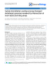
- Retrovirology BioMed Central Open Access Short report Generation of H9 T-cells stably expressing a membrane-bound form of the cytoplasmic tail of the Env-glycoprotein: lack of transcomplementation of defective HIV-1 virions encoding C-terminally truncated Env Denise Holtkotte, Tanya Pfeiffer and Valerie Bosch* Address: Forschungsschwerpunkt Infektion und Krebs, F020, Deutsches Krebsforschungszentrum, Im Neuenheimer Feld 242, 69120 Heidelberg, Germany Email: Denise Holtkotte - d.holtkotte@dkfz.de; Tanya Pfeiffer - t.pfeiffer@dkfz.de; Valerie Bosch* - v.bosch@dkfz.de * Corresponding author Published: 16 May 2006 Received: 21 April 2006 Accepted: 16 May 2006 Retrovirology 2006, 3:27 doi:10.1186/1742-4690-3-27 This article is available from: http://www.retrovirology.com/content/3/1/27 © 2006 Holtkotte et al; licensee BioMed Central Ltd. This is an Open Access article distributed under the terms of the Creative Commons Attribution License (http://creativecommons.org/licenses/by/2.0), which permits unrestricted use, distribution, and reproduction in any medium, provided the original work is properly cited. Abstract H9-T-cells do not support the replication of mutant HIV-1 encoding Env protein lacking its long cytoplasmic C-terminal domain (Env-CT). Here we describe the generation of a H9-T-cell population constitutively expressing the HIV-1 Env-CT protein domain anchored in the cellular membrane by it homologous membrane-spanning domain (TMD). We confirmed that the Env- TMD-CT protein was associated with cellular membranes, that its expression did not have any obvious cytotoxic effects on the cells and that it did not affect wild-type HIV-1 replication. However, as measured in both a single-round assay as well as in spreading infections, replication competence of mutant pNL-Tr712, lacking the Env-CT, was not restored in this H9 T-cell population. This means that the Env-CT per se cannot transcomplement the replication block of HIV-1 virions encoding C-terminally truncated Env proteins and suggests that the Env-CT likely exerts its function only in the context of the complete Env protein. nomena. Thus, for example, the HIV-Env-CT has been Findings In contrast to most other enveloped viruses, the surface reported to bind to calmodulin and to inhibit calmodu- Env glycoproteins of lentiviruses, including HIV-1, con- lin-regulated proteins [2,3]. Furthermore, yeast 2 hybrid tain very long C-terminal cytoplasmic tails (CTs). In the screenings have identified further potential cellular inter- action partners of the HIV-Env-CT. These are α-catenin, case of HIV-1, the Env-CT has a conserved length of about 150 amino acids (aa) and mutant viruses, encoding trun- which is involved in cellular adhesion [4,5], and p115- cated Env proteins, are unable to replicate in most T cell- RhoGEF, which regulates actin stress fiber formation and lines [1]. However, despite its undoubted importance for activates the serum response factor (SRF) [6]. It is possible the HIV-1 life cycle, the biological mechanism by which that these interactions with cellular processes, or others the long Env-CT facilitates virus replication is still not presently unknown, are important for viral replication. fully understood. Numerous studies employing HIV Env- For example, it is conceivable that the Env-CT itself CT mutants have addressed the potential roles of various accesses signal transduction pathways to alter cellular motifs and features within the Env-CT. Of relevance for gene expression and facilitate virus replication or, alterna- this study is the fact the Env-CT may impact cellular phe- tively, membrane-bound Env-CT itself may recruit essen- Page 1 of 5 (page number not for citation purposes)
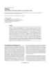
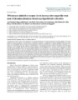
- Retrovirology 2006, 3:27 http://www.retrovirology.com/content/3/1/27 A Env -TMD -CT 3’ LTR/SIN 5’ LTR IRES GFP Figure 1 of, and virus replication in, an H9 T-cell population stably expressing HIV-Env-TMD-CT Generation Generation of, and virus replication in, an H9 T-cell population stably expressing HIV-Env-TMD-CT. A. schematic representa- tion of the SIN lentiviral vector pWPI-Env-TMD-CT employed. An internal human EF1-α promoter drives expression of a tran- scriptional unit consisting of the Env-TMD-CT gene, an IRES element and the gene for GFP. The composition of the Env-TMD- CT gene is described in the text. SP; signal peptide sequence of tissue plasminogen activator, Env-TMD; membrane anchor of HIV-Env, CT; cytoplasmic domain of HIV-Env. B. FACS for GFP expression (left panels) and indirect immunofluorescence anal- yses for Env-TMD-CT expression (right panels) of H9 T-cell populations stably transduced with pWPI (H9-GFP) and pWPI- Env-TMD-CT vector particles (H9-CT). Immunofluorescence of paraformaldehyde-fixed permeabilised H9 cells was per- formed with rabbit anti-gp160 serum shown to contain antibodies against the Env CT, followed by biotinylated goat anti-rabbit IgG and streptavidin phycoerythrin. Identical exposure times were used to generate the images from the H9-GFP and the H9- CT cells. C. Western blot analysis of H9 cells stably transduced with pWPI (H9-GFP) and pWPI-Env-TMD-CT (H9-CT) (left panel) and of cytosolic (C) and membrane (M) fractions of H9-CT cells (right panel) with gp41 Mab Chessie 8 [16] as indicated. After stripping, the right blot was reprobed with rabbit antibodies specific for the cytoplasmic protein 14-3-3 γ (C-16) (Santa Cruz Biotechnology). D. Replication kinetics of Wt-pNL-4-3 (Wt) (filled-in symbols) and pNL-Tr712 virions (Tr712) (empty symbols) in H9 cells (circles), H9-GFP cells (triangles) and H9-CT cells (squares). Infections were initiated with 100 ng virus per 106 cells, produced by transfection of the respective plasmids in 293T cells. 5 h p.i., the cells were thoroughly washed and the course of infections followed by measurement of newly released HIV-CA in the supernatant by ELISA. E. Western blot analyses of equalised amounts (by CA-ELISA of culture supernatants) of lysates of H9-GFP and H9-CT cells infected with pNL-∆ Env (∆), pNL-Wt (Wt) and pNL-Tr712 (712) (left) and of equalised amounts (by CA-ELISA of ultracentrifuged particles) of the respective virions released into the media (right). The top portions of the filters have been probed with anti-gp120 serum and the bottom portion with anti-CT antibodies (Chessie 8). Page 2 of 5 (page number not for citation purposes)
- Retrovirology 2006, 3:27 http://www.retrovirology.com/content/3/1/27 tial cellular proteins to cellular membranes sites and thus radation products, respectively, were detectable. In order facilitate virus assembly and release. to confirm localisation to cellular membranes, cytosolic (C) and membrane (M) fractions from H9-CT cells were In this study, we have generated and characterised H9 T- prepared employing published procedures [9]. Western cells which stably express a membrane-bound version of blot analysis of equivalent amounts of these fractions the Env-CT and examined if the presence of this region demonstrated that the Env-TMD-CT protein was localised alone might be sufficient for the transcomplementation of predominantly in the membrane fraction and only a originally non-infectious HIV-1 virions encoding for trun- minor amount remained in the cytosolic fraction (Fig. 1C, cated Env glycoproteins. right panels). Reprobing the blot with antibodies to the 30 kDa cytosolic protein 14-3-3 γ (C-16) [10] confirmed pWPI-Env-TMD-CT, depicted in Fig 1A, encodes a mem- the authenticity of the membrane/cytosol separation. In brane-bound form of the Env-CT and is based on the summary, these results point to functional membrane bicistronic lentiviral vector pWPI (obtained from D. insertion of the Env-TMD-CT protein. Trono, University of Geneva, Switzerland). pWPI-Env- TMD-CT and pWPI additionally express green fluorescent There were no obvious cytotoxic effects on the H9-CT cells protein (GFP) downstream of an internal ribosomal entry as a result of expression of the Env-TMD-CT protein. Thus site (IRES). The Env-TMD-CT gene consists of the signal cell growth was not reduced in comparison to H9-GFP peptide (SP) sequence from tissue plasminogen activator cells and cell morphology was unaffected (data not (tPA) (tPA amino acids (aa) 1–35) fused via a 4aa spacer shown). We then went on to examine the replication of to the membrane-spanning (TMD) and CT domains of wild-type HIV (pNL4-3, referred to as pNL-Wt) and the HIV BH10-Env protein (aa 684–851). The 4aa spacer mutant pNL-Tr712, encoding truncated Env protein in consists of 2 HIV-Env aa (Thr, Glu) C-terminal to the HIV- which only 7aa of the 151 aa long Env-CT remain [11], in SP cleavage site and 2 HIV-Env aa N-terminal to the TMD H9, H9-GFP and H9-CT cells. The viruses were generated (Ile, Lys). by transfection of the respective proviral plasmids in 293T cells and amounts equivalent to 100 ng CA/106 cells, as Lentiviral vector particles were generated by cotransfec- determined by enzyme-linked immunosorbant assay tion of 293T cells with pWPI-Env-TMD-CT or, as a con- (ELISA) (Innogenetics, Ghent, Belgium) were used to ini- trol, pWPI plus the packaging construct pCMV∆R8.91 [7] tiate infection of the H9 cell populations. After removing and the VSV-G expression plasmid pMD.G [8]. Vector par- input virus and thorough washing, the course of the infec- ticles, concentrated from the culture supernatant by ultra- tions was monitored by determining, via CA-ELISA, the centrifugation, were employed to transduce H9 cells. amounts of released virions in the respective culture Transduced cell populations were then sorted for maxi- supernatants over time. As shown in Fig. 1D, pNL-Wt mum GFP expression and expanded. The resulting trans- virus replicated efficiently in all the H9 cell populations duced populations were designated H9-CT cells but with a slight delay in both H9-CT and H9-GFP cells. (expressing Env-TMD-CT and GFP) and H9-GFP cells The basis for this slight delay in replication kinetics is not (expressing only GFP). As shown in Fig. 1B, both popula- known. As had been shown previously [1], pNL-Tr712 tions were over 90% positive for GFP expression although cannot give rise to a spreading infection in H9 cells nor, as clearly the fluorescence intensity of the H9-CT population to be expected, was this the case in H9-GFP cells. In the was lower than that of the H9-GFP population. This may H9-CT cell population, spreading infection of pNL-Tr712 be a result of GFP expression being decreased when pre- virus also does not occur despite the presence of the Env- ceded by the Env-TMD-CT gene. Expression of the Env- CT region anchored at the cellular membrane by its TMD-CT protein in the sorted H9-CT population was first homologous TMD. analysed by indirect immunofluorescence of paraformde- hyde-fixed, permeabilised cells employing rabbit anti- In order to generate virions for further analyses, the gp160 serum which we have previously demonstrated to respective H9 cell populations were infected with VSV-G pseudotyped pNL-Wt, pNL-Tr712 virions and pNL-∆ Env contain antibodies against the Env-CT. As shown in Fig. 1B, right panels, in comparison to H9-GFP cells, virtually virions using procedures previously described [12] and, all of the cells in the H9-CT culture were positive for Env- after removal of input virus, the respective newly gener- TMD-CT expression. Western blot analysis confirmed ated virions were collected. The infectivities of the virions expression of a specific Env-TMD-CT protein band migrat- in the supernatants of the infected H9 cells were analysed ing at about the position of its calculated molecular in a single-round assay in Tzm-bl reporter cells [13-15]. weight (18.8 kDa after removal of the SP) (Fig. 1C, left pNL-Tr712 virions exhibited reduced but still significant panel). In addition, some minor species migrating slightly infectivity in comparison to pNL-Wt virions but the extent slower or faster than the major Env-TMD-CT protein, of the reduction was independent of whether the virions which may represent species still containing the SP or deg- were produced in H9-GFP cells or in H9-CT cells express- Page 3 of 5 (page number not for citation purposes)
- Retrovirology 2006, 3:27 http://www.retrovirology.com/content/3/1/27 ing membrane-bound Env-TMD-CT (data not shown). in the H9 cells was not sufficient to transcomplement the This shows that the expression of the Env-TMD-CT pro- replication block of virions encoding C-terminally trun- tein in producer H9 T-cells does not result in an increase cated Env proteins. Although other reasons may account of the infectivity of released pNL-Tr712 virions. In order for this lack of transcomplementation, the most likely to examine if the Env-TMD-CT protein, expressed in the explanation is that the Env-TMD-CT has to be part of the H9-CT cells, was incorporated into released virions, the full-length Env protein in order to fulfill its essential func- respective virions were concentrated by ultracentrifuga- tion(s). Nevertheless, H9 CT cells (and control H9-GFP tion from the media of infected H9-CT cells or H9-GFP cells) may still be useful tools to study possible effects of cells and lysates of infected cells and virions examined in the Env-TMD-CT protein on cellular processes such as sig- Western blot (Fig. 1E). Virally-expressed gp160, gp120 nal transduction phenomena/cellular gene expression. and gp41 were detectable in lysates of pNL-Wt infected cells and truncated gp160 (gp140) and gp120 in lysates of Competing interests pNL-Tr712 infected cells. The truncated gp41 species The author(s) declare that they have no competing inter- (gp28) expressed by pNL-Tr712 was not detected since the ests. antibodies employed (Chessie 8 [16]) bind to an epitope in the Env-CT missing in this protein. In the lysates of all Authors' contributions the infected H9-CT cultures, constitutively expressed Env- DH carried out the replication kinetics, participated in the TMD-CT protein was detectable. Its expression level was cloning of pWPI-TMD-Env-CT and the generation of the similar to that of the gp41 protein expressed after infec- stable H9 cell lines and was involved in drafting the man- tion with pNL-Wt. In Fig. 1E, right panel, analysis of uscript. TP carried out FACS, Western blot and membrane equalised amounts (by CA-ELISA) of virions concentrated fractionation analyses. VB participated in the design of the from the supernatants of the respective infected cultures is study and in drafting the manuscript. All authors read and shown. Gp120 and gp41 proteins were detectable in pNL- approved the final manuscript. Wt virions and gp120 protein was detectable in pNL- Tr712 virions (again the truncated gp28 band cannot be Acknowledgements detected). This observation of gp120 incorporation into We thank Wolfram Hildebrandt for participation in the generation of the stably transduced H9 cells and Matthias T. Dittmar for discussion. The fol- pNL-Tr712 virions stands in contrast to two studies in the lowing reagent was obtained through the AIDS Research and Reference literature [1,17] which report that Env incorporation into Reagent Program, Division of AIDS, NIAID, NIH: HIV-gp41 hybridoma pNL-Tr712 virions is defective when these are produced in Chessie 8 from Dr. G. Lewis. Plasmid pWPI plasmid was provided by D. non-permissive cells. However, we consistently observe Trono, Geneva, Switzerland. This work was supported by the Deutsche gp120 incorporation into pNL-Tr712 and have recently Forschungsgemeinschaft, grant BO 517/5-1. reported that this is also the case with another mutant HIV encoding Env with a different C-terminal truncation [12]. References The reason for this discrepancy is presently unknown. Of 1. Murakami T, Freed EO: The long cytoplasmic tail of gp41 is required in a cell type-dependent manner for HIV-1 enve- interest in the context of this report is the fact that, lope glycoprotein incorporation into virions. Proc Natl Acad Sci although the respective virions have incorporated gp120/ U S A 2000, 97:343-348. gp41, the constitutively expressed Env-TMD-CT protein 2. Srinivas SK, Srinivas RV, Anantharamaiah GM, Segrest JP, Compans RW: Membrane interactions of synthetic peptides corre- was not detectable in any of the released virions. The phe- sponding to amphipathic helical segments of the human nomena which determine if particular cellular and viral immunodeficiency virus type-1 envelope glycoprotein. J Biol Chem 1992, 267:7121-7127. proteins are incorporated into virions or not are not 3. Srinivas SK, Srinivas RV, Anantharamaiah GM, Compans RW, Segrest understood in depth. Thus also in this case, we can only JP: Cytosolic domain of the human immunodeficiency virus speculate that perhaps the Env-TMD-CT protein may not envelope glycoproteins binds to calmodulin and inhibits cal- modulin-regulated proteins. J Biol Chem 1993, 268:22895-22899. be localised at the cellular sites of virus assembly or may 4. Kim EM, Lee KH, Kim JW: The cytoplasmic domain of HIV-1 not appropriately interact with cellular proteins influenc- gp41 interacts with the carboxyl-terminal region of alpha- catenin. Mol Cells 1999, 9:281-285. ing localisation/incorporation. 5. Kim JT, Kim EM, Lee KH, Choi JE, Jhun BH, Kim JW: Leucine zipper domain of HIV-1 gp41 interacted specifically with alpha-cat- In summary, in this report we describe a cell population enin. Biochem Biophys Res Commun 2002, 291:1239-1244. 6. Zhang H, Wang L, Kao S, Whitehead IP, Hart MJ, Liu B, Duus K, Burr- in which the majority, and likely all of the cells express a idge K, Der CJ, Su L: Functional interaction between the cyto- native i.e. untagged version of the HIV-Env-CT domain plasmic leucine-zipper domain of HIV-1 gp41 and p115- anchored in their cellular membranes by its homologous RhoGEF. Curr Biol 1999, 9:1271-1274. 7. Zufferey R, Nagy D, Mandel RJ, Naldini L, Trono D: Multiply atten- membrane anchor. We envisage that the expressed Env- uated lentiviral vector achieves efficient gene delivery in TMD-CT protein likely adopts its native conformation vivo. Nat Biotechnol 1997, 15:871-875. 8. Naldini L, Blomer U, Gallay P, Ory D, Mulligan R, Gage FH, Verma IM, although we cannot formally rule out the possibility that Trono D: In vivo gene delivery and stable transduction of non- this may require the presence of the Env ectodomain. The dividing cells by a lentiviral vector. Science 1996, 272:263-267. presence of the membrane-bound Env-TMD-CT protein 9. Resh MD, Erikson RL: Highly specific antibody to Rous sarcoma virus src gene product recognizes a novel population of Page 4 of 5 (page number not for citation purposes)
- Retrovirology 2006, 3:27 http://www.retrovirology.com/content/3/1/27 pp60v-src and pp60c-src molecules. J Cell Biol 1985, 100:409-417. 10. Morrison D: 14-3-3: modulators of signaling proteins? Science 1994, 266:56-57. 11. Wilk T, Pfeiffer T, Bosch V: Retained in vitro infectivity and cytopathogenicity of HIV-1 despite truncation of the C-ter- minal tail of the env gene product. Virology 1992, 189:167-177. 12. Holtkotte D, Pfeiffer T, Pisch T, Bosch V: Selection and character- ization of a replication-competent human immunodefi- ciency virus type 1 variant encoding C-terminally truncated env. AIDS Res Hum Retroviruses 2006, 22:57-65. 13. Derdeyn CA, Decker JM, Sfakianos JN, Zhang Z, O'Brien WA, Ratner L, Shaw GM, Hunter E: Sensitivity of human immunodeficiency virus type 1 to the fusion inhibitor T-20 is modulated by core- ceptor specificity defined by the V3 loop of gp120. J Virol 2000, 74:8358-8367. 14. Platt EJ, Wehrly K, Kuhmann SE, Chesebro B, Kabat D: Effects of CCR5 and CD4 cell surface concentrations on infections by macrophagetropic isolates of human immunodeficiency virus type 1. J Virol 1998, 72:2855-2864. 15. Wei X, Decker JM, Liu H, Zhang Z, Arani RB, Kilby JM, Saag MS, Wu X, Shaw GM, Kappes JC: Emergence of resistant human immu- nodeficiency virus type 1 in patients receiving fusion inhibi- tor (T-20) monotherapy. Antimicrob Agents Chemother 2002, 46:1896-1905. 16. Abacioglu YH, Fouts TR, Laman JD, Claassen E, Pincus SH, Moore JP, Roby CA, Kamin-Lewis R, Lewis GK: Epitope mapping and topol- ogy of baculovirus-expressed HIV-1 gp160 determined with a panel of murine monoclonal antibodies. AIDS Res Hum Retro- viruses 1994, 10:371-381. 17. Akari H, Fukumori T, Adachi A: Cell-dependent requirement of human immunodeficiency virus type 1 gp41 cytoplasmic tail for Env incorporation into virions. J Virol 2000, 74:4891-4893. Publish with Bio Med Central and every scientist can read your work free of charge "BioMed Central will be the most significant development for disseminating the results of biomedical researc h in our lifetime." Sir Paul Nurse, Cancer Research UK Your research papers will be: available free of charge to the entire biomedical community peer reviewed and published immediately upon acceptance cited in PubMed and archived on PubMed Central yours — you keep the copyright BioMedcentral Submit your manuscript here: http://www.biomedcentral.com/info/publishing_adv.asp Page 5 of 5 (page number not for citation purposes)
CÓ THỂ BẠN MUỐN DOWNLOAD

-
Báo cáo y học: "MRI bone oedema scores are higher in the arthritis mutilans form of psoriatic arthritis and correlate with high radiographic scores for joint damage"
 9 p |
9 p |  122
|
122
|  7
7
-
Báo cáo y học: " Interactions among type I and type II interferon, tumor necrosis factor, and -estradiol in the regulation of immune response-related gene expressions in systemic lupus erythematosus"
10 p | 88
| 5
-
Báo cáo y học: " Implication of granulocyte-macrophage colony-stimulating factor induced neutrophil gelatinase-associated lipocalin in pathogenesis of rheumatoid arthritis revealed by proteome analysis"
12 p | 109
| 5
-

Báo cáo y học: "Reduced levels of two modifiers of epigenetic gene silencing, Dnmt3a and Trim28, cause increased phenotypic nois"
10 p | 73
| 4
-
Báo cáo y học: "Introduction of medical emergency teams in Australia and New Zealand: a multicentre study"
2 p | 113
| 4
-
Báo cáo y học: "Effect of bladder volume on measured intravesical pressure:"
6 p | 109
| 4
-

Báo cáo y học: " Influence of the cystic fibrosis transmembrane conductance regulator on expression of lipid metabolism-related genes in dendritic cells"
15 p | 85
| 4
-

Báo cáo y học: " Arsenic trioxide, a potent inhibitor of NF-κB, abrogates allergen-induced airway hyperresponsiveness and inflammation"
12 p | 95
| 3
-

Báo cáo y học: ": Immunostaining of modified histones defines high-level features of the human metaphase epigenome"
14 p | 81
| 3
-

Báo cáo y học: "Rapid chromosome territory relocation by nuclear motor activity in response to serum removal in primary human fibroblasts"
0 p | 94
| 3
-
Báo cáo y học: " GE Rotterdam, the Netherlands. †Department of Human Genetics"
18 p | 68
| 3
-
Báo cáo y học: "The electronic version of this article is the complete one and can be found online"
6 p | 88
| 3
-
Báo cáo y học: "ontinuity, psychosocial correlates, and outcome of problematic substance use from adolescence to young adulthood in a community sample"
1 p | 79
| 3
-
Báo cáo y học: " Vgf is a novel biomarker associated with muscle weakness in amyotrophic lateral sclerosis (ALS), with a potential role in disease pathogenesis"
8 p | 95
| 3
-
Báo cáo y học: "Staffing level: a determinant of late-onset ventilator-associated pneumonia"
3 p | 106
| 3
-
Báo cáo y học: "TPO, but not soluble-IL-6 receptor, levels increase after anagrelide treatment of thrombocythemia in chronic myeloproliferative disorders"
5 p | 83
| 3
-
Báo cáo y học: "hese authors contributed equally to this work"
0 p | 84
| 2
-
Báo cáo y học: "Substantial deletion overlap among divergent Arabidopsis genomes revealed by intersection of short reads and tiling arrays"
0 p | 81
| 2


Chịu trách nhiệm nội dung:
Nguyễn Công Hà - Giám đốc Công ty TNHH TÀI LIỆU TRỰC TUYẾN VI NA
LIÊN HỆ
Địa chỉ: P402, 54A Nơ Trang Long, Phường 14, Q.Bình Thạnh, TP.HCM
Hotline: 093 303 0098
Email: support@tailieu.vn
Giấy phép Mạng Xã Hội số: 670/GP-BTTTT cấp ngày 30/11/2015 Copyright © 2022-2032 TaiLieu.VN. All rights reserved.

