


Báo cáo y học: "he effects of chemotherapeutics on cellular metabolism and consequent immune recognition"
lượt xem 3
download
 Download
Vui lòng tải xuống để xem tài liệu đầy đủ
Download
Vui lòng tải xuống để xem tài liệu đầy đủ
Tuyển tập báo cáo các nghiên cứu khoa học quốc tế ngành y học dành cho các bạn tham khảo đề tài: he effects of chemotherapeutics on cellular metabolism and consequent immune recognition...
Bình luận(0) Đăng nhập để gửi bình luận!
Nội dung Text: Báo cáo y học: "he effects of chemotherapeutics on cellular metabolism and consequent immune recognition"
- Journal of Immune Based Therapies and Vaccines BioMed Central Open Access Review The effects of chemotherapeutics on cellular metabolism and consequent immune recognition M Karen Newell*1, Robert Melamede1, Elizabeth Villalobos-Menuey1, Douglas Swartzendruber2, Richard Trauger3, Robert E Camley4 and William Crisp5 Address: 1Department of Biology, University of Colorado at Colorado Springs, Colorado Springs, CO 80933-7150, USA, 2Natural Sciences Division, Seaver College, Pepperdine University, Malibu, CA 90263, USA, 3Hollis Eden Pharmaceuticals, San Diego, CA 92121, USA, 4Department of Physics, University of Colorado at Colorado Springs, Colorado Ssprings, CO 80933-7150, USA and 5Cancer Research Institute, Arizona State University, Tempe, AZ 85287, USA Email: M Karen Newell* - mnewell@uccs.edu; Robert Melamede - rmelamed@uccs.edu; Elizabeth Villalobos-Menuey - emvillal@uccs.edu; Douglas Swartzendruber - douglas.swartzendruber@pepperdine.edu; Richard Trauger - rtrauger@holliseden.com; Robert E Camley - rcamley@uccs.edu; William Crisp - adcrc1@getnet.net * Corresponding author Published: 02 February 2004 Received: 29 December 2003 Accepted: 02 February 2004 Journal of Immune Based Therapies and Vaccines 2004, 2:3 This article is available from: http://www.jibtherapies.com/content/2/1/3 © 2004 Newell et al; licensee BioMed Central Ltd. This is an Open Access article: verbatim copying and redistribution of this article are permitted in all media for any purpose, provided this notice is preserved along with the article's original URL. chemotherapyimmune recognitionapoptosisFas (CD95)metabolism Abstract A widely held view is that oncolytic agents induce death of tumor cells directly. In this report we review and discuss the apoptosis-inducing effects of chemotherapeutics, the effects of chemotherapeutics on metabolic function, and the consequent effects of metabolic function on immune recognition. Finally, we propose that effective chemotherapeutic and/or apoptosis- inducing agents, at concentrations that can be achieved physiologically, do not kill tumor cells directly. Rather, we suggest that effective oncolytic agents sensitize immunologically altered tumor cells to immune recognition and immune-directed cell death. 4 or Fas Ligand on the T cell. We, and others, have Review reported that changes in the cell surface occur in drug- Do drugs kill tumor cells directly? Our laboratories have been investigating the conse- treated cells [4-10]. First, we observe changes and quences of chemotherapeutic agents on cell surface increases in cell surface expression of the B7 family mem- expression of immunologically important molecules, bers, CD80 and CD86, on drug-treated (adriamycin, 5- including Major Histocompatibility Complex (MHC) fluorouracil, or methotrexate-treataed) tumor cells. These encoded molecules (both MHC class I and II), B7.1 cell surface molecules have been extensively studied and (CD80), B7.2 (CD86), Fas (CD95), and Fas Ligand are now widely accepted as important in promoting the (CD95L) [1]. T cell activation requires recognition of anti- immunogenicity of tumor cells by providing costimula- gens associated with MHC molecules [2] and a second sig- tion for T cells [5]. Second, we, and others, have observed nal provided by co-stimulation [3] provided by the that most of the drugs we have used increase cell surface interaction of molecules including B7.1 or B7.2 or Fas expression of Fas (CD95) and sensitize the Fas-bearing (CD95) on the cell being recognized and CD28 or CTLA- tumor to Fas-induced death [1,7,9]. In the present report, Page 1 of 6 (page number not for citation purposes)
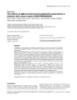
- Journal of Immune Based Therapies and Vaccines 2004, 2 http://www.jibtherapies.com/content/2/1/3 we discuss our working model that the concert of meta- Fas and yet remain insensitive to Fas-induced death bolic interference with the ability of the tumor to be more (including most dividing, regenerating, and self-renewing readily "seen" by the immune system may be the basis for cells) exhibit a metabolic phenotype characterized by effectiveness of many currently effective strategies or the high rate, cytosolic glycolysis. This "respiratory defi- basis for developing novel therapeutic approaches to ciency" is the result of a metabolic change in tumor cells treating cancers. that was first observed by Warburg in 1926 [27]. The co- incidence of increased cytosolic glycolysis and increased We first explore one of the relevant immunological cell Fas expression on tumor cells (and other dividing cells) surface receptors, Fas (CD95). Fas is a member of the provided the basis for examining a causal link between Fas tumor necrosis receptor (TNFR) family. The cytoplasmic expression and the use of glucose as a primary, glycolytic tail of Fas contains a death domain able to trigger intrac- source of fuel. ellular caspase cascades that culminate in apoptotic cell death [11-13]. Fas can induce apoptosis when ligated by Our experiments have demonstrated that the distribution its cognate ligand (FasL, CD95L) in Fas sensitive cells and levels of expression of Fas are altered in response to [11,12]. Paradoxically, Fas, like other members of its fam- changing concentrations of glucose in many cell lines and ily, can transduce growth-enhancing signals as well as in freshly isolated cells from a variety of tissues. Limited death signals [14-18]. In chemo-sensitive leukemia and glucose supplementation is known to enhance prolifera- solid tumors, anti-cancer drugs have been shown to tion of tumor cells and has been used for topical applica- induce apoptosis and for many tumors the pathways tions to accelerate wound healing in vivo [28,29]. Some of involved include, but are not limited to, Fas and FasL [19- our recent results suggest that glucose availability and 21]. consequent production of intracellular reactive oxygen species may regulate the striking change in the results In an attempt to reflect in vitro the concentrations of drugs from Fas engagement that promotes proliferation to Fas that can be achieved physiologically in vivo, we were sur- engagement that promotes death. Supporting this obser- prised to observe that tumor cells from many tissue ori- vation is the recent report that increasing glucose concen- gins were not dead at such concentrations. However, we trations can induce increased free radical production [30] found (and continue to find with a broad spectrum of and increases in reactive oxygen or free radicals are known agents) that the drugs have several important conse- to cause Fas engagement to result in cell death [31-33]. In quences. Our results have shown that chemotherapeutic addition, we have observed and reported that drug resist- agents sensitize Fas-bearing, Fas-insensitive tumors to Fas- ant cells appear to readily utilize the carbons derived from susceptibility and Fas-induced death [1]. Consistent with beta oxidation of fatty acids and exhibit a consequent loss these observations, cross-resistance to Fas/FasL and onco- of cell surface Fas. Taken together these observations sup- lytic agents has been reported by our group and others port the notion that Fas expression and function are inter- [1,8,10,22]. While much of our work has involved Fas and twined with glucose metabolism and the potential for FasL, other members of "death inducing" receptor-ligand changes in reactive intermediates in tissues or cells exhib- pairs likely perform similarly in the presence of effective iting changes in glucose metabolism. The fact that selec- oncolytic agents [23]. tion in drugs results in loss of Fas and in metabolic changes that may protect the cells from free radical dam- Together these data indicated that an important mecha- age will be important in designing novel cancer therapies. nism of chemotherapeutic agents may be to sensitize tumor cells to immune-directed death. Implied by these We have performed experiments to examine the correla- results is the importance of identifying and preserving tion between cell surface Fas expression and glucose (from death by high dose chemotherapy) the FasL (or metabolism. As a prototype for the Fas positive and Fas other ligand)-bearing cells to facilitate immunological negative cells we have used the L1210 cell and the destruction of drug-treated tumor cells. L1210DDP as Fas positive and Fas negative, respectively, Figure 1. In these experiments, we directly measured the rates of glucose utilization and oxidation of L1210 and How do chemotherapeutic agents sensitize the tumor cells L1210DDP [34]. to immune-mediated death? Our efforts at understanding the molecular mechanisms by which chemotherapeutic agents affect metabolism and L1210 DDP cells express no cell surface Fas [1]. To address immune recognition have been focused primarily on the the possibility that Fas is expressed, but has been targeted expression and function of Fas on the cell surface of tumor to a subcellular organelle, we permeabilized and stained cells. Fas is expressed on most rapidly dividing cells, L1210 and L1210DDP cells with fluorochrome conju- including tumor cells, hepatocytes, epithelial cells, and gated anti-Fas antibody (J02.2, Pharmingen). The cells lymphocytes [24-26]. Interestingly, tissues that express were examined by flow cytometry. Our data indicate that Page 2 of 6 (page number not for citation purposes)
- Journal of Immune Based Therapies and Vaccines 2004, 2 http://www.jibtherapies.com/content/2/1/3 Intracellular FAS Surface FAS L1210/0 L1210/DDP Key FAS Expression Isotype Control Figure 1 Distribution and Level of Fas in L1210/0 and L1210/DDP Cells Distribution and Level of Fas in L1210/0 and L1210/DDP Cells. Expression of cell-surface Fas, leftmost panels, and intracellular Fas, right most panels in L1210/0, upper two panels, and L1210/DDP cells, lower two panels. The levels of cell sur- face Fas (dark lines) were determined using fluorochrome conjugated anti-Fas antibodies (Pharmingen Inc.) and flow cytometry. The levels of intracellular Fas were determined subsequent to cellular permeabilization and fixation. The Fas levels are meas- ured relative to staining for fluorochrome-conjugated isotype control (grey lines). L1210 DDP cells express no cell surface Fas; however, the cell death unless the intracellular pool can be redistrib- cells do express intracellular Fas. Fluorochrome-conju- uted to the cell surface and potentially re-wired to "death- gated isotype matched antibody was used as control, and inducing" machinery. specific antibody stains were confirmed as specific. These data demonstrate that the Fas negative, apoptosis resistant It is known that T cells require two signals for activation cells, express intracellular Fas, Figure 1 below. The rele- [3]. One of these signals involves the binding of the pro- vance of internal Fas in drug-selected, drug resistant teins CD28 or CTLA-4, which are constitutively expressed tumor cells is that the cell is rendered Fas-insensitive to on most resting T cells, with the proteins B7.1 (CD80) or Page 3 of 6 (page number not for citation purposes)
- Journal of Immune Based Therapies and Vaccines 2004, 2 http://www.jibtherapies.com/content/2/1/3 B7.2 (CD86). T cell activation through CD28 binding, results in a proliferative T cell response, enhanced T cell survival and cytokine release [35]. Conversely, CTLA-4 engagement induces powerful inhibitory signals in T cell activation resulting in the negative regulation of T cell responses [36]. Collins et al. recently showed that B7.1 favors CTLA-4 over CD28 engagement [37]. This is still controversial, nonetheless is raises the possibility that co- stimulatory receptor/ligand pairs are multifunctional. We propose that co-stimulatory interactions between B7 family members and CD28 or CTLA4-bearing T cells and the resulting cytokines directly impact the subcellular dis- tribution of Fas and the ultimate outcome of Fas engage- ment on tumor cells. In Figure 2 we show that B7.2 levels in HL60 (human leukemic cells) also increase after treatment with 10-8 M of Adriamycin. We note that HL60 is a human cell line and that the drug is different than that in previous figures. This Figure 2 Adriamycin Induced Increase in B7.2 Expression Adriamycin Induced Increase in B7.2 Expression. figure is representative of many experiments with other Expression of the cell-surface co-stimulatory molecule B7.2 cell lines and additional drugs that include methotrexate, as a function of treatment with adriamycin. The level of cell- adriamycin, and 5-fluorouracil. While we have not tested surface B7.2 was determined using fluorochrome conjugated the ability of all drugs to promote immunogenicity, these anti-B7.2 antibodies and flow cytometry. The B7.2 levels are resuts may imply that the increase in the co-stimulatory sig- measured relative to staining for fluorochrome-conjugated nal as a result of drug treatment is a general phenomenon. isotype control. Which immune cell can kill the tumor cell? The first attempts at cancer immunotherapy were made over 100 years ago on the assumption that tumor antigens might be recognized as foreign [38]. These studies gave mechanism for both phenomenons has been attributed to rise to animal tumor models using syngeneic tumors, T cell receptor recognition and effector functions that spontaneously arising tumors, and xenografts into immu- occur only when MHC molecules and antigen are recog- nodeficient hosts. The collective of these studies resulted nized by the T cell receptor for antigen. Cells implicated in in a variety of immunotherapeutic protocols including tumor cell death include CD4+ T cells, CD8+ T cells, natu- ral killer (NK) cells, or more recently, gamma delta (γδ) T adjuvant therapy, cytokines, NK cell activation, macro- phages, and attempts to stimulate tumor antigen specific cells [38]. Immune recognition and destruction of alloge- B and/or T cell responses against tumor antigens. Some neic tumor cells likely results from increased expression of approaches have had partial success, but what has become MHC antigens on the tumor cell surface, processing and clear is that tumor cells are, by definition, "immunologi- presentation of tumor antigens, and expression of costim- cally privileged" and successfully evade effective tumori- ulatory molecules on the tumor cell. Rejection of tumor cidal immune recognition [38]. An alternate possibility is cells following drug treatment, therefore, may be directly suggested by the premise which Prehn has postulated that related to "recognition" of a cell which has changed in cell effective chemotherapies may result from suppressing a surface expression of immunologically important cell sur- particular type of immune response that supports tumor face receptors and that has been metabolically "rewired" cell growth [39]. An example of this notion would be T by chemotherapeutic agents. cell-produced cytokines which have been reported to sup- port neural regeneration [40]. Conclusion Thus, we suggest that a drug-treated tumor cell is made MHC encoded molecules were defined by Peter Gorer and susceptible by drugs or radiation to "death-inducing" George Snell as surface molecules responsible for the receptor/ligand pairs, including, but not limited to, Fas rejection of tumor cells between genetically distinct mem- and FasL expressed on candidate immune cells, such as bers of the same species [41]. These molecules are also CD4+ T cells, CD8+ T cells, gamma delta T cells, and NK responsible for graft rejection and T cell activation. The cells. We propose that selective identification of the Page 4 of 6 (page number not for citation purposes)
- Journal of Immune Based Therapies and Vaccines 2004, 2 http://www.jibtherapies.com/content/2/1/3 immunocytes proliferating in the tumor-bearing lymph ity. We suggest identifying cells responding to the tumor node as a key element in personalizing and selectively in the node (unsuccessfully or not) so that drug-sensitized sensitizing an individual's tumor cells to chemo-, radio-, tumor cells can be killed rather than supported by the and immunotherapy. identified immune cells. Minimally we suggest that a re- evaluation of the mechanism of tumor cell death and While the potential of immune-directed cytotoxicity of therapeutic approaches be experimentally and clinically drug-treated tumor cells may provide an important new considered. perspective, the question arises as to how to reconcile this idea with the accepted notion that chemotherapy can be Declaration of Competing Interests immunosuppressive. The key factor in resolving this None declared. seeming paradox may be the dose of the agent or the nature of a given chemotherapeutic agent. Clearly, there Authors' Contributions are cases where drugs at high doses have immunosuppres- This paper is distinct because it is an opinion paper. How- sive effects (perhaps by direct cytotoxicity of the immune ever, each author contributed uniquely to the manuscript. cells). In contrast, decreased doses have recently been Author 1, MKN, provided the conceptual framework for shown to be more effective in the clinic. Taken together, the model presented in this paper. Author 2, RM, partici- both views suggest that "less may be more" effective for pated in discussions and drafts of the manuscript. Author chemotherapy [45,46]. We propose that an in depth eval- 3, EVM, performed the flow cytometric data provided in uation of the effects of popular chemotherapeutic agents this manuscript. Author 4, DS, provided discussion about on induction of immunologically relevant molecules on a supportive role for T-Cells in the growth of a tumor. the tumor be rigorously evaluated. Author 5, RT, participated in discussions and drafts of the manuscript. Author 6, WC, contributed the effects of sur- Considering the potential importance of cells of the gery on tumor growth. Author 7, RC, participated in dis- immune system in controlling cancer growth, with or cussions and drafts of the manuscript. without chemotherapy, an important question is raised. Should lymph nodes, the local "home" too many Acknowledgements immune cells, be removed as therapy? Although axillary We greatly acknowledge Jaimi Kupperman and Jeff Rogers for their assist- ance with this work. node removal is still a standard regime for treatment of invasive breast cancer, it is clear that regional lymph References nodes have biological significance for being more than 1. Bhushan A, Kupperman JL, Stone JE, Kimberly PJ, Calman NS, Hacker just anatomical filters. The regional lymph node is the MP, Birge RB, Tritton TR, Newell MK: Drug resistance results in heart of our immunologic defense system and the present alterations in expression of immune recognition molecules and failure to express Fas (CD95). Immunology and Cell Biology routine practice of partial resection of the regional nodes 1998, 76:350-356. where they are easiest to remove undoubtedly has an Yague J, White J: The T-cell receptor: The α and β chains define 2. effect on immunological and physiological function. idiotype, and antigen and MHC specificity. Cell 1985, 42:81-87. 3. Bretscher P: The two-signal model of lymphocyte activation twenty-one years later. Immunology Today 1992, 13:74-76. Macroscopically involved lymph nodes should possibly 4. Delabie J, Ceuppens JL, Vandenberghe P, Coorevits L, De Wolf- Peeters C: The B7/BB1 antigen is expressed by Reed-Stern- be removed for prognosis [42] and for the identification berg cells of Hodgkin's disease and contributes to the stimu- of the immune cells involved in tumor recognition, but lation capacity of Hodgkin's disease derived cell lines. Blood the routine removal of lymph nodes is questioned as 1993, 82:2845-2852. 5. Guinan EC, Gribben JG, Boussiotis VA, Freeman GJ, Nadle L: Pivotal noted above by our group and others [43]. It is becoming role of the B7:CD28 pathway in transplantation tolerance clear that many patients can be spared axillary node dis- and tumor immunity. Blood 1994, 84(10):3261-3282. 6. Baskar S, Clements VK, Glimcher LH, Nabavi N, Ostrand-Rosenberg section without adversely affecting outcome [44]. As we S: Rejection of MHC class II transfected tumor cells requires begin to better understand the inter-relationships of sur- induction of tumor encoded B7-1 and/or B7-2 costimulatory gery, tumor cell kinetics, chemotherapy, and the host molecules. Journal of Immunology 1996, 156:3821-3827. 7. Cai Z, Stancou R, Korner M, Chouaib S: Impairment of Fas-anti- immune response, new paradigms are developing. These gen expression in adriamycin-resistant but not TNF resist- include the notion that routine surgical removal of axil- ant MCF7 tumor cells. International Journal of Cancer 1996, lary nodes provides no additional benefit and could be 68(4):535-546. 8. Fulda S, Los M, Friesen C, Debatin KM: Chemosensitivity of solid omitted to spare the patient unnecessary axillary node tumor cells in vitro is related to activation of the CD95 removal [43]. system. Int J Cancer 1998, 76(1):105-114. 9. Landowski TH, Gleason-Guzman MC, Dalton MS: Selection for drug resistance results in resistance to Fas-mediated In summary, we suggest a novel perspective be applied to apoptosis. Blood 1997, 89:1854-1861. the clinical diagnosis and treatment of tumors. Maxi- 10. Los M, Herr I, Friesen C, Fulda S, Schulze-Osthoff K, Debatin KM: Cross-resistance of CD95- and drug-induced apoptosis as a mally, we suggest that each tumor be screened for the consequence of deficient activation of caspases (ICE/Ced-3 effects of potential chemotherapeutics on immunogenic- proteases). Blood 1997, 90(8):3118-3129. Page 5 of 6 (page number not for citation purposes)
- Journal of Immune Based Therapies and Vaccines 2004, 2 http://www.jibtherapies.com/content/2/1/3 11. Nagata S, Golstein P: The Fas death factor. Science 1995, 32. Kasahara Y, Iwai K, Yachie A, Ohta K, Konno A, Seki H, Miyawaki T, 267:1449-1456. Taniguchi N: Involvement of reactive oxygen intermediates in 12. Yonehara S, Ishii A, Yonehara M: A cell-killing monoclonal anti- spontaneous and CD95 (Fas/APO-1)-mediated apoptosis of body (anti-Fas) to a cell surface antigen co-downregulated neutrophils. Blood 1997, 89(5):1748-1753. with the receptor of tumor necrosis factor. J Exp Med 1989, 33. Stassi G, DeMaria R, Trucco G, Rudert W, Testi R, Galluzzo A, 169:1747-1756. Giordano C, Trucco M: Nitric Oxide Primes Pancreatic B cells 13. Itoh N, Yonehara S, Ishii A, Yonehara M, Mizushima S, Sameshima M, for Fas-mediated Destruction in Insulin-dependent Diabetes Hase A, Seto Y, Nagata S: The polypeptide encoded by the Mellitus. Journal of Experimental Medicine 1997, 186:1193-1200. cDNA for human cell surface antigen Fas can mediate 34. Harper ME, Antoniou A, Villalobos-Menuey E, Russo A, Trauger R, apoptosis. Cell 1991, 66:233-243. Vendemelio M, George A, Bartholmew R, Carlo D, Shaikh A, Kupper- 14. Alderson MR, Armitage RJ, Maraskovsky E, Tough TW, Roux E, man J, Newell EW, Bespalov I, Wallace SS, Liu Y, Rogers J, Gibbs GL, Schooley K, Ramsdell F, Lynch DH: Fas transduces activation sig- Leahy JL, Camley RE, Melamede R, Newell MK: Characterization nals in normal human T lymphocytes. J Exp Med 1993, of a novel metabolic strategy used by drug-resistant tumor 178:2231-2235. cells. FASEB 2002, 16:1550-1557. 15. Freiberg RA, Armitage RJ, Maraskovsky E, Tough TW, Roux E, Schoo- 35. Noelle RJ, McCann J, Marshall L, Bartlett WC: Cognate interac- ley K, Ramsdell F, Lynch DH: Fas signal transduction triggers tions between helper T cells and B cells. III. Contact-depend- either proliferation or apoptosis in human fibroblasts. J Invest ent, lymphokine-independent induction of B cell cycle entry Dermatol 1997, 108(2):215-219. by activated helper T cells. Journal of Immunology 1989, Desbarats J: Dichotomy between naïve and memory CD4+ T 16. 143:1807-1814. cell responses to Fas (CD95) engagement. Proc Natl Acad Sci 36. Krummel MF, Allison JP: CD28 and CTLA-4 have opposing USA 1999, 96:8104-8109. effects on the response of T cells to stimulation. J Exp Med 17. Desbarats J, Newell MK: Fas engagement accelerates liver 1995, 182:459-463. regeneration after partial hepatectomy. Nature Medicine 2000, 37. Collins AV, Brodie DW, Gilbert RJ: The interaction properties of 6(8):920-923. costimulatory molecules revisited. Immunity 2002, 18. Desbarats J: Fas engagement induces neurite outgrowth 17(2):201-210. through ERK activation and p35 upregulation. Nature Cell 38. Ben-Efraim S: One hundred years of cancer immunotherapy: a Biology 2003, 5(2):91-102. critical appraisal. Tumor Biology 1999, 20:1-24. 19. Osagawara J, Watanabe-Fukunaga M, Adachi A, Matsuzawa T, Kita- 39. Prehn R: Stimulatory effects of immune reactions upon the mura N, Itoh N, Suda T, Nagata S: Lethal effects of the anti-Fas growth of untransplanted tumors. Cancer Research 1994, antibody in mice. Nature 1993, 364:806-809. 54(4):908-914. 20. Fulda S, Sieverts H, Friesen C, Herr I, Debatin KM: The CD95 40. Schwartz M: T cell mediated neuroprotection is a physiologi- (APO-1/Fas) system mediated drug-induced apoptosis in cal response to central nervous system insults. Journal of Molec- neuroblastoma cells. Cancer Research 1997, 57:3823-3829. ular Medicine 2001, 78(11):594-597. 21. Muller M, Strand S, Hug H, Heineman EM, Walczak H, Hofmann WJ, 41. Snell G: Studies in Histocompatibility. Science 1981, Stremmel W, Krammer PH, Galle PR: Drug-induced Apoptosis in 213:172-177. Hepatoma Cells is Mediated by the CD95 (APO-1/Fas) 42. Sim F: A prospective randomized study of the efficiency of Receptor/Ligand System and Involves Activation of Wild- routine elective lymphadenectomy in management of malig- Type p53. Journal of Clinical Investigation 1997, 99(3):403-413. nant melanoma. Cancer 1978, 41:948-956. 22. Trauth BC, Klas C, Peters AMJ, Matzku S, Moller P, Falk W, Debatin 43. Santin A: Routine Lymph Node Dissection in the Treatment KM, Krammer PH: Monoclonal antibody-mediated tumor of Early Stage Cancer: Are we doing the right thing? Gyneco- regression by induction of apoptosis. Science 1989, 245:301-305. logic Oncology 1998, 68:1-3. 23. Schulze-Osthoff K, Bakker AC, Vanhaesebroeck B, Beyaert R, Jacob 44. Holmgren L, O'Reilly M, Folkman J: Dormancy of micrometas- WA, Fiers W: Cytotoxic activity of tumor necrosis factor is tases: Balanced proliferation and apoptosis in the presence mediated by early damage of mitochondrial functions. Evi- of angiogenesis suppression. Nature Medicine 1995, 1:149-153. dence for the involvement of mitochondrial radical 45. Klement G, Huang P, Mayer B, Green SK, Man S, Bohlen P, Hicklin D, generation. J Biol Chem 1992, 267(8):5317-23. Kerbel RS: Differences in therapeutic indexes of combination 24. Leithauser F, Dhein J, Mechtersheimer G, Koretz K, Bruderlein S, metronomic chemotherapy and an anti-VEGFR-2 antibody Henne C, Schmidt S, Debatin KM, Krammer PH, Moller P: Constitu- in multidrug resistant human breast cancer xenografts. Clini- tive and induced expression of APO-1, a new member of the cal Cancer Research 2002, 8(1):221-32. nerve growth factor/tumor necrosis factor receptor super- 46. Crewsdon J, Peres J: Cancer-drug treatment: Less might prove family, in normal and neoplastic cells. Lab Invest 1993, more. Chicago Tribune . April 2, 2000 69(4):415-429. 25. Dhein J, Walczak H, Baumler C, Debatin KM, Krammer PH: Auto- crine T-cell suicide mediated by APO-1/(Fas/CD95). Nature 1995, 373:438-440. 26. French LE, Hahne M, Viard I, Radlgruber G, Zanone R, Becker K, Muller C, Tschopp T: Fas and Fas ligand in embryos and adult mice: ligand expression in several immune privilege tissues and coexpression in adult tissues characterized by apoptotic cell turnover. Journal of Cell Biology 1996, 133(2):335-343. 27. Warburg O, Wind F: Uber den Stoffwechsel von Tumorim Publish with Bio Med Central and every Korper. Klin Woch 1926, 5:829-832. scientist can read your work free of charge 28. Tanner AG: Successful treatment of chronically infected wounds with sugar past. Eur J Clin Microbiol Infect Disease 1988, "BioMed Central will be the most significant development for 7(4):524-525. disseminating the results of biomedical researc h in our lifetime." 29. Seal DV, Middleton K: Healing of cavity wounds with sugar. Lan- Sir Paul Nurse, Cancer Research UK cet 1991, 8766:571-572. 30. Nishikawa T, Edelstein D, Du XL, Yamagishi SI, Matsumura T, Kane- Your research papers will be: doa Y, Yorek MA, Beebe D, Oates PJ, Hammes HP, Giardano I, available free of charge to the entire biomedical community Brownlee M: Normalizing mitochondrial superoxide produc- tion blocks three pathways of hyperglycaemic damage. peer reviewed and published immediately upon acceptance Nature 2000, 404:787-790. cited in PubMed and archived on PubMed Central 31. Brand KA, Hermfisse U: Aerobic glycolysis by proliferating cells: a protective strategy against reactive oxygen species. FASEB J yours — you keep the copyright 1997, 11(5):388-95. BioMedcentral Submit your manuscript here: http://www.biomedcentral.com/info/publishing_adv.asp Page 6 of 6 (page number not for citation purposes)
CÓ THỂ BẠN MUỐN DOWNLOAD

-

Báo cáo y học: "he combined effect of gender and age on post traumatic stress disorder: do men and women show differences in the lifespan distribution of the disorder"
 12 p |
  45
|
45
|  5
5
-

Báo cáo khoa học: "he Effect of Transdermal Delivery of Fentanyl on Activity in Growing Pigs"
9 p | 58
| 4
-
Báo cáo y học: "he effects of IgM-enriched immunoglobulin preparations in patients with severe sepsis [ISRCTN28863830]."
6 p | 65
| 4
-

Báo cáo y học: "he effect of carbon dioxide on near-death experiences in out-of-hospital cardiac arrest survivors: a prospective observational study"
7 p | 61
| 4
-

Báo cáo y học: " The effects of low and high glycemic index foods on exercise performance and beta-endorphin responses"
11 p | 44
| 4


Chịu trách nhiệm nội dung:
Nguyễn Công Hà - Giám đốc Công ty TNHH TÀI LIỆU TRỰC TUYẾN VI NA
LIÊN HỆ
Địa chỉ: P402, 54A Nơ Trang Long, Phường 14, Q.Bình Thạnh, TP.HCM
Hotline: 093 303 0098
Email: support@tailieu.vn
Giấy phép Mạng Xã Hội số: 670/GP-BTTTT cấp ngày 30/11/2015 Copyright © 2022-2032 TaiLieu.VN. All rights reserved.

