


Báo cáo y học: " HIV-1 associated dementia: symptoms and causes"
lượt xem 2
download
 Download
Vui lòng tải xuống để xem tài liệu đầy đủ
Download
Vui lòng tải xuống để xem tài liệu đầy đủ
Tuyển tập các báo cáo nghiên cứu về y học được đăng trên tạp chí y học quốc tế cung cấp cho các bạn kiến thức về ngành y đề tài: HIV-1 associated dementia: symptoms and causes
Bình luận(0) Đăng nhập để gửi bình luận!
Nội dung Text: Báo cáo y học: " HIV-1 associated dementia: symptoms and causes"
- Retrovirology BioMed Central Open Access Review HIV-1 associated dementia: symptoms and causes Mohammad Ghafouri1, Shohreh Amini2, Kamel Khalili1 and Bassel E Sawaya*1 Address: 1Department of Neuroscience, Center for Neurovirology, Temple University School of Medicine, Pennsylvania 19122, USA and 2Department of Biology, College of Science and Technology, Temple University, Philadelphia, Pennsylvania 19122, USA Email: Mohammad Ghafouri - ghafouri@temple.edu; Shohreh Amini - shohreh.amini@temple.edu; Kamel Khalili - kamel.khalili@temple.edu; Bassel E Sawaya* - sawaya@temple.edu * Corresponding author Published: 19 May 2006 Received: 18 March 2006 Accepted: 19 May 2006 Retrovirology 2006, 3:28 doi:10.1186/1742-4690-3-28 This article is available from: http://www.retrovirology.com/content/3/1/28 © 2006 Ghafouri et al; licensee BioMed Central Ltd. This is an Open Access article distributed under the terms of the Creative Commons Attribution License (http://creativecommons.org/licenses/by/2.0), which permits unrestricted use, distribution, and reproduction in any medium, provided the original work is properly cited. Abstract Despite the use of highly active antiretroviral therapy (HAART), neuronal cell death remains a problem that is frequently found in the brains of HIV-1-infected patients. HAART has successfully prevented many of the former end-stage complications of AIDS, however, with increased survival times, the prevalence of minor HIV-1 associated cognitive impairment appears to be rising among AIDS patients. Further, HIV-1 associated dementia (HAD) is still prevalent in treated patients as well as attenuated forms of HAD and CNS opportunistic disorders. HIV-associated cognitive impairment correlates with the increased presence in the CNS of activated, though not necessarily HIV-1-infected, microglia and CNS macrophages. This suggests that indirect mechanisms of neuronal injury and loss/death occur in HIV/AIDS as a basis for dementia since neurons are not themselves productively infected by HIV-1. In this review, we discussed the symptoms and causes leading to HAD. Outcome from this review will provide new information regarding mechanisms of neuronal loss in AIDS patients. zfeldt-Jakob disease), bacteria in syphilis and borrelia, Definition and causes Dementia cannot be considered as a disease by itself but parasites in toxoplasmosis, cryptococcosis and neurocyst- it is the term used to describe a set of symptoms resulting icercosis [2], however viral agents are the leading cause of from damages and disorders affecting the brain. These infection related dementia. Among the viruses infecting symptoms can be caused by a multitude of diseases and the brain, human immunodeficiency virus type 1 (HIV-1) depend upon the specific brain regions affected. These is the most common cause of dementia, other CNS viral symptoms appear as a variety of cognitive, behavioral, infection implying herpes simplex virus type I, Varicella affective, motor, and psychiatric disorders. Dementia can zoster virus, cytomegalovirus, Epstein-Barr virus cause be caused by a variety of diseases, known as neurodegen- encephalitis and severe brain dysfunction. The collection erative diseases resulting from protein aggregation in the of viral agent infecting the CNS and producing viral brain [1]. These diseases include Alzheimer's, Lewy bod- encephalitis includes also arboviruses, rabies viruses, ies, Huntington and Parkinson [1]. Infectious diseases polyomaviruses and enteroviruses [3]. Finally, dementia affecting the central nervous system (CNS) may lead to could also be caused by vascular disorders (e.g. multiple- dementia. These infections can be caused by different infarct dementia), drug addiction, hydrocephalus, and agents such as: abnormal protein in prion diseases (Creut- injury or brain tumors [4,5]. Despite the variability of Page 1 of 11 (page number not for citation purposes)
- Retrovirology 2006, 3:28 http://www.retrovirology.com/content/3/1/28 symptoms with the disease causing dementia there is common in HIV patients [14]. In this condition, memory overlap, potentially because of the involvement of com- loss and the reduction of cognitive and computational mon neural pathways and the nature of the damage. How- functions are much less pronounced. Recently it has been ever, the time of appearance, the severity, and type of estimated that nearly 30% of adults infected with HIV are symptoms allow, in most cases, to help making the dis- affected by MCMD. However, HAD is far from being con- tinction between diseases. There are however cases of trolled by HAART; in the setting of HAART the HIV-1 coexistence of clinical and/or pathological features where infection become chronic and recent studies show a rise in more than one disease is manifested in one individual, the incidence of the HAD [3], it is noteworthy that HAART and which might be due to co-occurrence of common dis- is not designed to target the inflammatory cascade under- eases within the individual [6]. In elderly populations, lying the HAD. In addition some of the HIV-1 infected Alzheimer's disease is the most frequent cause of demen- population develop resistance against HAART and an tia, while neuroAIDS is the major cause of dementia in important fraction of AIDS patients, especially in develop- younger population (less than 60 years old). In the United ing countries, have not access to HAART. In the United States, HIV-1 infection is the most common cause of States, HIV-1 infection is the most common cause of dementia in young adults [7,8]. Since many diseases and dementia in young adults [7,8]. viral infection lead to dementia, we focused our review on HIV-1 associated dementia, its symptoms and causes. The HIV-1 associated neuropathology is characterized by the infiltration of macrophages into the CNS; the forma- tion of microglial nodules; and multinucleated giant cells Neuropathology of AIDS HIV-1 is the causative agent of acquired immunodefi- which result possibly from virus-induced fusion of micro- ciency syndrome (AIDS), which is a multi-system disorder glia and/or macrophages in central white and deep gray including the CNS. Neurological impairment affects matter; astrocyte activation and damage; neuronal loss approximately 60% of HIV-infected patients [9]. HIV-1 particularly in hippocampus, basal ganglia and caudate enters the CNS at the early phase of infection [10], persists nucleus. In addition, a variable degree of white matter in that system for decades and induces multiple symp- pathology with evidence of broad range of myelin damage toms of motor, cognitive dysfunction and behavioral ranging from pallor to widespread breakdown and loss changes. Many factors can contribute to the neuropathol- leading to accumulation of lipid macrophages in extreme ogy of AIDS, particularly opportunistic brain infections cases, with axonal damage in the latter cases, and the pres- such as cryptococcus, Toxoplasma gondii, JC virus, ence of HIV-1 in the cerebral spinal fluid (CSF) has been cytomegalovirus, Epstein-Barr virus, Varicella zoster virus, reported [13,15]. These neuropathological consequences and human herpes virus type 6 [2]. In the absence of of infection are collectively termed HIV-1 associated opportunistic infections, major clinical symptoms include encephalitis (HIVE). impaired short term-memory coupled with reduced abil- ity of mental concentration, leg weakness, slowness of Clinical observations, using MRI, confirm that HIV infec- hand movement and gait as well as depression [11,12]. tion is associated with progressive cortical atrophy within These symptoms are often accompanied by behavioral the gray and white matter in the brain, particularly in the symptoms such as personality changes, apathy and social later stage of the disease [16-19]. These studies report a withdrawal. The terms AIDS dementia complex (ADC), correlation between the deterioration of cognitive func- and HIV-1 associated dementia (HAD), are used to tion and the reduction in volume of certain brain struc- describe these neurological and psychiatric symptoms tures including the basal ganglia and caudate nucleus. caused by HIV-1 infection [11,12]. An effective therapy for Volumetric MRI analysis has shown that cortical atrophy HIV/AIDS became available in 1995, generally known as associated with HIV infection might be caused by neuro- highly active antiretroviral therapy (HAART). This therapy nal loss and demyelination. The degree of atrophy is cor- consists of a combination of at least three drugs blocking related to the degree of cognitive motor dysfunction in different aspect of viral replication, markedly reverse tran- both cross-sectional and longitudinal cohorts [16,19,20]. scriptase inhibitors and protease inhibitors. HAART has Quantitative MRI shows a correlation between cerebral the capability of restoring immune function; suppressing atrophy and neuropsychological performance. Over time, viral replication to nearly undetectable level, conse- the correlation persists between an increase in atrophy quently ameliorating HIV related symptoms in the CNS and worsening in certain cognitive functions [16]. and preventing opportunistic conditions. Before the intro- duction of HAART, nearly 30% of the infected population Viral entry and replication developed HAD at the late stage of HIV/AIDS. With the HIV-1 targets the lymphoid and nervous systems by use of HAART this rate is reduced to 10% [13]. However, infecting cells containing major HIV-1 receptors, CD4 and a more subtle form of CNS dysfunction, known as minor CD8, and various chemokine receptors considered as cognitive motor disorder (MCMD), has become more HIV-1 co-receptors. These receptors help the attachment Page 2 of 11 (page number not for citation purposes)
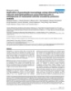
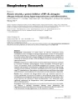
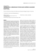
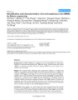
- Retrovirology 2006, 3:28 http://www.retrovirology.com/content/3/1/28 of the virus to the cell and the fusion of their membrane tent viral genome persists. The restricted infection implies resulting in the entry of the virus into the cell [21]. HIV-1- that efficient HIV-1 replication might be blocked at differ- specific CD4+ helper T lymphocytes and CD8+ cytotoxic T ent stage of virus life cycle, including virus entry, reverse lymphocytes have been detected within 4-6 weeks after transcription, nucleo-cytoplasmic HIV-1 RNA transport, HIV-1 inoculation [22]. Infected CD4+ T cells and mono- translation of viral DNA, and maturation of progeny vir- cytes, which circulate in the blood, are the potential ion. Studies of different astrocytes cell lines, which are source of CNS infection [14]. The mechanisms of entry of known to be non-productively infected, demonstrated a these cells into the CNS are discussed in the next section. cytoplasmic presence of Rev up to seven time more ele- Among the chemokine receptors expressed on human vated than in productively infected cells [33,34]. These cells, CXCR4 appears to be the most important for HIV-1 observations lead to the hypothesis that restricted HIV-1 entry into lymphocytes and CCR5 for monocytes, macro- production in astrocyte may be partly due to a cell deter- phages and microglia [23]. Because of the variability of mined block in nucleo-cytoplasmic Rev shuttling causing HIV-1 phenotypes, these strains of virus are defined by the nuclear retention of Rev-dependent HIV-1 mRNA their usage of the CCR5 or CXCR4 co-receptors, and des- classes where they are degraded [35,32]. Changes in cell ignated as R5- and X4- viruses respectively [23]. Following environment, like the elevation in the level of cytokines such as TNF-α and IL-1β, might reactivate virus produc- entry into the cell, the virus undergoes reverse transcrip- tion of its RNA genome to form a double-stranded DNA, tion [10,36]. a pre-integration complex of viral DNA with integrase and other viral protein including Vpr and matrix protein is Neuroinvasion of HIV-1 transported to the nucleus. The pre-integration complex The role of blood-brain barrier (BBB), which is a continu- facilitates the integration of the HIV-1 DNA genome into ous cellular layer of tightly linked brain microvascular host chromatin. The integration of viral DNA into the endothelial cells, is to separate the CNS from the periph- host cell genome generates the provirus that allows the ery (Figure 1). The BBB is selectively permeable and regu- production of HIV-1. In addition, high levels of viral DNA lates the trafficking of cells and substances between the remain non-integrated in the nucleus and are capable of brain parenchyma and the bloodstream [14,15]. The CSF directing expression of viral transcript [24-26]. The gener- is also separated from the periphery by the blood-CSF bar- ation of infectious virus particles involves the production rier of the choroids-plexus epithelium. In order to enter of viral transcripts and proteins and viral assembly, release the brain, HIV-1 must cross the BBB using mechanisms and maturation. During the production phase, first the that remain unclear. Numerous studies have used animal vial regulatory factors Nef, Tat and Rev are generated, and models and in vitro experimentation to understand the viral structural proteins and the RNA genome are pro- mechanisms of HIV-1 introduction into the CNS through duced in a later phase. In the assembly phase, Gag and BBB [14]. The generally accepted model, with most com- Gag-Pol Polyproteins, envelope proteins and viral RNA pelling evidence, is the "Trojan Horse hypothesis" genomes are assembled into immature virus particles at [37,38]. According to this model, HIV-1 and other lentivi- the cell membrane and released from the host cell. The ruses enter the CNS as a passenger in cells trafficking to cleavage of Gag and Gag-Pol Polyproteins by the HIV-1 the brain (Figure 1). Many CD4+ cells, such as T cells and protease results in the production of mature virus [27-29]. monocytes are infected by HIV-1, these cells circulate in the blood and can cross the BBB and propagate the infec- The intracellular environment plays a major role in HIV-1 tion within the CNS [37]. This model was confirmed by in virus replication [30]. HIV-1 infected cells are classified as situ hybridization and immunohistochemical analysis highly active producers and low or non-producers of that brought evidence of virus accumulation in perivascu- viruses, known as "productive" and "restricted" infection, lar regions [39-41]. Though BBB abnormalities due to respectively. Both types of infections occur in the CNS. HIV-1 infection have been observed, however, the mech- Productively infected cells support productive viral repli- anisms of endothelial cells infection and the expression of cation and participate in the transmission of the infection conventional HIV receptors in these cells remain a contro- and the rapid evolution of viral genome in the human versial issue. Although some studies suggest that human host and die ultimately. Restricted infection is only detect- brain microvascular endothelial cells lack CD4 receptors able by highly sensitive methods showing the presence of [42], other studies have found that CD4 was expressed in HIV-1 DNA or RNA. However, in the absence of structural isolated endothelial cells and microvessels of HIV-1 viral protein expression, it has been reported that acces- infected children's brains [43,44], moreover the expres- sory/regulatory protein such as Rev and Nef have been sion of HIV-1 co-receptors such as CCR5 and CXCR4 have expressed [31,32]. Restrictedly infected cells are permis- also being reported on isolated primary human brain's sive to infection by HIV-1 strains but are refractory to effi- microvascular endothelial cells [45]. An alternative cient virus expression, they restrict the HIV-1 replication hypothesis of HIV-1 neuro-invasion proposes the entry of and survive as virus reservoir in which replication-compe- free HIV-1 by migration between or, transcytosis of Page 3 of 11 (page number not for citation purposes)
- Retrovirology 2006, 3:28 http://www.retrovirology.com/content/3/1/28 Figure 1 HIV-1 neuroinvasion HIV-1 neuroinvasion. 1) According to the "Trojan Horse hypothesis" entry of HIV-1 into the brain takes place by the migration of infected monocytes which differentiate into perivascular macrophage. 2) The passage of infected CD4+ T cells can be another source of infection in the brain. Other probable causes of CNS infection might be: 3) the direct entrance of the virus or 4) entrance of HIV-1 by transcytosis of brain microvascular endothelial cells. Once the virus is in the brain it infects produc- tively macrophages and microglia. Astrocyte infection is known to be restricted. The infection of oligodendrocytes and spe- cially neurons is questionable. endothelial cells [10,14,46,47]. Theoretically all the main of its proximity to the interface with the periphery. This cell types of the CNS, astrocytes, oligodendrocytes, neu- replenishment that takes place by the migration of mono- rons, perivascular macrophage and microglia, can be cytes into the CNS has the side effect of opening the door infected by HIV-1 since they possess the receptors and/or to the intracellular pathogen. As the monocytes take resi- co-receptors for HIV-1 entry, but only the latter two are dency in the CNS they differentiate into macrophages. the most commonly infected cells by HIV-1 [14]. Microglia and monocyte-derived macrophage are consid- ered to be the main sources of productive HIV-1 infection in the brain [48,49]. One of the characteristics of HIVE is Macrophage and microglia Perivascular macrophage, microglia, and astrocytes are the presence of multinucleated giant cells expressing CD4. the cells coming into direct contact with infected cells in These cells are assumed to be infected monocytes differen- perivascular region. The two first types of cells are the res- tiated into macrophage after entering the brain or arising ident immunocompetent cells of the brain and their from the fusion of infected microglia [50]. It has been major role is to respond to all types of insults. Peripheral shown that in the primate Simian Immunodeficiency macrophage population is replenished through the Virus (SIV) model the spread of the virus from perivascu- lifespan with a relatively fast turnover, probably because lar cells to the parenchymal microglia does not occur [51], Page 4 of 11 (page number not for citation purposes)
- Retrovirology 2006, 3:28 http://www.retrovirology.com/content/3/1/28 however this issue remains controversial and has not been Oligodendrocytes confirmed for SIV and HIV-1. In contrast, many studies In vivo, Oligodendrocytes infection by HIV-1 remains con- suggest the opposite for HIV-1. Immunostaining has troversial. While some studies have detected viral nucleic revealed HIV-1 infection of parenchymal microglia, in acids by in situ PCR [63,64], other studies have reported some cases the infection is widespread, but in other cases the absence of HIV-1 markers in oligodendrocytes [33]. In it is restricted to the perivascular compartment [52]. It is vitro studies, using human oligodendrocytes indicates not clear whether the HIV-1 immunopositive microglia restricted infection by R5 and X4 strains of the virus [68]. consists of an influx of infected cells from the blood or Some studies have reported a reduced expression of spe- results from long-term infection in the CNS. In-vitro stud- cific oligodendrocyte markers, such as MBP and CNPase, ies have demonstrated that HIV-1 replication takes place in mice expressing HIV-1 Nef [69]. Oligodendrocytes do in primary microglia isolated from adults [53,54], infants not possess CD4 receptors and the mechanisms of their [55], and fetal brain [56,57]. HIV-1 infection in Microglia potential infection remain unclear. can be associated with cytopathology, including the for- mation of syncytia [54]. The study of the course of HIV-1 Neurons infection in purified primary cultures of human microglia Most studies have indicated an absence of in vivo infection shows that productive infection was more readily estab- in neurons, however a few studies have reported the pres- lished by R5-tropic strains of HIV-1 than by an X4-tropic ence of HIV-1 DNA and proteins in neurons [63,64]. It strain [55]. Microglial cells similar to macrophage express, has been suggested that the detection of infected neurons CD4/CCR5, major receptors/co-receptors used by HIV-1 in the brain might be complicated by the loss of the [58-60]. Other chemokine receptors, e.g. CCR3, CCR2b, infected neuronal populations [14]. In vitro studies have CCR8, CXCR6, and CX3CR1, are also expressed by these reported restricted infection of primary neurons [70], and cells but less efficiently used by HIV-1 [60,61]. In vitro neuronal cell lines by X5 and R4 viruses [71,72]. studies have shown that long-lived mixed microglial cul- tures isolated from human brain, when infected with R5 Mechanisms of neurodegeneration in HIV- HIV-1, retain replication competent viruses for up to 2.5 associated dementia months with low level virus replication, providing an acti- The absence of significant neuronal infection by HIV-1 vating condition can result in productive virus replication contrasts with the extensive neuropathological damage [62]. observed in HAD, therefore different mechanisms involv- ing the HIV-1 infection of perivascular macrophages, microglia, and possibly astrocytes might play the princi- Astrocytes Astrocytes do not have the CD4 receptor, which plays an pal role in neuronal injury and the disruption of normal important role in the infection of immune system cells, neurological function. The neuronal injury can result but they express CXCR4 and possibly other HIV-1 co- from a direct mechanism by interaction with viral pro- receptors including CCR5 [32]. However, several studies teins, such as gp120, Tat (Transcriptional transactivator) have reported the infection of astrocytes by HIV-1 and Vpr (viral protein R) produced by infected cells, or by although the mechanisms of viral attachment to astro- an indirect effect resulting from the inflammatory process cytes remain unclear. Immunopositivity of astrocytes for involving activated monocytes, macrophages and astro- HIV-1 structural proteins has occasionally been reported cytes (Figure 2). [35]. However, in situ hybridization, or in situ PCR have revealed the presence of HIV-1-specific nucleic acids in HIV-1 Tat astrocytes [40,63,64]. Other studies reported the presence The viral protein Tat, which is mainly active in the of the viral DNA and HIV-1 Nef protein in astrocytes [65]. nucleus, was shown to be secreted at high-level in vitro. Secreted Tat can cause direct or indirect injury to neurons, HIV-1 infection was studied using primary human fetal therefore it has been suggested that Tat contributes to astrocytes and tumor derived cell lines, several HIV-1 iso- HAD neuropathogenesis [73]. The neurotoxicity of Tat lates, namely X4-using T-cell line adapted (NL4-3, 1 MB, involves prolonged increase in intracellular calcium fol- SF2), R5-using, macrophage tropic (JR-FL, SF162) strains lowed by an increase of reactive oxygen species and cas- and primary isolates from blood [32,66,67]. The partici- pase activation of apoptotic pathway [73,74], in addition pation of astrocytes in productive infection has not been it has been shown that the up-regulation of caspase-8 by reported, though virus production in persistently infected HIV-1 Tat expression in CD4 T cell lines may contribute to cells can be transiently activated by the treatment with the increased apoptosis and sensitivity to apoptotic sig- inflammatory cytokines [32,66,67]. nals [75]. Tat is shown to alter the expression distribution of tight junction proteins, claudin-1 and claudin-5 in cer- ebral microvascular endothelial cells [76]. By affecting endothelial permeability, Tat contributes to the disrup- Page 5 of 11 (page number not for citation purposes)
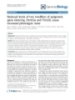
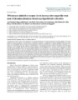
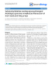
- Retrovirology 2006, 3:28 http://www.retrovirology.com/content/3/1/28 Figure 2 of neuropathogenesis Mechanism Mechanism of neuropathogenesis. Two components of this mechanism are: A) the direct effect of the HIV-1 infection, including HIV-1 proteins and B) the indirect consequence of infection comprising the secretion of cytokines and neurotoxins. The infected macrophages and microglia participate actively in the neurodegeneration by: 1) shedding viral proteins and 2) releasing significant amount of cytokines and neurotoxins into the CNS. 3) Tat and TNF-α contribute to the disruption of the blood brain barrier, which in turn become more permeable to infected monocytes and cytokines present in the periphery. The secreted pro-inflammatory cytokines activates 4) microglia and 5) astrocytes which in turn secrete neurotoxins, moreover the alteration of astrocytes function results in an increase in the level of neurotoxicity in the brain. 6) Multifactorial neuronal injury: neurotoxins released from several sources, as the direct and indirect consequences of HIV-1 infection, lead to neuronal injury. tion of the BBB that leads to infiltration of inflammatory HIV-1 Vpr cells into the CNS [76,77]. Further, Tat participates in the The regulatory protein Vpr might also be a player in the HAD associated inflammatory cascade by promoting direct mechanism of neuronal damage (reviewed in [78]). TNF-α and interleukin IL-1 production by monocytes and Vpr has been found in the CSF of HAD patients [79]. Vpr macrophages, and stimulates the production of several induces cell cycle arrest at G2/M phase, which leads to cell cytokines and chemokines, including IL-8, RANTES, death [80], a recent model of Vpr mediated induction of MCP-1 and TNF-α in astrocytes, which leads to neurotox- apoptosis, in CD4+ cells, proposes that Vpr expression icity [73]. activates cancer-associated protein BRCA1 and up-regu- Page 6 of 11 (page number not for citation purposes)
- Retrovirology 2006, 3:28 http://www.retrovirology.com/content/3/1/28 lates the expression of DNA damage-45 protein α can have neuro-protective effects [49,87]. The role of (GADD45α) [81]. It has been reported that Vpr also alters CXCR4 in the gp120 mediated neurotoxicity can be direct, mitochondrial permeability, which can cause cytochrome through the activation of neuronal receptors by gp120, or c release and eventually lead to apoptosis [82], however indirect through the stimulation of glial cells leading to this issue needs to be confirmed. Furthermore, another release of neurotoxic factors. Several studies have shown study of Vpr-mitochondria interaction has shown that Vpr that T tropic (X4) and dual tropic (X4/R5) gp120 induce targets HAX-1, an antiapoptotic mitochondrial protein, apoptosis in primary neurons and in neuronal cell lines Vpr associates physically to that protein and Vpr over- [96,97]. In contrast to the neuroprotective role of RANTES/CCL5 and MIP-1β against gp120, in mixed neu- expression leads to dislocation of HAX-1 from its normal rons/glial cultures, it has been shown that SDF-1α/CXCL2 mitochondrial residence and causes mitochondrial insta- bility and apoptosis [83]. Recent studies have demon- not only failed to provide neuro-protection from gp120, strated that both intracellular and extra cellular Vpr can but induced apoptosis in its absence [49]. Beside its direct induce apoptosis of human neuronal-precursor cells and neurotoxic effect, the viral protein gp120 has a significant mature, differentiated neurons by increasing the activa- role in the indirect mechanisms of neurodegenertion by tion of caspase-8 [84]. Finally, Tat and Vpr mediated- acting on macrophages, microglia or astrocytes [87,96]. apoptosis could increase significantly by co-exposure of Gp120 interaction with astrocytes stimulates the induci- cells to ethanol [84,85]. ble form of nitric oxide synthase and increases the release of arachidonic acid from astrocytes, which leads to the inhibition of glutamate uptake by astrocytes and neurons HIV-1 gp120 HIV-1 envelope glycoprotein gp160 is shown to have neu- [98]. As a result the extracellular concentration of gluta- rotoxic effect. This protein can be cleaved into two prod- mate increases and could lead to neurotoxicity via activa- ucts that remain non-covalently associated: gp120 and tion of excitatory amino acid receptors on neurons [73]. gp41. The soluble viral envelope protein gp120, which is By acting on monocytes and macrophages gp120 induces the production of TNF-α, IL-1 and arachidonic acid released in large quantities by HIV infected cells, might be involved in neuronal injury. The toxic effect of gp120 on metabolites which are implicated in HIV-1 neuropatho- neuronal population was demonstrated by many studies genesis. [86,87], dopaminergic neurons might be more suscepti- ble to gp120 neurotoxicity [88]. It has been shown that HIV-1 associated chemokines transgenic mice overexpressing gp120 had neuropatho- The chemokines and their receptors are considered to be logical features similar to abnormalities in brains of HAD involved in the pathogenesis of a number of neurological patients [89]. Neurodegeneration induced by gp120 can diseases including HAD, multiple sclerosis, Alzheimer's be direct through interaction with NMDA (N-Methyl-D- disease, and prion infection. The over-expression of some Aspartate) receptor or indirect by interaction with chem- chemokines in specific brain areas might contribute to the okine receptors [90,91]. Further, it has been shown that pathological condition. The chemokines and their recep- the presence of p53 is essential for gp120-induced neuro- tors are the gate of entrance of HIV into the CNS [99]. nal apoptosis [92]. Furthermore, both gp120 and Tat have Because of the alterations and abnormalities in the expres- been shown to disrupt neuronal calcium homeostasis by sion of chemokines and their receptors in the HIV infected perturbing calcium-regulating systems in the plasma CNS cells, and the role of chemokines in several neurode- membrane and endoplasmic reticulum, which leads to generative diseases, they have been the focus of attention neuronal death [93]. Recently, it has been described that in studies of HAD pathogenesis [100]. All members of the SDF-1α and gp120 induced a similar level of neuronal CXCR family are expressed, mainly by neurons, in the apoptosis, but by activating different intracellular path- brains of individuals affected by HAD [101]. Semiquanti- ways. SDF-1α enhanced NMDA activity indirectly via Src tative immunohistochemical analysis of the brain of HIV- phosphorylation, whereas gp120 probably activated the 1 infected individual, investigating the expression of four NMDA receptor directly and phosphorylated JNK [94]. HIV-1 co-receptors CCR2, CCR3, CCR5 and CXCR4 has These results are in accord with other studies, where shown that the hippocampal neurons were positive for gp120 was shown to induce neuronal dysfunction and CCR2, CCR3, and CXCR4 [102]. In other regions of the death through actions at p38 mitogen-activated protein brain, neurons, as well as glial cells were positive for kinase, while Tat kills neurons through actions that are CCR2, CCR3, and CXCR4, whereas only primary micro- independent of p38 or c-jun-N-terminal kinase mitogen- glial cells were positive for CCR5. The areas of highest activated protein kinase, or through the concurrent activa- expression seem to be subcortical regions and the limbic tion of multiple pro-apoptotic pathways [95]. system. The role of limbic system in memory and other cognitive functions, and the presence of CXCR4 on a sub- Some chemokine receptors are considered to act as a population of neuron from this system might explain cog- direct conduit for gp120 neurotoxicity, whereas others nitive and memory dysfunction in HAD. The presence of Page 7 of 11 (page number not for citation purposes)
- Retrovirology 2006, 3:28 http://www.retrovirology.com/content/3/1/28 chemokines and chemokine receptors increases in the lead to leukoencephalopathy and ultimately neuronal brain tissues of HIVE patients, particularly in areas of neu- apoptosis [111,112]. Some of these neurotoxins include TNF-α, arachidonic acid, platelet activating factors (PAF), roglial reaction, where they might be involved in the recruitment of inflammatory infiltrates and formation of nitric oxide (NO), and quinolinic acid (QUIN). NO is microglial nodules. The levels of expression of CCR1, synthesized by endothelial cells, macrophages and neu- CCR3, CCR5 and CXCR4 are especially elevated in the rons and might be associated with the NMDA type gluta- microglial nodules [59,103]. Moreover, CCR3 and mate associated neurotoxicity. A high level of inducible CXCR4 are highly expressed in the pyramidal neurons of NO synthase has been found in the brain of HAD patients hippocampus, and in the enthorinal cortex for CCR3. [113]. In HIV-1 patients who also are/were drug addicted Compared to AIDS patients without HAD, the brain tissue (e.g. cocaine, heroine), a 40-fold increase in expression of of patients with HAD shows an over-expression of CX3C NO synthase in neurons of temporal lobes was reported [114]. TNF-α is released by HIV-1 infected macrophage chemokine, fractalkine/CX3CL1 [104,105]. The upregula- tion of fractalkine/CX3CL1 was found in neurons in microglia and particularly affects oligodendrocytes [115]. It has been shown that TNF-α mRNA level in the subcor- brains of pediatric patients [104]. In contrast, fractalkine/ CX3CL1 was found to be over-expressed in astrocytes in tical regions of HAD patients' CNS are higher than in adult patients [105]. The level of chemokines in the CSF AIDS patients without neurological symptoms [116]. In addition, TNF-α can damage the BBB, as shown in an in- of HIV-infected patients with and without HAD has been determined in several studies. The results show that CSF vivo model, which could facilitate entry into the brain of chemokine concentration of MCP-1/CCL2, MIP-1α/ HIV-1 protein(s) and cytokines secreted in the periphery CCL3, MIP-1β/CCL4, RANTES/CCL5, IL-8/CXCL8 and [117]. Not only the level of pro-inflammatory cytokines, such as TNF-α, IL-1 and IFN-γ, anti-inflammatory fractalkine/CX3CL1 is positively correlated with the sever- cytokines including TGF-β and IL-6, and soluble cytokine ity of dementia and the viral load, indicating HIV induced brain damage. The role of CCR5, which is expressed by receptors is elevated in AIDS patients, but the cytokine neurons, microglia and astrocytes in the brain, seems production is correlated with the gravity of the neuropa- more controversial in the pathogenesis of HAD. The acti- thology [118,119]. vation of CCR5 by RANTES or MIP-1α/β, in in-vitro stud- ies, is shown to offer neuro-protection against gp120 This review is a summary of some of the current data sup- induced apoptosis [87,106]. However, in vitro observa- porting both the direct and indirect mechanisms by which tions indicate that neuro-virulent strains of HIV are essen- neuronal death may occur during infection with HIV-1. tially M-tropic with increased affinity for CCR5 [107]. It HAD is a complex phenomenon, which could be the has also been shown that CCR5 activation via its specific result of several mechanisms caused by players using dif- ligand induced apoptosis in neuroblastoma but not in ferent pathways. Some of these players, mechanisms, and fibroblast cell lines [108]. Therefore, it can be assumed pathways were mentioned in this review and some of that CCR5 might act as a death receptor in neurons and them are either un-identified or left out e.g. MCP-1, cellu- participate in HIV-1 induced neuropathology. lar proteins involved in the regulation of HIV-1 gene expression, Ca++ induction, HIV-1 activated apoptotic In brief, cognitive, motor decline and behavioral disorders programs (reviewed in [120]). Finally, more strategies are in HAD can be explained by significant neuronal cell needed for treating or preventing HAD by targeting spe- death that has been reported as a consequence of HIV-1 cific neurotoxic mechanisms used by the above-men- infection in the brain [109,110]. However, very few trace tioned viral proteins. of infection has been found in neurons of HAD patients' brains. Therefore the neuronal loss might be caused by the Competing interests release of neurotoxic factors by HIV infected microglia The author(s) declare that they have no competing inter- and astrocytes and/or by neurotoxic HIV-1 proteins. ests. Authors' contributions The inflammatory cascade The indirect mechanisms of AIDS neuropathogenesis also MG wrote the manuscript, SA and KK shared ideas and include the effect of the inflammation resulting from the discussion, BES conceived of the plan for the manuscript modification of extracellular secretory functions of micro- and coordinated its preparation. All authors read and glia and brain macrophages and inflammatory cytokine approved the final manuscript. production in the CNS (Figure 2). Following entry to the brain, monocytes, lymphocytes, activated macrophage, Acknowledgements microglia and astrocytes release cytokines, reactive oxygen We thank past and present members of the Center for Neurovirology for their insightful discussions and sharing of ideas. species, and other neurotoxins that disrupt normal cellu- lar functioning, modify neurotransmitter action, and may This review was made possible by Grants awarded by NIH to B.E.S. Page 8 of 11 (page number not for citation purposes)
- Retrovirology 2006, 3:28 http://www.retrovirology.com/content/3/1/28 References 25. Wu Y, Marsh JW: Gene transcription in HIV infection. Microbes Infect 2003, 5(11):1023-1027. 1. Forman MS, Trojanowski JQ, Lee VM: Neurodegenerative dis- 26. Kilzer JM, Stracker T, Beitzel B, Meek K, Weitzman M, Bushman FD: eases: a decade of discoveries paves the way for therapeutic Roles of host cell factors in circularization of retroviral DNA. breakthroughs. Nat Med 2004, 10(10):1055-1063. Virology 2003, 314(1):460-467. 2. Almeida OP, Lautenschlager NT: Dementia associated with 27. Bukrinskaya AG: HIV-1 assembly and maturation. Arch Virol infectious diseases. Int Psychogeriatr 2005, 17(Suppl 1):S65-S77. 2004, 149(6):1067-1082. 3. Wang T, Rumbaugh JA, Nath A: Viruses and the brain from 28. Nielsen MH, Pedersen FS, Kjems J: Molecular strategies to inhibit inflammation to dementia. Clin Sci (Lond) 2006, 110(4):393-407. HIV-1 replication. Retrovirology 2005, 2(1):10-15. 4. Starkstein SE, Jorge R: Dementia after traumatic brain injury. 29. Seelamgari A, Maddukuri A, Berro R, de la Fuente C, Kehn K, Deng Int Psychogeriatr 2005, 17(Suppl 1):S93-S107. L, Dadgar S, Bottazzi ME, Ghedin E, Pumfery A, Kashanchi F: Role of 5. Hulse GK, Lautenschlager NT, Tait RT, Almeida OP: Dementia viral regulatory and accessory proteins in HIV-1 replication. associated with alcohol and other drug use. Int Psychogeriatr Front Biosci 2004, 9:2388-2413. 2005, 17(Suppl 1):S109-S127. 30. Trkola A: HIV-host interactions: vital to the virus and key to 6. Armstrong RA, Lantos PL, Cairns NJ: Overlap between neurode- its inhibition. Curr Opin Microbiol 2004, 7(5):555-559. generative disorders. Neuropathology 2005, 25(2):111-124. 31. Ranki A, Nyberg M, Ovod V, Haltia M, Elovaara I, Raininko R, Haa- 7. Janssen RS: Epidemiology of human immunodeficiency virus pasalo H, Krohn K: Abundant expression of HIV Nef and Rev infection and the neurologic complications of the infection. proteins in brain astrocytes in vivo is associated with demen- Semin Neurol 1992, 12(1):10-17. tia. AIDS 1995, 9(9):1001-1008. 8. McArthur JC, Sacktor N, Selnes O: Human immunodeficiency 32. Gorry PR, Ong C, Thorpe J, Bannwarth S, Thompson KA, Gatignol A, virus-associated dementia. Semin Neurol 1999, 19(2):129-150. Vesselingh SL, Purcell DF: Astrocyte infection by HIV-1: mecha- 9. Fischer-Smith T, Rappaport J: Evolving paradigms in the patho- nisms of restricted virus replication, and role in the patho- genesis of HIV-1-associated dementia. Expert Rev Mol Med genesis of HIV-1-associated dementia. Curr HIV Res 2003, 2005, 7(27):1-26. 1(4):463-473. 10. Kramer-Hammerle S, Rothenaigner I, Wolff H, Bell JE, Brack-Werner 33. Neumann M, Afonina E, Ceccherini-Silberstein F, Schlicht S, Erfle V, R: Cells of the central nervous system as targets and reser- Pavlakis GN, Brack-Werner R: Nucleocytoplasmic transport in voirs of the human immunodeficiency virus. Virus Res 2005, human astrocytes: decreased nuclear uptake of the HIV Rev 111(2):194-213. shuttle protein. J Cell Sci 2001, 114(Pt 9):1717-1729. 11. Janssen RS, Nwanyanwu OC, Selik RM, Stehr-Green JK: Epidemiol- 34. Ludwig E, Silberstein FC, van Empel J, Erfle V, Neumann M, Brack- ogy of human immunodeficiency virus encephalopathy in the Werner R: Diminished rev-mediated stimulation of human United States. Neurology 1992, 42(8):1472-1476. immunodeficiency virus type 1 protein synthesis is a hall- 12. Reger M, Welsh R, Razani J, Martin DJ, Boone KB: A meta-analysis mark of human astrocytes. J Virol 1999, 73(10):8279-8289. of the neuropsychological sequelae of HIV infection. J Int Neu- 35. Brack-Werner R: Astrocytes: HIV cellular reservoirs and ropsychol Soc 2002, 8(3):410-424. important participants in neuropathogenesis. AIDS 1999, 13. Lawrence DM, Major EO: HIV-1 and the brain: connections 13(1):1-22. between HIV-1-associated dementia, neuropathology and 36. Gorry P, Purcell D, Howard J, McPhee D: Restricted HIV-1 infec- neuroimmunology. Microbes Infect 2002, 4(3):301-308. tion of human astrocytes: potential role of nef in the regula- 14. Gonzalez-Scarano F, Martin-Garcia J: The neuropathogenesis of tion of virus replication. J Neurovirol 1998, 4(4):377-386. AIDS. Nat Rev Immunol 2005, 5(1):69-81. 37. Haase AT: Pathogenesis of lentivirus infections. Nature 1986, 15. Gendelman HE, Lipton SA, Tardieu M, Bukrinsky MI, Nottet HS: The 322(6075):130-136. neuropathogenesis of HIV-1 infection. J Leukoc Biol 1994, 38. Peluso R, Haase A, Stowring L, Edwards M, Ventura P: A Trojan 56(3):389-398. Horse mechanism for the spread of visna virus in monocytes. 16. Hall M, Whaley R, Robertson K, Hamby S, Wilkins J, Hall C: The cor- Virology 1985, 147(1):231-236. relation between neuropsychological and neuroanatomic 39. Wiley CA, Schrier RD, Nelson JA, Lampert PW, Oldstone MB: Cel- changes over time in asymptomatic and symptomatic HIV- lular localization of human immunodeficiency virus infection 1-infected individuals. Neurology 1996, 46(6):1697-1702. within the brains of acquired immune deficiency syndrome 17. Dal Pan GJ, McArthur JH, Aylward E, Selnes OA, Nance-Sproson TE, patients. Proc Natl Acad Sci U S A 1986, 83(18):7089-7093. Kumar AJ, Mellits ED, McArthur JC: Patterns of cerebral atrophy 40. Takahashi K, Wesselingh SL, Griffin DE, McArthur JC, Johnson RT, in HIV-1-infected individuals: results of a quantitative MRI Glass JD: Localization of HIV-1 in human brain using polymer- analysis. Neurology 1992, 42(11):2125-2130. ase chain reaction/in situ hybridization and immunocyto- 18. Stout JC, Ellis RJ, Jernigan TL, Archibald SL, Abramson I, Wolfson T, chemistry. Ann Neurol 1996, 39(6):705-711. McCutchan JA, Wallace MR, Atkinson JH, Grant I: Progressive cer- 41. Fischer-Smith T, Croul S, Adeniyi A, Rybicka K, Morgello S, Khalili K, ebral volume loss in human immunodeficiency virus infec- Rappaport J: Macrophage/microglial accumulation and prolif- tion: a longitudinal volumetric magnetic resonance imaging erating cell nuclear antigen expression in the central nerv- study. Arch Neurol 1998, 55(2):161-168. ous system in human immunodeficiency virus 19. Aylward EH, Henderer JD, McArthur JC, Brettschneider PD, Harris encephalopathy. Am J Pathol 2004, 164(6):2089-2099. GJ, Barta PE, Pearlson GD: Reduced basal ganglia volume in 42. Petito CK, Cash KS: Blood-brain barrier abnormalities in the HIV-1-associated dementia: results from quantitative neu- acquired immunodeficiency syndrome: immunohistochemi- roimaging. Neurology 1993, 43(10):2099-2104. cal localization of serum proteins in postmortem brain. Ann 20. Tucker KA, Robertson KR, Lin W, Smith JK, An H, Chen Y, Aylward Neurol 1992, 32(5):658-666. SR, Hall CD: Neuroimaging in human immunodeficiency virus 43. Stins MF, Shen Y, Huang SH, Gilles F, Kalra VK, Kim KS: Gp120 acti- infection. J Neuroimmunol 2004, 157(1–2):153-162. vates children's brain endothelial cells via CD4. J Neurovirol 21. Zaitseva M, Peden K, Golding H: HIV coreceptors: role of struc- 2001, 7(2):125-134. ture, posttranslational modifications, and internalization in 44. Stins MF, Pearce D, Di Cello F, Erdreich-Epstein A, Pardo CA, Sik Kim viral-cell fusion and as targets for entry inhibitors. Biochim Bio- K: Induction of intercellular adhesion molecule-1 on human phys Acta 2003, 1614(1):51-61. brain endothelial cells by HIV-1 gp120: role of CD4 and 22. Lichterfeld M, Yu XG, Le Gall S, Altfeld M: Immunodominance of chemokine coreceptors. Lab Invest 2003, 83(12):1787-1798. HIV-1-specific CD8(+) T-cell responses in acute HIV-1 infec- 45. Mukhtar M, Harley S, Chen P, BouHamdan M, Patel C, Acheampong tion: at the crossroads of viral and host genetics. Trends Immu- E, Pomerantz RJ: Primary isolated human brain microvascular nol 2005, 26(3):166-171. endothelial cells express diverse HIV/SIV-associated chem- 23. Moore JP, Kitchen SG, Pugach P, Zack JA: The CCR5 and CXCR4 okine coreceptors and DC-SIGN and L-SIGN. Virology 2002, coreceptors-central to understanding the transmission and 297(1):78-88. pathogenesis of human immunodeficiency virus type 1 infec- 46. Bomsel M: Transcytosis of infectious human immunodefi- tion. AIDS Res Hum Retroviruses 2004, 20(1):111-126. ciency virus across a tight human epithelial cell line barrier. 24. Wu Y, Marsh JW: Selective transcription and modulation of Nat Med 1997, 3(1):42-47. resting T cell activity by preintegrated HIV DNA. Science 2001, 293(5534):1503-1506. Page 9 of 11 (page number not for citation purposes)
- Retrovirology 2006, 3:28 http://www.retrovirology.com/content/3/1/28 47. Banks WA, Freed EO, Wolf KM, Robinson SM, Franko M, Kumar VB: lated from brain tissue with HIV-1 encephalitis by laser cap- Transport of human immunodeficiency virus type 1 pseudo- ture microdissection. Brain Pathol 2003, 13(2):144-154. viruses across the blood-brain barrier: role of envelope pro- 66. Sabri F, Tresoldi E, Di Stefano M, Polo S, Monaco MC, Verani A, Fiore teins and adsorptive endocytosis. J Virol 2001, JR, Lusso P, Major E, Chiodi F, Scarlatti G: Nonproductive human 75(10):4681-4691. immunodeficiency virus type 1 infection of human fetal 48. Anderson E, Zink W, Xiong H, Gendelman HE: HIV-1-associated astrocytes: independence from CD4 and major chemokine dementia: a metabolic encephalopathy perpetrated by virus- receptors. Virology 1999, 264(2):370-384. infected and immune-competent mononuclear phagocytes. 67. Wang Z, Trillo-Pazos G, Kim SY, Canki M, Morgello S, Sharer LR, Gel- J Acquir Immune Defic Syndr 2002, 31(Suppl 2):S43-S54. bard HA, Su ZZ, Kang DC, Brooks AI, Fisher PB, Volsky DJ: Effects 49. Kaul M, Garden GA, Lipton SA: Pathways to neuronal injury and of human immunodeficiency virus type 1 on astrocyte gene apoptosis in HIV-associated dementia. Nature 2001, expression and function: potential role in neuropathogene- 410(6831):988-994. sis. J Neurovirol 2004, 10(Suppl 1):25-32. 50. Dickson DW: Multinucleated giant cells in acquired immuno- 68. Albright AV, Strizki J, Harouse JM, Lavi E, O'Connor M, Gonzalez- deficiency syndrome encephalopathy. Origin from endog- Scarano F: HIV-1 infection of cultured human adult oli- enous microglia? Arch Pathol Lab Med 1986, 110(10):967-968. godendrocytes. Virology 1996, 217(1):211-219. 51. Williams KC, Corey S, Westmoreland SV, Pauley D, Knight H, 69. Radja F, Kay DG, Albrecht S, Jolicoeur P: Oligodendrocyte-spe- deBakker C, Alvarez X, Lackner AA: Perivascular macrophages cific expression of human immunodeficiency virus type 1 Nef are the primary cell type productively infected by simian in transgenic mice leads to vacuolar myelopathy and alters immunodeficiency virus in the brains of macaques: implica- oligodendrocyte phenotype in vitro. J Virol 2003, tions for the neuropathogenesis of AIDS. J Exp Med 2001, 77(21):11745-11753. 193(8):905-915. 70. Ensoli F, Cafaro A, Fiorelli V, Vannelli B, Ensoli B, Thiele CJ: HIV-1 52. Morris A, Marsden M, Halcrow K, Hughes ES, Brettle RP, Bell JE, Sim- infection of primary human neuroblasts. Virology 1995, monds P: Mosaic structure of the human immunodeficiency 210(1):221-225. virus type 1 genome infecting lymphoid cells and the brain: 71. Obregon E, Punzon C, Fernandez-Cruz E, Fresno M, Munoz-Fernan- evidence for frequent in vivo recombination events in the dez MA: HIV-1 infection induces differentiation of immature evolution of regional populations. J Virol 1999, neural cells through autocrine tumor necrosis factor and 73(10):8720-8731. nitric oxide production. Virology 1999, 261(2):193-204. 53. Albright AV, Shieh JT, O'Connor MJ, Gonzalez-Scarano F: Charac- 72. Mizrachi Y, Rodriguez I, Sweetnam PM, Rubinstein A, Volsky DJ: HIV terization of cultured microglia that can be infected by HIV- type 1 infection of human cortical neuronal cells: enhance- 1. J Neurovirol 2000, 6(Suppl 1):S53-S60. ment by select neuronal growth factors. AIDS Res Hum Retrovi- 54. Watkins BA, Dorn HH, Kelly WB, Armstrong RC, Potts BJ, Michaels ruses 1994, 10(12):1593-1596. F, Kufta CV, Dubois-Dalcq M: Specific tropism of HIV-1 for 73. Nath A: Human immunodeficiency virus (HIV) proteins in microglial cells in primary human brain cultures. Science 1990, neuropathogenesis of HIV dementia. J Infect Dis 2002, 249(4968):549-553. 186(Suppl 2):S193-S198. 55. Ioannidis JP, Reichlin S, Skolnik PR: Long-term productive human 74. Song L, Nath A, Geiger JD, Moore A, Hochman S: Human immun- immunodeficiency virus-1 infection in human infant micro- odeficiency virus type 1 Tat protein directly activates neuro- glia. Am J Pathol 1995, 147(5):1200-1206. nal N-methyl-D-aspartate receptors at an allosteric zinc- 56. McCarthy M, He J, Wood C: HIV-1 strain-associated variability sensitive site. J Neurovirol 2003, 9(3):399-403. in infection of primary neuroglia. J Neurovirol 1998, 4(1):80-89. 75. Bartz SR, Emerman M: Human immunodeficiency virus type 1 57. Sundar KS, Kamaraju LS, Dingfelder J, McMahon J, Gollapudi S, Wilson Tat induces apoptosis and increases sensitivity to apoptotic WH, Kong LY, Hong JS, Weiss JM, Lee JE: beta-Endorphin signals by up-regulating FLICE/caspase-8. J Virol 1999, enhances the replication of neurotropic human immunode- 73(3):1956-1963. ficiency virus in fetal perivascular microglia. J Neuroimmunol 76. Toborek M, Lee YW, Flora G, Pu H, Andras IE, Wylegala E, Hennig B, 1995, 61(1):97-104. Nath A: Mechanisms of the blood-brain barrier disruption in 58. Jordan CA, Watkins BA, Kufta C, Dubois-Dalcq M: Infection of HIV-1 infection. Cell Mol Neurobiol 2005, 25(1):181-199. brain microglial cells by human immunodeficiency virus type 77. Andras IE, Pu H, Deli MA, Nath A, Hennig B, Toborek M: HIV-1 Tat 1 is CD4 dependent. J Virol 1991, 65(2):736-742. protein alters tight junction protein expression and distribu- 59. Vallat AV, De Girolami U, He J, Mhashilkar A, Marasco W, Shi B, Gray tion in cultured brain endothelial cells. J Neurosci Res 2003, F, Bell J, Keohane C, Smith TW, Gabuzda D: Localization of HIV- 74(2):255-265. 1 co-receptors CCR5 and CXCR4 in the brain of children 78. Le Rouzic E, Benichou S: The Vpr protein from HIV-1: distinct with AIDS. Am J Pathol 1998, 152(1):167-178. roles along the viral life cycle. Retrovirology 2005, 2(1):11. 60. Albright AV, Shieh JT, Itoh T, Lee B, Pleasure D, O'Connor MJ, Doms 79. Levy DN, Refaeli Y, Weiner DB: The vpr regulatory gene of HIV. RW, Gonzalez-Scarano F: Microglia express CCR5, CXCR4, and Curr Top Microbiol Immunol 1995, 193:209-336. CCR3, but of these, CCR5 is the principal coreceptor for 80. Stewart SA, Poon B, Song JY, Chen IS: Human immunodeficiency human immunodeficiency virus type 1 dementia isolates. J virus type 1 vpr induces apoptosis through caspase activa- Virol 1999, 73(1):205-213. tion. J Virol 2000, 74(7):3105-3111. 61. Martin-Garcia J, Kolson DL, Gonzalez-Scarano F: Chemokine 81. Andersen JL, Zimmerman ES, DeHart JL, Murala S, Ardon O, Blackett receptors in the brain: their role in HIV infection and patho- J, Chen J, Planelles V: ATR and GADD45alpha mediate HIV-1 genesis. AIDS 2002, 16(13):1709-1730. Vpr-induced apoptosis. Cell Death Differ 2005, 12(4):326-334. 62. Albright AV, Vos RM, Gonzalez-Scarano F: Low-level HIV replica- 82. Jacotot E, Ravagnan L, Loeffler M, Ferri KF, Vieira HL, Zamzami N, tion in mixed glial cultures is associated with alterations in Costantini P, Druillennec S, Hoebeke J, Briand JP, et al.: The HIV-1 the processing of p55(Gag). Virology 2004, 325(2):328-339. viral protein R induces apoptosis via a direct effect on the 63. Nuovo GJ, Becker J, Burk MW, Margiotta M, Fuhrer J, Steigbigel RT: mitochondrial permeability transition pore. J Exp Med 2000, In situ detection of PCR-amplified HIV-1 nucleic acids in 191(1):33-46. lymph nodes and peripheral blood in patients with asympto- 83. Yedavalli VS, Shih HM, Chiang YP, Lu CY, Chang LY, Chen MY, matic HIV-1 infection and advanced-stage AIDS. J Acquir Chuang CY, Dayton AI, Jeang KT, Huang LM: Human immunode- Immune Defic Syndr 1994, 7(9):916-923. ficiency virus type 1 Vpr interacts with antiapoptotic mito- 64. Bagasra O, Lavi E, Bobroski L, Khalili K, Pestaner JP, Tawadros R, chondrial protein HAX-1. J Virol 2005, 79(21):13735-13746. Pomerantz RJ: Cellular reservoirs of HIV-1 in the central nerv- 84. Pomerantz RJ: Effects of HIV-1 Vpr on neuroinvasion and neu- ous system of infected individuals: identification by the com- ropathogenesis. DNA Cell Biol 2004, 23(4):227-238. bination of in situ polymerase chain reaction and 85. Acheampong E, Mukhtar M, Parveen Z, Ngoubilly N, Ahmad N, Patel immunohistochemistry. AIDS 1996, 10(6):573-585. C, Pomerantz RJ: Ethanol strongly potentiates apoptosis 65. Trillo-Pazos G, Diamanturos A, Rislove L, Menza T, Chao W, Belem induced by HIV-1 proteins in primary human brain microv- P, Sadiq S, Morgello S, Sharer L, Volsky DJ: Detection of HIV-1 ascular endothelial cells. Virology 2002, 304(2):222-234. DNA in microglia/macrophages, astrocytes and neurons iso- Page 10 of 11 (page number not for citation purposes)
- Retrovirology 2006, 3:28 http://www.retrovirology.com/content/3/1/28 86. Dreyer EB, Kaiser PK, Offermann JT, Lipton SA: HIV-1 coat pro- brain-derived human immunodeficiency virus type 1 enve- tein neurotoxicity prevented by calcium channel antago- lope genes differs between demented and nondemented nists. Science 1990, 248(4953):364-367. AIDS patients. J Virol 1998, 72(11):9045-9053. 87. Kaul M, Lipton SA: Chemokines and activated macrophages in 108. Cartier L, Hartley O, Dubois-Dauphin M, Krause KH: Chemokine HIV gp120-induced neuronal apoptosis. Proc Natl Acad Sci U S A receptors in the central nervous system: role in brain inflam- 1999, 96(14):8212-8216. mation and neurodegenerative diseases. Brain Res Brain Res Rev 88. Bennett BA, Rusyniak DE, Hollingsworth CK: HIV-1 gp120- 2005, 48(1):16-42. induced neurotoxicity to midbrain dopamine cultures. Brain 109. Masliah E, Achim CL, Ge N, DeTeresa R, Terry RD, Wiley CA: Spec- Res 1995, 705(1–2):168-176. trum of human immunodeficiency virus-associated neocorti- 89. Cioni C, Annunziata P: Circulating gp120 alters the blood-brain cal damage. Ann Neurol 1992, 32(3):321-329. barrier permeability in HIV-1 gp120 transgenic mice. Neuro- 110. Petito CK, Roberts B: Effect of postmortem interval on in situ sci Lett 2002, 330(3):299-301. end-labeling of DNA oligonucleosomes. J Neuropathol Exp Neu- 90. Barks JD, Liu XH, Sun R, Silverstein FS: gp120, a human immuno- rol 1995, 54(6):761-765. deficiency virus-1 coat protein, augments excitotoxic hip- 111. Boven LA, van der Bruggen T, Sweder van Asbeck B, Marx JJ, Nottet pocampal injury in perinatal rats. Neuroscience 1997, HS: Potential role of CCR5 polymorphism in the develop- 76(2):397-409. ment of AIDS dementia complex. FEMS Immunol Med Microbiol 91. Corasaniti MT, Strongoli MC, Piccirilli S, Nistico R, Costa A, Bilotta 1999, 26(3–4):243-247. A, Turano P, Finazzi-Agro A, Bagetta G: Apoptosis induced by 112. Panek RB, Benveniste EN: Class II MHC gene expression in gp120 in the neocortex of rat involves enhanced expression microglia. Regulation by the cytokines IFN-gamma, TNF- of cyclooxygenase type 2 and is prevented by NMDA recep- alpha, and TGF-beta. J Immunol 1995, 154(6):2846-2854. tor antagonists and by the 21-aminosteroid U-74389G. Bio- 113. Adamson DC, Wildemann B, Sasaki M, Glass JD, McArthur JC, Chris- chem Biophys Res Commun 2000, 274(3):664-669. tov VI, Dawson TM, Dawson VL: Immunologic NO synthase: ele- 92. Garden GA, Guo W, Jayadev S, Tun C, Balcaitis S, Choi J, Montine TJ, vation in severe AIDS dementia and induction by HIV-1 Moller T, Morrison RS: HIV associated neurodegeneration gp41. Science 1996, 274(5294):1917-1921. requires p53 in neurons and microglia. FASEB J 2004, 114. Minagar A, Shapshak P, Fujimura R, Ownby R, Heyes M, Eisdorfer C: 18(10):1141-1143. The role of macrophage/microglia and astrocytes in the 93. Haughey NJ, Mattson MP: Calcium dysregulation and neuronal pathogenesis of three neurologic disorders: HIV-associated apoptosis by the HIV-1 proteins Tat and gp120. J Acquir dementia, Alzheimer disease, and multiple sclerosis. J Neurol Immune Defic Syndr 2002:S55-S61. Sci 2002, 202(1–2):13-23. 94. Geeraerts T, Deiva K, M'sika I, Salim H, Hery C, Tardieu M: Effects 115. Wilt SG, Milward E, Zhou JM, Nagasato K, Patton H, Rusten R, Griffin of SDF-1alpha and gp120(IIIB) on apoptotic pathways in SK- DE, O'Connor M, Dubois-Dalcq M: In vitro evidence for a dual N-SH neuroblastoma cells. Neurosci Lett 2006 in press. role of tumor necrosis factor-alpha in human immunodefi- 95. Singh IN, El-Hage N, Campbell ME, Lutz SE, Knapp PE, Nath A, Hauser ciency virus type 1 encephalopathy. Ann Neurol 1995, KF: Differential involvement of p38 and JNK MAP kinases in 37(3):381-394. HIV-1 Tat and gp120-induced apoptosis and neurite degen- 116. Wesselingh SL, Takahashi K, Glass JD, McArthur JC, Griffin JW, Grif- eration in striatal neurons. Neuroscience 2005, 135(3):781-790. fin DE: Cellular localization of tumor necrosis factor mRNA 96. Lipton SA, Brenneman DE, Silverstein FS, Masliah E, Mucke L: gp120 in neurological tissue from HIV-infected patients by com- and neurotoxicity in vivo. Trends Pharmacol Sci 1995, bined reverse transcriptase/polymerase chain reaction in 16(4):122-130. situ hybridization and immunohistochemistry. J Neuroimmunol 97. Pandey V, Bolsover SR: Immediate and neurotoxic effects of 1997, 74(1–2):1-8. HIV protein gp120 act through CXCR4 receptor. Biochem Bio- 117. Fiala M, Rhodes RH, Shapshak P, Nagano I, Martinez-Maza O, Diagne phys Res Commun 2000, 274(1):212-215. A, Baldwin G, Graves M: Regulation of HIV-1 infection in astro- 98. Lipton SA: AIDS-related dementia and calcium homeostasis. cytes: expression of Nef, TNF-alpha and IL-6 is enhanced in Ann N Y Acad Sci 1994, 747:205-224. coculture of astrocytes with macrophages. J Neurovirol 1996, 99. Li W, Galey D, Mattson MP, Nath A: Molecular and cellular 2(3):158-166. mechanisms of neuronal cell death in HIV dementia. Neuro- 118. Yoshioka M, Bradley WG, Shapshak P, Nagano I, Stewart RV, Xin KQ, tox Res 2005, 8(1–2):119-134. Srivastava A, Nakamura S: Role of immune activation and 100. Dou H, Kingsley JD, Mosley RL, Gelbard HA, Gendelman HE: Neu- cytokine expression in HIV-1-associated neurologic diseases. roprotective strategies for HIV-1 associated dementia. Neu- Adv Neuroimmunol 1995, 5(3):335-358. rotox Res 2004, 6(7–8):503-521. 119. Griffin DE: Cytokines in the brain during viral infection: clues 101. Brandimarti R, Khan MZ, Fatatis A, Meucci O: Regulation of cell to HIV-associated dementia. J Clin Invest 1997, cycle proteins by chemokine receptors: A novel pathway in 100(12):2948-2951. human immunodeficiency virus neuropathogenesis? J Neuro- 120. Gougeon ML: Apoptosis as an HIV strategy to escape immune virol 2004, 10(Suppl 1):108-112. attack. Nat Rev Immunol 2003, 3(5):392-404. 102. Vander Meer P, Ulrich AM, Gonzalez-Scarano F, Lavi E: Immunohis- tochemical analysis of CCR2, CCR3, CCR5, and CXCR4 in the human brain: potential mechanisms for HIV dementia. Exp Mol Pathol 2000, 69(3):192-201. 103. Sanders VJ, Pittman CA, White MG, Wang G, Wiley CA, Achim CL: Chemokines and receptors in HIV encephalitis. AIDS 1998, 12(9):1021-1026. Publish with Bio Med Central and every 104. Tong N, Perry SW, Zhang Q, James HJ, Guo H, Brooks A, Bal H, Kin- scientist can read your work free of charge near SA, Fine S, Epstein LG, Dairaghi D, Schall TJ, Gendelman HE, Dewhurst S, Sharer LR, Gelbard HA: Neuronal fractalkine "BioMed Central will be the most significant development for expression in HIV-1 encephalitis: roles for macrophage disseminating the results of biomedical researc h in our lifetime." recruitment and neuroprotection in the central nervous sys- Sir Paul Nurse, Cancer Research UK tem. J Immunol 2000, 164(3):1333-1339. 105. Pereira CF, Middel J, Jansen G, Verhoef J, Nottet HS: Enhanced Your research papers will be: expression of fractalkine in HIV-1 associated dementia. J available free of charge to the entire biomedical community Neuroimmunol 2001, 115(1–2):168-175. 106. Meucci O, Fatatis A, Simen AA, Bushell TJ, Gray PW, Miller RJ: peer reviewed and published immediately upon acceptance Chemokines regulate hippocampal neuronal signaling and cited in PubMed and archived on PubMed Central gp120 neurotoxicity. Proc Natl Acad Sci U S A 1998, 95(24):14500-14505. yours — you keep the copyright 107. Power C, McArthur JC, Nath A, Wehrly K, Mayne M, Nishio J, Lan- BioMedcentral gelier T, Johnson RT, Chesebro B: Neuronal death induced by Submit your manuscript here: http://www.biomedcentral.com/info/publishing_adv.asp Page 11 of 11 (page number not for citation purposes)
CÓ THỂ BẠN MUỐN DOWNLOAD

-
Báo cáo y học: "MRI bone oedema scores are higher in the arthritis mutilans form of psoriatic arthritis and correlate with high radiographic scores for joint damage"
 9 p |
9 p |  122
|
122
|  7
7
-
Báo cáo y học: " Interactions among type I and type II interferon, tumor necrosis factor, and -estradiol in the regulation of immune response-related gene expressions in systemic lupus erythematosus"
10 p | 88
| 5
-
Báo cáo y học: " Implication of granulocyte-macrophage colony-stimulating factor induced neutrophil gelatinase-associated lipocalin in pathogenesis of rheumatoid arthritis revealed by proteome analysis"
12 p | 109
| 5
-
Báo cáo y học: "Reduced levels of two modifiers of epigenetic gene silencing, Dnmt3a and Trim28, cause increased phenotypic nois"
10 p | 73
| 4
-
Báo cáo y học: "Introduction of medical emergency teams in Australia and New Zealand: a multicentre study"
2 p | 113
| 4
-
Báo cáo y học: "Effect of bladder volume on measured intravesical pressure:"
6 p | 109
| 4
-
Báo cáo y học: " Influence of the cystic fibrosis transmembrane conductance regulator on expression of lipid metabolism-related genes in dendritic cells"
15 p | 85
| 4
-
Báo cáo y học: " Arsenic trioxide, a potent inhibitor of NF-κB, abrogates allergen-induced airway hyperresponsiveness and inflammation"
12 p | 95
| 3
-
Báo cáo y học: ": Immunostaining of modified histones defines high-level features of the human metaphase epigenome"
14 p | 81
| 3
-
Báo cáo y học: "Rapid chromosome territory relocation by nuclear motor activity in response to serum removal in primary human fibroblasts"
0 p | 94
| 3
-
Báo cáo y học: " GE Rotterdam, the Netherlands. †Department of Human Genetics"
18 p | 68
| 3
-
Báo cáo y học: "The electronic version of this article is the complete one and can be found online"
6 p | 88
| 3
-
Báo cáo y học: "ontinuity, psychosocial correlates, and outcome of problematic substance use from adolescence to young adulthood in a community sample"
1 p | 79
| 3
-
Báo cáo y học: " Vgf is a novel biomarker associated with muscle weakness in amyotrophic lateral sclerosis (ALS), with a potential role in disease pathogenesis"
8 p | 95
| 3
-
Báo cáo y học: "Staffing level: a determinant of late-onset ventilator-associated pneumonia"
3 p | 106
| 3
-
Báo cáo y học: "TPO, but not soluble-IL-6 receptor, levels increase after anagrelide treatment of thrombocythemia in chronic myeloproliferative disorders"
5 p | 83
| 3
-
Báo cáo y học: "hese authors contributed equally to this work"
0 p | 84
| 2
-
Báo cáo y học: "Substantial deletion overlap among divergent Arabidopsis genomes revealed by intersection of short reads and tiling arrays"
0 p | 81
| 2


Chịu trách nhiệm nội dung:
Nguyễn Công Hà - Giám đốc Công ty TNHH TÀI LIỆU TRỰC TUYẾN VI NA
LIÊN HỆ
Địa chỉ: P402, 54A Nơ Trang Long, Phường 14, Q.Bình Thạnh, TP.HCM
Hotline: 093 303 0098
Email: support@tailieu.vn
Giấy phép Mạng Xã Hội số: 670/GP-BTTTT cấp ngày 30/11/2015 Copyright © 2022-2032 TaiLieu.VN. All rights reserved.

