


Báo cáo y học: " Intracellular assembly and budding of the Murine Leukemia Virus in infected cells"
lượt xem 3
download
 Download
Vui lòng tải xuống để xem tài liệu đầy đủ
Download
Vui lòng tải xuống để xem tài liệu đầy đủ
Tuyển tập các báo cáo nghiên cứu về y học được đăng trên tạp chí y học quốc tế cung cấp cho các bạn kiến thức về ngành y đề tài: Intracellular assembly and budding of the Murine Leukemia Virus in infected cells
Bình luận(0) Đăng nhập để gửi bình luận!
Nội dung Text: Báo cáo y học: " Intracellular assembly and budding of the Murine Leukemia Virus in infected cells"
- Retrovirology BioMed Central Open Access Research Intracellular assembly and budding of the Murine Leukemia Virus in infected cells Laurent Houzet, Bernard Gay, Zakia Morichaud, Laurence Briant and Marylène Mougel* Address: Laboratoire Infections Rétrovirales et Signalisation Cellulaire, CNRS UMR5121, UMI, IFR122, Institut de Biologie, Montpellier, France Email: Laurent Houzet - lhouzet@univ-montp1.fr; Bernard Gay - bgay@univ-montp1.fr; Zakia Morichaud - zmorichaud@univ-montp1.fr; Laurence Briant - lbriant@univ-montp1.fr; Marylène Mougel* - mmougel@univ-montp1.fr * Corresponding author Published: 10 February 2006 Received: 23 December 2005 Accepted: 10 February 2006 Retrovirology2006, 3:12 doi:10.1186/1742-4690-3-12 This article is available from: http://www.retrovirology.com/content/3/1/12 © 2006Houzet et al; licensee BioMed Central Ltd. This is an Open Access article distributed under the terms of the Creative Commons Attribution License (http://creativecommons.org/licenses/by/2.0), which permits unrestricted use, distribution, and reproduction in any medium, provided the original work is properly cited. Abstract Background: Murine Leukemia Virus (MLV) assembly has been long thought to occur exclusively at the plasma membrane. Current models of retroviral particle assembly describe the recruitment of the host vacuolar protein sorting machinery to the cell surface to induce the budding of new particles. Previous fluorescence microscopy study reported the vesicular traffic of the MLV components (Gag, Env and RNA). Here, electron microscopy (EM) associated with immunolabeling approaches were used to go deeply into the assembly of the "prototypic" MLV in chronically infected NIH3T3 cells. Results: Beside the virus budding events seen at the cell surface of infected cells, we observed that intracellular budding events could also occur inside the intracellular vacuoles in which many VLPs accumulated. EM in situ hybridization and immunolabeling analyses confirmed that these latter were MLV particles. Similar intracellular particles were detected in cells expressing MLV Gag alone. Compartments containing the MLV particles were identified as late endosomes using Lamp1 endosomal/lysosomal marker and BSA-gold pulse-chase experiments. In addition, infectious activity was detected in lysates of infected cells. Conclusion: Altogether, our results showed that assembly of MLV could occur in part in intracellular compartments of infected murine cells and participate in the production of infectious viruses. These observations suggested that MLV budding could present similarities with the particular intracellular budding of HIV in infected macrophages. ticles. The standard model for retrovirus production Background Retroviruses consist of an enveloped capsid containing a describes the budding of particles at the plasma mem- dimer of genomic RNA. Genomic RNA contains genes brane [1]. Addressing of Gag to the plasma membrane is encoding Gag, Gag/Pol and Env precursor proteins. The promoted by the Matrix domain [2] and release of the polyprotein Gag is sufficient for driving virus particle pro- newly formed particles from the cellular membrane is duction by promoting assembly of immature capsid to the driven by the conserved "late domain" present in the Gag cellular membrane, budding, and release of the virus par- polyprotein of all retroviruses [3]. Integrity of the L- Page 1 of 9 (page number not for citation purposes)
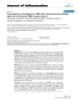
- Retrovirology 2006, 3:12 http://www.retrovirology.com/content/3/1/12 latter, the fusion of the endosomes with the plasma mem- brane leads to virus particles release in the extracellular space [12]. Using fluorescence microscopy, several works reported the traffic of MLV Gag and Env proteins [11,14-16]] and viral genomic RNA [14] in endosomes of transfected or chronically infected cells. Here, we investigated virus assembly in NIH3T3 cells chronically infected with the replication-competent MLV using electron microscopy (EM). We showed that intracellular virus budding could arise and that numerous VLPs containing MLV genomic RNA accumulated in the Lamp-1 positive vacuoles. The absence of VLPs in lysosomal degradative compartments and the detection of intracellular infectious activity sug- gested that these intracellular virus particles could partici- pate in the MLV infection. Figure MLV-infected NIH3T3 cells Electron1microscopy analysis of VLPs assembly in vacuoles of Results Electron microscopy analysis of VLPs assembly in Intravacuolar virion-like particles in cells infected with the vacuoles of MLV-infected NIH3T3 cells. EM analysis of replication-competent MLV epon embedded NIH3T3 cells chronically infected with MLV. MLV assembly was investigated by EM analysis in chroni- A) Virus budding at the plasma membrane. B) Numerous cally infected NIH3T3 cells producing 105-106 FFU per ml mature (arrows) and immature (arrowheads) particles inside the intracellular endosomes. C) Budding particle into a vacu- of cell culture supernatant. The use of chronically infected ole. cells precludes reinfection with virion entry and ensures that only late phases of the viral cycle were observed. For cell morphology analysis, the cells were included in epon domain sequences is required for the late membrane fis- as previously described [17]. Rare budding viruses were sion event and the final pinching off of the budding virus detected at the plasma membrane (Fig. 1A), with only two [4-6]. In the last few years, late domain sequences were budding events for hundred of analyzed cell-sections. In found to direct the interaction between the Gag proteins contrast, a large amount of particles with virus-like mor- and some cellular factors involved in the protein sorting phology were detected in intracellular vacuoles (Fig. 1B). process and the vesicle formation during the multivesicu- The average size of these particles (90 nm diameter) cor- lar bodies (MVB) biogenesis [7,8]. MVB are late endo- responds to MLV particles. Moreover, the presence of dark somal compartments accumulating internal vesicles electron dense ring or circle in these particles is typical of produced from intracisternal invagination of the endo- assembled MLV particles and corresponds respectively to somal membrane. These internal vesicles are released immature and mature forms of capsids [18]. Noteworthy, either in lysosomes to allow associated protein and lipid several intravesicular buddings were also observed (Fig. degradation or in the extracellular space as exosomes for 1C), with similar frequency as that observed for the exter- intercellular communication [9]. Internal vesicles produc- nal budding (2 events for hundreds of observed infected tion and virus budding are topologically similar processes cells). These results indicated that intracellular budding of consisting of budding away from the cytosol. Moreover, VLPs did occur in intracellular compartments of chroni- vacuolar protein sorting factors are involved in both cally infected cells. events. These observations support the hypothesis that the virus hijacks the MVB production system to direct the Identification of the intracellular VLPs by EM budding and the release of virus particles [10]. immunolabeling To further characterize intracellular VLPs in the infected Recently, it was shown that HIV and MLV Gag polypro- cells, we used EM approach coupled to immunolabeling teins can lead to the formation of virus-like particles with an anti-Gag antibody on lowicryl embedded sec- (VLPs) in late endosomes [11]. Interestingly, intracellular- tions. To estimate the frequency and intensity of the labe- formed particles are the principal source of infectious HIV ling on particles, we quantitated the number of labeled particles in infected macrophages [12]. These observa- particles with the associated gold dots. Particles were iden- tions have led to the actual consideration of two pathways tified by their size (between 90 and 100 nm diameter) and for HIV production: the standard budding at the plasma electron density criteria. membrane and a new endosomal pathway [13]. In this Page 2 of 9 (page number not for citation purposes)
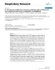
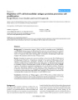
- Retrovirology 2006, 3:12 http://www.retrovirology.com/content/3/1/12 detected in the intravacuolar VLPs (Fig. 2B, C). Despite the lower quality inherent in the immunolabeling procedure, all these Gag-labeled particles displayed the size and elec- tron density characteristic of MLV particles and correlated to the VLPs observed before in epon embedded samples (Fig. 1B). Quantification of the labeling showed that 88% of the analyzed intravacuolar particles were labeled, with an intensity of 4 gold dots per particle, close to the labe- ling intensity observed for released viruses (Table 1). The unlabeled 12% could probably correspond to cellular ves- icles which were identified by mistake as VLPs because of the non-optimal resolution in these assays. A weak Gag labeling was also observed on the vacuolar delimiting membrane (Fig. 2C), supporting our observation that the intravacuolar particles originated from budding of the delimiting membrane. These results indicated that intrac- ellular VLPs observed in MLV-infected cells contained MLV Gag proteins and then corresponded to MLV virion- like particles. Encapsidation of the viral RNA genome in vesicular VLPs To go further in the analysis of the vesicular VLPs, pres- ence of the genomic RNA was investigated by EM in situ hybridization with a specific DIG-labeled riboprobe. One difficulty of the labeling consists of the accessibility of the target sequence complementary to the probe, which must be exposed at the section surface to allow riboprobe hybridization. Among the 62 intracellular VLPs analyzed, 27 (44%) were labeled with the antisense riboprobe, showing that at least half of the internal VLPs contained genomic RNA (Fig. 3A). As expected, no particle labeling Figure 2 MLV-infected cells and progeny viruses Immunoelectron microscopy analysis of Gag distribution in was observed with the sense riboprobe used as control Immunoelectron microscopy analysis of Gag distri- (Fig. 3B). These results clearly showed that the viral RNA bution in MLV-infected cells and progeny viruses. Gag genome was packaged into the intracellular VLPs. was detected by immunogold labeling in lowicryl embedded sections of MLV-infected cells. A) Extracellular virus particle Characterization of the VLP-containing vacuoles (black arrow) released from the plasma membrane (black In order to characterize the compartment which included arrowhead) labeled with 5 nm gold particles. B) VLPs (black the VLPs, immunolabeling experiments with an antibody arrows) present in intracellular vacuoles (white arrows) directed against the late endosomal/lysosomal marker were labeled with similar intensity as extracellular viruses. C) Magnification of intravacuolar Gag-labeled VLPs (arrows). Lamp-1 were undertaken. A weak labeling was observed Weak labeling was also observed on the vacuolar delimiting along the membrane of the vacuoles containing the VLPs, membrane (arrowhead). which is typical of a late endosome labeling (Fig. 4A) [12]. In addition, some VLPs present in these vacuoles also exhibited some low labeling (Fig. 4A and 4B). We noted Due to the experimental procedure, very few extracellular that other non viral structures were labeled inside the vac- virus particles were detected (7 for hundreds of infected uoles (Fig. 4A), which could correspond to the intracister- cells). As expected, all were labeled for Gag antigens (Fig nal vesicles of the MVB. To discriminate between late 2A), with an average intensity of 3,3 gold dots per virus endosomes and lysosomes, lysosomal compartments (Table 1). Since virus particles were all immature and were labeled by BSA-gold endocytosis. Infected cells were located in close vicinity of the plasma membrane, they pulsed 4 hours with conjugates of BSA and 13-nm colloi- probably have been just released from the plasma mem- dal gold, chased for 20 hours to label lysosomes [19], and brane. prepared for EM analysis. As expected, gold-labeled BSA exclusively accumulated in lysosomal compartments In the cells, very few labeled-Gag localized individually at which appeared as white electron-light vacuoles (Figures the plasma membrane and most of the Gag proteins were 5-A and C). Clearly, the lysosome morphology differed Page 3 of 9 (page number not for citation purposes)
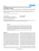
- Retrovirology 2006, 3:12 http://www.retrovirology.com/content/3/1/12 Table 1: Quantification of anti-Capsid signal labeled particles (%) Average labeling intensity (gold dots per particle) extracellular 100 3,3 ± 1,5 intracellular 88 4,0 ± 2,5 The number of gold dots present on particles that displayed the typical morphology and size of virus particles was determined. Due to their very low abundance, only 7 external particles were analyzed, while a total of 52 intracellular particles were examined. from that of other vacuoles containing VLPs (Compare Fig 5A and 5B). More than hundred cells were analyzed and the colocalisation of the gold-labeled BSA and the VLPs was never observed in the same vacuole. Altogether, these results indicated that the intracellular compartments where the VLPs accumulated corresponded to the late endosomes related to MVB, and that no VLPs could be detected in lysosomes. Search for infectious activity in the lysate of MLV-infected cells Previous study reported that intracellular HIV particles in macrophages could harbor some infectious ability [20]. In order to test whether the intracellular MLV VLPs could Detection of the viral RNA genome in intracellular VLPs Figure 3 also display some infectious ability, we undertook freeze Detection of the viral RNA genome in intracellular VLPs. The MLV genomic RNA was specifically detected by and thaw experiments to release MLV related particles EM in situ hybridization and was visualized by 10 nm gold from the chronically infected cells. After drastic washes particles. VLPs (black arrows) inside the intracellular vacu- with cold PBS, 5 × 10⁶ cells were lysed by several freeze- oles (white arrows) were labeled (arrowheads) with the spe- thaw cycles followed by a sonication step to release intra- cific antisense probe (A), while no signal was detected with cellular particles as described in Materials and Methods. the control sense riboprobe (B). Cellular debris were removed by centrifugation and filtra- tion and the clarified cell lysate was used to infect target Dunni cells. Infectivity of intracellular particles was mon- itored by focal immunofluorescence assay (FIA) using an released VLPs can be obtained upon cellular expression of antibody specific to the MLV Env protein (Fig. 6A). As a the sole Gag polyprotein [21] or can be assembled from control of wash efficiency, the same procedure was per- purified Gag under certain conditions in vitro [22]. To formed with the last wash supernatant of same cells left investigate whether Gag may also promote the formation intact. The results are presented in Figure 6. Very little of the vesicular MLV particles, immunolabeling of Gag infectious activity was detected in the control assay that protein and EM analysis were conducted in derivative might result from residual contamination with external human HT1080 cells that expressed Gag/Gag-Pol alone virus particles. However, a marked increase of the infec- (HT-Fly cells). As expected, a Gag-labeling of the external tious activity was obtained when cells were submitted to VLPs, recently detached from the plasma membrane was the freeze-thaw and sonication lysis. The level of this detected (Fig. 7A). Interestingly, we also noted the pres- intracellular activity is probably underestimated since ence of intracellular VLPs displaying similar Gag-labeling virus particles contained in cell lysate could be damaged (Fig. 7B). As observed with intravacuolar MLV particles in by the lysis procedure. Lysate obtained from mock- infected cells (Fig. 2B), these internal VLPs were concen- infected NIH3T3 did not show any infectious activity trated (with some other unidentified vesicles) in intracel- (data not shown). These results indicated the presence of lular compartments with a morphology that might intracellular infectious MLV particles in the chronically correspond to this of MVB. Analysis of several cell sections infected cells. revealed that Gag-VLP budded more frequently at the plasma membrane than at intracellular membrane. These observations differ somewhat with that observed in the Gag is sufficient to assemble vesicular VLPs The Gag polyprotein is the basic component in the mak- context of chronic infection where frequencies of budding ing of virion particles at the plasma membrane. Indeed, at the plasma membrane or in endosomes were similar. In Page 4 of 9 (page number not for citation purposes)
- Retrovirology 2006, 3:12 http://www.retrovirology.com/content/3/1/12 Figure 4 VLP-containing vacuoles and their VLPs are positive for Lamp-1 VLP-containing vacuoles and their VLPs are positive for Lamp-1. Lamp1 was detected in lowicryl embedded sections by immunogold labeling (5nm gold particles). A) Low labeling was observed on the periphery of MLV-VLPs containing vacuoles and on other intravacuolar components (white arrowheads). Gold particles could sometimes be found on individual intracellu- lar MLV-VLP (black arrowhead). B) Magnification of the boxed area in A showed a Lamp1 positive MLV-VLP. conclusion, these results indicated that Gag alone was suf- cellular budding events mediated by Gag. Furthermore, ficient to generate not only the extracellular budding but since the VLP-containing vacuoles were labeled by the late also the formation of VLPs in intracellular compartment. endosomal/lysosomal marker Lamp-1 and not by the BSA-gold, these latter could be identified as late endo- somes. These results correlate with the visualization of Discussion During the last years, most of the studies of endosomal MLV Gag in late endosomes by fluorescence microscopy traffic of retrovirus components were undertaken using [11,14]. fluorescence microscopy. Here we decided to take advan- tage of the high resolution of the EM to evaluate the We observed infectious activity in lysates of infected cells, assembly of the replication-competent MLV in chronically indicating that infectious MLV particles were present infected cells. Analysis of cellular content clearly showed inside the cells. It is tempting to speculate that these infec- that intracellular VLPs appeared very abundant in vacu- tious virus particles corresponded to the MLV-VLPs we olar compartments. This first observation substantiates observed in late endosomes. Existence of intraendosomal previous study reporting VLPs in intracellular compart- virus budding and presence of many immature virus par- ments in MLV-infected cells [23]. Using EM immunolabe- ticles among these intracellular particles strongly sug- ling experiments, we identified them as MLV related VLPs. gested that these virus particles came from direct budding Several immunofluorescence studies reported that Env in the endosomal vacuoles. The frequency of intracellular and Gag colocalized in intracellular compartments budding events appeared low (2 for hundred of analyzed [11,14-16] and that a viral genomic RNA pool reaches the cell-sections) compared to the numerous particles accu- plasma membrane bound to Gag and Env tethered at the mulated in endosomes. However, in agreement with these cytoplasmic face of the endosomal membrane [14]. The results, Hansen et al [23] reported one intracellular VLP high resolution of the EM brings more precise results budding event for 22 analyzed cell-sections of MLV chron- which clearly showed that, in the context of infection, a ically infected cells. Similar observations were previously part of this genomic RNA pool was already encapsidated reported for the well documented intracellular HIV bud- inside the intravesicular MLV particles and likely via intra- ding in macrophages where about 100 virions per vacuole Page 5 of 9 (page number not for citation purposes)
- Retrovirology 2006, 3:12 http://www.retrovirology.com/content/3/1/12 Figure 5 Specific labeling of lysosomal compartments by pulses of BSA-gold Specific labeling of lysosomal compartments by pulses of BSA-gold. Representative pictures of infected cells incu- bated with the BSA-gold. The BSA-gold accumulated in VLP-free lysosomes (arrowhead) (A). VLPs (large arrow) and budding event (little arrow) were shown in unlabeled vacuole (B). Absence of colocalization of VLP (arrow) and BSA-gold (arrowhead) (C). were observed with only occasional budding events. This lar medium by fusion of the endosomal membrane with accumulation suggested that budding detection was prob- the plasma membrane and participate in MLV infection, ably dependent of the budding rates which drastically dif- as proposed for HIV in macrophages [12,20]. Thus, the fers among the viruses [24,25] and which should be faster virus particles production might occur from two different than the rate of the particles release. but non exclusive ways in MLV-infected NIH3T3 cells: the classical budding at the plasma membrane, and the bud- The intraendosomal budding observed in the present ding into MVB. These budding events were both detected study, in the context of the infection with the replication- in cells expressing only Gag, suggesting the recruitment of competent MLV, might be a common alternative process a similar mechanism promoted by Gag. shared by all retroviruses, since it was also documented in HIV infected macrophages [12]. It is not clear what determines the incidence of intracellu- lar versus cell surface assembly. Indeed, numerous EM Because late endosomes/MVB are directly linked to degra- analyses performed with transfected MLV Gag or reconsti- dative pathway by fusion with lysosomal compartments, tuted viruses, usually in human (293T), monkey (Cos), or intraendosomal virus particles could be directly routed for hamster (BHK21) cell lines, described exclusive budding degradation and not participate in virus production proc- at the plasma membrane [26-28]. At the opposite, only ess. But the absence of detectable particles or viral compo- two studies (ours and [23]) showed the coexistence of nents in lysosomes suggests that virus particles could intracellular and cell surface in chronically infected cells. escape the degradation pathway. Then, one can speculate One possible explanation for these different results is the that intracellular particles could be released in extracellu- experimental system : transient versus stable expression Page 6 of 9 (page number not for citation purposes)
- Retrovirology 2006, 3:12 http://www.retrovirology.com/content/3/1/12 some-like virus particles in the MVB and their release into the cell culture supernatant. Our report of similar alterna- tive of intraendosomal budding in MLV-infected cells par- ticipates to a better understanding of the fundamental process involved in this late phase of retroviral infection. Moreover, MLV infection could constitute a new valuable model to evaluate in vivo the effect of new therapeutic agents directed against intraendosomal virus production. Materials and methods Cell culture and infection NIH3T3, Dunni, and Fly (a kind gift from FL Cosset) cells were cultured in Dulbecco's modified Eagle's medium (DMEM) supplemented with glutamine (2 mM), penicil- lin, streptomycin and 10% heat-inactivated fetal calf serum at 37°C. Infections were performed with Friend- MLV viral stocks with average titer of 5 × 10⁵ focus-form- ing units per ml (FFU/ml) as previously described [34]. MLV-infected NIH3T3 were maintained 1 month after infection and considered as chronically infected. EM and immuno-EM For conventional EM, MLV-infected NIH3T3 cell samples were processed and embedded in epon (Embed-812, Elec- tron Microscopy Sciences Inc.) according to a previously described method [17]. For immuno-EM, cells were fixed Figure 7 Gag alone can promote intracellular VLPs formation in 2,5% formaldehyde in 0.1M phosphate-buffered saline Gag alone can promote intracellular VLPs formation. Gag was detected by immunogold labeling in lowicryl embed- (PBS), pH7.4 for 90 minutes, washed in PBS + 0.05M ded sections of Fly packaging cells which expressed the Gag ammonium chloride one hour, gathered in fibrin clot, and Pol proteins only. A) Extracellular VLP (arrow) released and embedded in methacrylate resin (Lowicryl K4M, Che- from the plasma membrane (arrowhead) labeled with 5 nm mische Werke Lowi). Ultrathin sections were cut with a gold particles. B) VLPs (arrows) present in intracellular vacu- Reichert OMU2 ultramicrotome and collected with gold ole (arrowhead). grids 300 mesh. After blocking 20 minutes in Tris buffered saline (TBS) proteined (20 mM Tris-HCl pH 8,2, 20 mM sodium azide, 0,1% Tween 20, 1% goat serum, 1% bovine systems. The chronic phase of infection could favor the serum albumin), immunogold labeling was performed by intracellular assembly as reported by Orenstein et al who incubating sections overnight at 4°C with primary anti- compared HIV assembly during acute and chronic phases body diluted in proteined TBS and one hour at room tem- of infection [29]. Nevertheless, a recent work of Sherer et perature with diluted gold labeling secondary antibody. al showed that even in Gag-MLV transfected 293T and Then, the grids were stained 20 minutes with 2% uranyl HeLa cells, MLV-VLPs can bud both at the plasma mem- acetate in water, air dried, and examined on a Hitachi brane and at the late endosomal membrane [11]. In addi- H1700 electron microscope. The following antibodies tion, it cannot be excluded that the incidence of MLV were used: rat monoclonal anti-Gag antibody (H187, a intracellular versus cell surface assembly is also largely kind gift from B. Chesebro) or rat monoclonal anti-lamp1 dependent of the cell lines as well documented in the case antibody (clone 1D4B, a kind gift from M. Vidal) with of HIV. In this latter, the plasma membrane budding was goat anti-rat antibody coupled to 5 nm gold particles predominantly observed in T cells whereas virions accu- (British Biocell International, Cardiff, UK). mulated in MVB in macrophages (see reviews [30-32]). EM in situ hybridization Recently, the observation of HIV intraendosomal budding The Digoxigenin labeled RNA probes were prepared from and the discovery of the impact of endosomal proteins a linearized Bluescript plasmid containing a 652 bp MLV sorting pathway in retroviral budding have lead to the genomic fragment (position 1181 to 1833 bp). In vitro Trojan exosome hypothesis [33]. This original hypothesis transcription was performed in the sense or anti-sense ori- proposes that retroviruses hijack the cellular exosomal entation using a DIG RNA labeling kit (Roche). Digoxi- production machinery leading to the production of exo- genin labeled RNA were quantified as instructed by the Page 7 of 9 (page number not for citation purposes)
- Retrovirology 2006, 3:12 http://www.retrovirology.com/content/3/1/12 goat anti-mouse antibody labeled with 10 nm colloidal gold particle (British Biocell International, Cardiff, UK). The procedure was the same as described above for immuno-EM, except that the incubation with primary antibody was 90 minutes at room temperature. Labeling of lysosomes by BSA-gold endocytosis Colloidal gold (13 nm) was prepared by trisodium citrate reduction of gold chloride [35]. The colloid was adjusted to pH 6.0 with 0,2 M K2CO3 and conjugated to sufficient BSA to afford protection from NaCl-induced flocculation. BSA-gold was harvested using ultracentrifugation proto- cols which yielded monodisperse preparations free of aggregates and unbound protein. The preparations were dialyzed against PBS and adjusted to an A520 of 1.5 with DMEM. For lysosomes labeling, infected cells grown to 70% con- fluence in 6 wells plate were starved 2 hours in DMEM. After cells incubation at 37°C in 3 ml of DMEM contain- ing 150 µl of BSA-gold solution for 4 hours, the cells were washed 3 times with PBS and incubated in conjugate-free medium for 20 hours as previously described [19], prior to fixation and processing for EM. Detection of intracellular infectious activity Figure 6 and sonication treatment Infectivity of intracellular particles released by freeze-thaw For each experiment, 5 × 10⁶ chronically infected cells, Infectivity of intracellular particles released by producing viral supernatant with average titer of 5 × 10⁵ freeze-thaw and sonication treatment. FIA was used to FFU/ml, were washed 2 times with 10 ml of ice-cold PBS, quantitate infectious particles present in the cell lyzed by scraped with a rubber policeman and transferred to centri- freeze-thaw and sonication (cell lysate) or in the last wash of fuge tubes. Cells were washed 3 more times with 20 ml cells left intact (control). A) One typical FFU labeled with cold PBS, resuspended in 100 µl of PBS and subjected to anti-Env antibody and detected in FIA. Insert: magnification of 4 freeze-thaw cycles followed by 2 times 30 sec sonication. the boxed area showing infected (arrow) and non infected Total cell disruption was microscopically validated using (arrowhead) Dunni cells. B) Results of the FIA expressed as trypan blue staining. As a control for wash efficiency, the the total number of infectious FFU detected in the total lysate of 5 × 10⁶ cells. Lysis and infectivity experiments were same procedure was performed with the last wash of same performed at least 3 times and each infection test was per- cells left intact. The samples (cell lysate or control) were formed in triplicate. Bars, the standard error of the mean of then centrifuged at 2400 rpm for 10 min at 4°C and the each series. supernatants of the centrifugation were added to 6 ml of culture medium and filtrated (0,45µm). For infections, serial dilutions of samples were used to infect target Dunni cells. Infectious particles were detected and quanti- manufacturer. After 10 minutes incubation in the pre- tated by FIA, using monoclonal antibody (H48, a kind gift hybridization buffer (4 × SSC + 50% formamide) at 37°C, from B. Chesebro) specific to Friend-MLV Env protein ultra-thin sections were incubated overnight at 37°C in [36]. moist chamber in hybridization solution (1 µg/ml Dig- labeled RNA probe in 40% formamide deionised, 10% Abbreviations sulfate dextran, 1 × Denhart solution, 4× SSC, 250 µg/ml EM, electron microscopy; HIV, human immunodeficiency tRNA, 250 µg/ml salmon sperm DNA). The grids were virus; MLV, murine leukemia virus; MVB, multi vesicular washed 5 minutes in 2 × SSC and washed three times 5 bodies; VLPs, virus-like particles; FIA, focal immunofluo- minutes in 0,2 × SSC/60 % formamide at 37°C and twice rescence assay; FFU, focus-forming unit. 5 minutes in 2 × SSC at room temperature. Competing interests Immunogold detection of the Dig-labeled riboprobe was The author(s) declare that they have no competing inter- performed using mouse anti-Dig antibody (Roche) and ests. Page 8 of 9 (page number not for citation purposes)
- Retrovirology 2006, 3:12 http://www.retrovirology.com/content/3/1/12 Acknowledgements Pr55gag virus-like particles from recombinant baculovirus- infected insect cells. Cell 1989, 59(1):103-112. We thank D. Muriaux for critical reading of the manuscript. The work was 22. Campbell S, Vogt VM: Self-assembly in vitro of purified CA-NC supported by grants to MM from the ANRS (n°03N60/0674), SIDACTION proteins from Rous sarcoma virus and human immunodefi- (AO15-2) and ACI (BCMS299). LH was supported by a fellowship from ciency virus type 1. J Virol 1995, 69(10):6487-6497. 23. Hansen M, Jelinek L, Jones RS, Stegeman-Olsen J, Barklis E: Assembly Fondation de France and SIDACTION. and composition of intracellular particles formed by Molo- ney murine leukemia virus. J Virol 1993, 67(9):5163-5174. References 24. Wills JW, Cameron CE, Wilson CB, Xiang Y, Bennett RP, Leis J: An 1. Chazal N, Gerlier D: Virus entry, assembly, budding, and mem- assembly domain of the Rous sarcoma virus Gag protein brane rafts. Microbiol Mol Biol Rev 2003, 67(2):226-237. required late in budding. J Virol 1994, 68(10):6605-6618. 2. Ono A, Orenstein JM, Freed EO: Role of the Gag matrix domain 25. Hermida-Matsumoto L, Resh MD: Localization of human immu- in targeting human immunodeficiency virus type 1 assembly. nodeficiency virus type 1 Gag and Env at the plasma mem- J Virol 2000, 74(6):2855-2866. brane by confocal imaging. J Virol 2000, 74(18):8670-8679. 3. Freed EO: Viral late domains. J Virol 2002, 76(10):4679-4687. 26. Choi G, Park S, Choi B, Hong S, Lee J, Hunter E, Rhee SS: Identifi- 4. Yuan B, Campbell S, Bacharach E, Rein A, Goff SP: Infectivity of cation of a cytoplasmic targeting/retention signal in a retro- Moloney murine leukemia virus defective in late assembly viral Gag polyprotein. J Virol 1999, 73(7):5431-5437. events is restored by late assembly domains of other retro- 27. Suomalainen M, Hultenby K, Garoff H: Targeting of Moloney viruses. J Virol 2000, 74(16):7250-7260. murine leukemia virus gag precursor to the site of virus bud- 5. Gottlinger HG, Dorfman T, Sodroski JG, Haseltine WA: Effect of ding. J Cell Biol 1996, 135(6 Pt 2):1841-1852. mutations affecting the p6 gag protein on human immuno- 28. Steidl S, Schule S, Muhlebach MD, Stitz J, Boller K, Cichutek K, Sch- deficiency virus particle release. Proc Natl Acad Sci U S A 1991, weizer M: Genetic engineering of onco/lentivirus hybrids 88(8):3195-3199. results in formation of infectious but not of replication-com- 6. Parent LJ, Bennett RP, Craven RC, Nelle TD, Krishna NK, Bowzard petent viruses. J Gen Virol 2004, 85(Pt 3):665-678. JB, Wilson CB, Puffer BA, Montelaro RC, Wills JW: Positionally 29. Orenstein JM, Meltzer MS, Phipps T, Gendelman HE: Cytoplasmic independent and exchangeable late budding functions of the assembly and accumulation of human immunodeficiency Rous sarcoma virus and human immunodeficiency virus Gag virus types 1 and 2 in recombinant human colony-stimulat- proteins. J Virol 1995, 69(9):5455-5460. ing factor-1-treated human monocytes: an ultrastructural 7. Garrus JE, von Schwedler UK, Pornillos OW, Morham SG, Zavitz KH, study. J Virol 1988, 62(8):2578-2586. Wang HE, Wettstein DA, Stray KM, Cote M, Rich RL, et al.: Tsg101 30. Resh MD: Intracellular trafficking of HIV-1 Gag: how Gag and the vacuolar protein sorting pathway are essential for interacts with cell membranes and makes viral particles. HIV-1 budding. Cell 2001, 107(1):55-65. AIDS Rev 2005, 7(2):84-91. 8. VerPlank L, Bouamr F, LaGrassa TJ, Agresta B, Kikonyogo A, Leis J, 31. Muriaux D, Darlix JL, Cimarelli A: Targeting the assembly of the Carter CA: Tsg101, a homologue of ubiquitin-conjugating human immunodeficiency virus type I. Curr Pharm Des 2004, (E2) enzymes, binds the L domain in HIV type 1 Pr55(Gag). 10(30):3725-3739. Proc Natl Acad Sci U S A 2001, 98(14):7724-7729. 32. Freed EO: HIV-1 and the host cell: an intimate association. 9. Thery C, Zitvogel L, Amigorena S: Exosomes: composition, bio- Trends Microbiol 2004, 12(4):170-177. genesis and function. Nat Rev Immunol 2002, 2(8):569-579. 33. Gould SJ, Booth AM, Hildreth JE: The Trojan exosome hypothe- 10. Pornillos O, Garrus JE, Sundquist WI: Mechanisms of enveloped sis. Proc Natl Acad Sci U S A 2003, 100(19):10592-10597. RNA virus budding. Trends Cell Biol 2002, 12(12):569-579. 34. Dejardin J, Bompard-Marechal G, Audit M, Hope TJ, Sitbon M, Mougel 11. Sherer NM, Lehmann MJ, Jimenez-Soto LF, Ingmundson A, Horner M: A novel subgenomic murine leukemia virus RNA tran- SM, Cicchetti G, Allen PG, Pypaert M, Cunningham JM, Mothes W: script results from alternative splicing. J Virol 2000, Visualization of retroviral replication in living cells reveals 74(8):3709-3714. budding into multivesicular bodies. Traffic 2003, 4(11):785-801. 35. Frens G: Controlled nucleation for the regulation of particle 12. Pelchen-Matthews A, Kramer B, Marsh M: Infectious HIV-1 size in monodisperse gold suspensions. Nature Phys Sci 1973, assembles in late endosomes in primary macrophages. J Cell 241(105):20-12. Biol 2003, 162(3):443-455. 36. Sitbon M, Nishio J, Wehrly K, Lodmell D, Chesebro B: Use of a focal 13. Amara A, Littman DR: After Hrs with HIV. J Cell Biol 2003, immunofluorescence assay on live cells for quantitation of 162(3):371-375. retroviruses: distinction of host range classes in virus mix- 14. Basyuk E, Galli T, Mougel M, Blanchard JM, Sitbon M, Bertrand E: Ret- tures and biological cloning of dual-tropic murine leukemia roviral genomic RNAs are transported to the plasma mem- viruses. Virology 1985, 141(1):110-118. brane by endosomal vesicles. Dev Cell 2003, 5(1):161-174. 15. Sandrin V, Cosset FL: Intracellular vs. cell surface assembly of retroviral pseudotypes is determined by the cellular localiza- tion of the viral glycoprotein, its capacity to interact with Gag and the expression of the Nef protein. J Biol Chem 2005. 16. Sandrin V, Muriaux D, Darlix JL, Cosset FL: Intracellular traffick- ing of gag and env proteins and their interactions modulate pseudotyping of retroviruses. J Virol 2004, 78(13):7153-7164. 17. Cartier C, Hemonnot B, Gay B, Bardy M, Sanchiz C, Devaux C, Briant L: Active cAMP-dependent protein kinase incorporated Publish with Bio Med Central and every within highly purified HIV-1 particles is required for viral scientist can read your work free of charge infectivity and interacts with viral capsid protein. J Biol Chem 2003, 278(37):35211-35219. "BioMed Central will be the most significant development for 18. Coffin J, Hughes S, Varmus H: Retroviruses Cold Spring Harbor Press, disseminating the results of biomedical researc h in our lifetime." Cold Spring Harbor, NY; 1997. Sir Paul Nurse, Cancer Research UK 19. Reaves BJ, Bright NA, Mullock BM, Luzio JP: The effect of wort- mannin on the localisation of lysosomal type I integral mem- Your research papers will be: brane glycoproteins suggests a role for phosphoinositide 3- available free of charge to the entire biomedical community kinase activity in regulating membrane traffic late in the endocytic pathway. J Cell Sci 1996, 109(Pt 4):749-762. peer reviewed and published immediately upon acceptance 20. Kramer B, Pelchen-Matthews A, Deneka M, Garcia E, Piguet V, Marsh cited in PubMed and archived on PubMed Central M: HIV interaction with endosomes in macrophages and den- dritic cells. Blood Cells Mol Dis 2005, 35(2):136-142. yours — you keep the copyright 21. Gheysen D, Jacobs E, de Foresta F, Thiriart C, Francotte M, Thines D, BioMedcentral De Wilde M: Assembly and release of HIV-1 precursor Submit your manuscript here: http://www.biomedcentral.com/info/publishing_adv.asp Page 9 of 9 (page number not for citation purposes)
CÓ THỂ BẠN MUỐN DOWNLOAD

-

Báo cáo y học: " The intracellular detection of MIP-1beta enhances the capacity to detect IFN-gamma mediated HIV-1-specific CD8 T-cell responses in a flow cytometric setting pro"
 13 p |
  52
|
52
|  4
4
-

Báo cáo y học: " Ultrastructural changes of the intracellular surfactant pool in a rat model of lung transplantation-related events"
10 p | 39
| 4
-
Báo cáo y học: ": Colocalization of endogenous TNF with a functional intracellular splice form of human TNF receptor type 2."
15 p | 29
| 4
-
Báo cáo y học: "IL-13-induced proliferation of airway epithelial cells: mediation by intracellular growth factor mobilization and ADAM17"
12 p | 51
| 4
-

Báo cáo y học: "Glucosamine affects intracellular signalling through inhibition of mitogen-activated protein kinase phosphorylation in human chondrocytes"
9 p | 32
| 3
-

Báo cáo y học: "Intracellular immunity to HIV-1: newly defined retroviral battles inside infected cells"
13 p | 53
| 3
-

Báo cáo y học: "Evaluation of recombinant invasive, non-pathogenic Eschericia coli as a vaccine vector against the intracellular pathogen, Brucella"
14 p | 44
| 3
-

Báo cáo y học: " Wild-type and central DNA flap defective HIV-1 lentiviral vector genomes: intracellular visualization at ultrastructural resolution levels"
7 p | 48
| 3
-

Báo cáo y học: " Intracellular HIV-1 Gag localization is impaired by mutations in the nucleocapsid zinc fingers"
12 p | 38
| 3
-

Báo cáo y học: "Intracellular interactions between APOBEC3G, RNA, and HIV-1 Gag: APOBEC3G multimerization is dependent on its association with RNA"
20 p | 42
| 3
-

Báo cáo y học: "Nicotine enhances murine airway contractile responses to kinin receptor agonists via activation of JNK- and PDE4-related intracellular pathways"
17 p | 44
| 3
-
Báo cáo y học: " Depletion of T-cell intracellular antigen proteins promotes cell proliferation"
0 p | 36
| 2
-

Báo cáo y học: "Phylogenomic evidence supports past endosymbiosis, intracellular and horizontal gene transfer in Cryptosporidium parvum"
15 p | 53
| 2
-
Báo cáo y học: "A pathway sensor for genome-wide screens of intracellular proteolytic cleavage"
11 p | 40
| 2
-

Báo cáo y học: "Intracellular expression of IRF9 Stat fusion protein overcomes the defective Jak-Stat signaling and inhibits HCV RNA replication";
12 p | 41
| 2
-

Báo cáo y học: " Elemental analysis of lung tissue particles and intracellular iron content of alveolar macrophages in pulmonary alveolar proteinosis"
7 p | 64
| 2
-

Báo cáo y học: " The conserved dileucine- and tyrosine-based motifs in MLV and MPMV envelope glycoproteins are both important to regulate a common Env intracellular trafficking"
19 p | 38
| 1


Chịu trách nhiệm nội dung:
Nguyễn Công Hà - Giám đốc Công ty TNHH TÀI LIỆU TRỰC TUYẾN VI NA
LIÊN HỆ
Địa chỉ: P402, 54A Nơ Trang Long, Phường 14, Q.Bình Thạnh, TP.HCM
Hotline: 093 303 0098
Email: support@tailieu.vn
Giấy phép Mạng Xã Hội số: 670/GP-BTTTT cấp ngày 30/11/2015 Copyright © 2022-2032 TaiLieu.VN. All rights reserved.

