


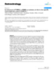
Báo cáo y học: " Involvement of claudin-7 in HIV infection of CD4(-) cells"
lượt xem 1
download
 Download
Vui lòng tải xuống để xem tài liệu đầy đủ
Download
Vui lòng tải xuống để xem tài liệu đầy đủ
Tuyển tập các báo cáo nghiên cứu về y học được đăng trên tạp chí y học quốc tế cung cấp cho các bạn kiến thức về ngành y đề tài: Involvement of claudin-7 in HIV infection of CD4(-) cells
Bình luận(0) Đăng nhập để gửi bình luận!
Nội dung Text: Báo cáo y học: " Involvement of claudin-7 in HIV infection of CD4(-) cells"
- Retrovirology BioMed Central Open Access Research Involvement of claudin-7 in HIV infection of CD4(-) cells Junying Zheng1, Yiming Xie2, Richard Campbell4, Jun Song1, Samira Massachi1, Miriam Razi1, Robert Chiu1, James Berenson4, Otto O Yang3, Irvin SY Chen2 and Shen Pang*1 Address: 1UCLA School of Dentistry, UCLA Dental Institute, and Jonsson Comprehensive Cancer Center, 10833 Le Conte Ave., Los Angeles, CA 90095, USA, 2Departments of Medicine and Microbiology & Immunology, and UCLA AIDS Institute, David Geffen School of Medicine at UCLA, 10833 Le Conte Ave., Los Angeles, CA 90095, USA, 3Department of Medicine, Div. of Infectious Diseases, David Geffen School of Medicine at UCLA, 10833 Le Conte Ave., Los Angeles, CA 90095, USA and 4Institute for Myeloma & Bone Cancer Research, 9201 Sunset Blvd., Suite 300, West Hollywood, CA90069, USA Email: Junying Zheng - zyzheng29@hotmail.com; Yiming Xie - yxie@ucla.edu; Richard Campbell - rcampbell@myelomasource.org; Jun Song - junsong@ucla.edu; Samira Massachi - shekar55@aol.com; Miriam Razi - MRazi1@aol.com; Robert Chiu - rchiu@dent.ucla.edu; James Berenson - cjames@myelomasource.org; Otto O Yang - OYang@mednet.ucla.edu; Irvin SY Chen - ichen@ucla.edu; Shen Pang* - shenp@dent.ucla.edu * Corresponding author Published: 20 December 2005 Received: 07 September 2005 Accepted: 20 December 2005 Retrovirology 2005, 2:79 doi:10.1186/1742-4690-2-79 This article is available from: http://www.retrovirology.com/content/2/1/79 © 2005 Zheng et al; licensee BioMed Central Ltd. This is an Open Access article distributed under the terms of the Creative Commons Attribution License (http://creativecommons.org/licenses/by/2.0), which permits unrestricted use, distribution, and reproduction in any medium, provided the original work is properly cited. Abstract Background: Human immunodeficiency virus (HIV) infection of CD4(-) cells has been demonstrated, and this may be an important mechanism for HIV transmission. Results: We demonstrated that a membrane protein, claudin-7 (CLDN-7), is involved in HIV infection of CD4(-) cells. A significant increase in HIV susceptibility (2- to 100-fold) was demonstrated when CLDN-7 was transfected into a CD4(-) cell line, 293T. In addition, antibodies against CLDN-7 significantly decreased HIV infection of CD4(-) cells. Furthermore, HIV virions expressing CLDN-7 on their envelopes had a much higher infectivity for 293T CD4(-) cells than the parental HIV with no CLDN-7. RT-PCR results demonstrated that CLDN-7 is expressed in both macrophages and stimulated peripheral blood leukocytes, suggesting that most HIV virions generated in infected individuals have CLDN-7 on their envelopes. We also found that CLDN-7 is highly expressed in urogenital and gastrointestinal tissues. Conclusion: Together these results suggest that CLDN-7 may play an important role in HIV infection of CD4(-) cells. lia covering the reproductive tract, the oral cavity, the gas- Background Human immunodeficiency virus (HIV) transmission trointestinal tract, or other tissues during viral through sexual intercourse accounts for the majority of transmission is poorly understood. This is an important infections. It must cross the epithelium during transmis- question to investigate, because the epithelium, which is sion, because the primary targets for HIV infection, composed of stratified CD4(-) epithelial cells, protects the CD4(+) cells, are protected by epithelial lining. However, interface between host and environment (e.g., urogenital, the mechanism by which the virus transverses the epithe- Page 1 of 12 (page number not for citation purposes)
- Retrovirology 2005, 2:79 http://www.retrovirology.com/content/2/1/79 gastrointestinal tract) or between organs and fluid spaces We used subtractive cDNA cloning to identify a gene spe- (prostate, kidney). cifically expressed in LNCaP cells but not in the 293T and HeLa cell lines, which are not susceptible to HIV infection HIV may not utilize the mechanism of binding between [20]. Here we characterize the role of this protein, claudin- gp120 on virions and CD4 molecules to infect epithelial 7 (CLDN-7), in gp120-independent HIV infection. cells, because these cells are CD4(-). One possible mecha- nism is that HIV utilizes lesions in the mucosal surface to As previously described [20], we generated Env(-) HIVNL4- invade underlying lymphoid cells [1,2]. Conversely, it has 3 by deleting a fragment of 581 bp from the env coding been shown that lesions need not be present for the virus region. This deletion truncates the gp120 envelope pro- to cross the epithelial barrier [3-5]. Therefore, it is likely tein and introduces a frameshift into downstream gp41, that HIV can penetrate epithelial layers by other mecha- thereby abrogating gp120 and gp41. The modified HIV nism(s). HIV may enter epithelial cells by a simple in-and- also contains a reporter gene, the enhanced green fluores- out means [6] or by transcytosis [7], whereby the cells cent protein (EGFP). Insertion of the EGFP gene enables passing across are not infected. However, recent reports direct and sensitive detection of HIV infection. Previous demonstrate that many types of epithelial cells can be reports have demonstrated that the substitution of the nef infected with HIV-1 [8-12], and that viral replication also gene with EGFP does not alter viral infectivity [23-25]. To occurs in infected epithelial cells. examine gp120-independent infection, gp120 and gp41 were deleted from the HIVNL4-3 genome, which eliminates Two possible mechanisms by which HIV infects CD4(-) the interference of viral envelope proteins. We have suc- cells have been proposed. Some reports suggest that the cessfully utilized this modified viral strain to study gp120- HIV gp120 surface glycoprotein binds to galactosylcera- independent infection, and therefore used this strain for mide (GalCer) [13-15] or chemokine receptors, including the studies described herein [20,22]. CXCR4 and CCR5, on the surface of CD4(-) cells [15-19], and that this binding initiates HIV entry into CD4(-) cells. Our previous studies demonstrated that a membrane pro- Therefore, gp120 would be a key protein for HIV infection tein, claudin-7 (CLDN-7), is expressed in the HIV-suscep- of CD4(-) cells. However, our previous results demon- tible cell line, LNCaP, but not in the HIV non-susceptible strated that HIV infects many types of CD4(-) cells, some cell line, 293T [26]. We studied the relationship of the without surface gp120 [20-22]. Therefore, CD4(-) cell expression of this protein and infection by HIV. In the infection can be gp120-independent; i.e., the presence of described study, we transfected 293T cells with cloned gp120 glycoprotein molecules on the viral surface is not CLDN-7, then characterized the infection of these cells crucial for CD4(-) cell infection. with EGFP-modified HIVNL4-3. An important approach to understanding gp120-inde- Results pendent HIV infection is to identify the elements involved The prostate cell line, LNCaP, is highly susceptible to in this mechanism of infection. To do so, we compared a gp120-independent HIV-1 infection CD4(-) cell line that is highly susceptible to HIV infection We previously reported that by employing a gp120-inde- to another cell line that has low susceptibility. By screen- pendent infection mechanism, HIV-1 virus infected sev- ing membrane proteins that are specifically expressed in eral CD4(-) cell lines, including oral cell lines Tu139 and the cell line highly susceptible to HIV, it is possible to Tu177, prostate cell lines LNCaP and DU145, and the identify the genes that are involved in HIV infection. fibroblast cell line, HT-1080. However, the infection lev- els of 293T cells by HIV are low [20]. The CD4(-) cell line, Our previous studies demonstrated that HIV efficiently LNCaP, demonstrated the highest HIV susceptibility [22]. When 104 LNCaP cells per well in a 24-well plate were infects the prostate cancer cell line, LNCaP [22]. When a viral load of approximately 100 ng p24 was used for infec- infected with virus at a concentration of 100 ng/ml p24, tion of 104 cells in culture, approximately 3–20% of more than 10% of the cells were infected by virus pre- LNCaP cancer cells were infected. The concentration of pared from either 293T cells (Figure 1A) or an oral epithe- 100 ng p24/0.5 ml is similar to the viral load found in lial cell line (Figure 1B). Because there is a partial gp120 patients in the acute phase of infection. Infection of sequence remaining (279 of 509 amino acid residues), it LNCaP cells is gp120-independent, because HIV with or was necessary to ascertain whether the truncated gp120 without gp120 on its envelope is equally infectious for has any effects upon infection of LNCaP cells. Antiserum these cells, and antibodies against gp120 do not block against gp120 or gp160, the precursor of gp120 and gp41, infection. It is expected that certain proteins specifically was used to block the interaction between the truncated expressed on the surface of this cell line are responsible for gp120 and its potential ligands. Infection of LNCaP cells gp120-independent HIV infection. with HIV-1 Env(-) virus was not significantly affected, sug- gesting that HIV infection of LNCaP cells is gp120-inde- Page 2 of 12 (page number not for citation purposes)
- Retrovirology 2005, 2:79 http://www.retrovirology.com/content/2/1/79 A. -AB 16 -gp41 14 -gp160RF (HT3-HT7) % of EGFP-positive Cells -gp120 T1-SP 12 -gp160B and RF 10 -gp120 SF2 8 6 4 2 0 HeLa-CD4 LNCaP LNCaP DU145 DU145 HIV Env(+) HIV Env(+) HIV Env(-) HIV Env(+) HIV Env(-) C. B. 1000 LNCaP 1000 LNCaP EGFP-Positive colonies/well EGFP-Positive colonies/well R11 HeLa-CD4 100 HT1080 100 - HeLa 10 10 PC3 1 1 0 0 5000 500 50 5 0.1 0.3 1.0 3.0 10.0 p24 of HIVNL4-3-EGFP-Env(+) (pg) p24 of HIVNL4-3-EGFP-Env(-) (ng) Figure 1 HIV infection of LNCaP cells HIV infection of LNCaP cells. LNCaP or HeLa-CD4 cells in 24-well culture plates (104 cells/well) were infected with HIV either with or without gp120 protein on its envelope [Env(-) and Env(+) HIV]. A) A significantly high percentage of EGFP-positive cells was demonstrated in the LNCaP cell cultures infected by HIV Env(-) virus (9–14%). Infection of HeLa-CD4 cells by HIV Env(+) was used as a positive control to assess anti-gp120 or -gp160 function of the antibodies. Infection of HeLa cells was performed as a negative control. The infections of HIV either with or without Env showed very low infectivity for HeLa cells, as demon- strated with no EGFP-positive cells in the infected culture, or occasionally there were one or two EGFP-positive colonies. In other experiments, we also infected HeLa-CD4 cells with Env(-) HIVNL4-3, and found that the Env(-) HIV strain did not infect HeLa-CD4. These results have been previously reported (22). B) Infection of CD4(-) cell lines by HIVNL4-3-Env(-)-EGFP virus prepared from an oral epithelial cell line derived from a patient. The cell line was established and maintained in our laboratory. C) Infection of the LNCaP and HeLa-CD4 cell lines by HIV at various concentrations. Because the figure is in log scale, the standard deviations do not appear clearly. These are: 1) LNCaP: 900 ± 38 (5 ng), 205 ± 11(1.5 ng), 24 ± 5.6 (0.5 ng), 8.5 ± 0.7 (0.15 ng), 2.5 ± 0.7 (50 pg); and 2) HeLa-CD4: 1114 ± 115 (5 ng), 638 ± 47 (1.5 ng), 80 ± 10 (0.5 ng), 14.5 ± 2.1 (0.15 ng), 3.5 ± 0.71 (50 pg), 1 ± 0 (15 pg). pendent. It should also be noted that the infection rate of cules on the cell surface of HeLa-CD4 cells, we would LNCaP cells by HIV was similar to that of HeLa-CD4, sug- expect that infection of LNCaP by HIV Env(-) virus would gesting that the infectivity of HIV-1 for some types of occur through binding of unknown proteins of the virus CD4(-) cells is high. Because 12% HIV infectivity of HeLa- and the cells, and the binding affinity should be compara- CD4 occurs through the binding of gp120 to CD4 mole- ble to that between gp120 and CD4 molecules. Page 3 of 12 (page number not for citation purposes)
- Retrovirology 2005, 2:79 http://www.retrovirology.com/content/2/1/79 A. B. Exp. 1 Exp. 2 300 200 EGFP Positive colonies 60 200 40 100 100 20 0 0 0 293T/ 293T/ 293T/ 293T/ 293T/ 293T/ 293T/ 293T 293T 293T CLDN-7158 CLDN-7211 CLDN-7158 CLDN-7211 pCDNA CLDN-7211 CLDN-7158 Figure 2 Effect of CLDN-7 molecules upon infection of 293T cells by HIVNL4-3 Effect of CLDN-7 molecules upon infection of 293T cells by HIVNL4-3. A) HIVNL4-3 Env(-) virus infection of either CLDN-7211- or CLDN-7158-modified 293T cells. In experiment 1, HIV Env(-) at 50 ng of p24 was used to infect 104 cells in each well; in experiment 2, 104 cells were infected by virus at 100 ng of p24. B) HIVNL4-3 Env(+) virus infection of either CLDN-7211- or CLDN-7158-modified 293T cells. Plasmid pCDNA3-transfected 293T cells were also used as a control. CLDN-7211 or CLDN-7158 increases susceptibility of CD4(-) To accurately compare the infectivity for LNCaP and HeLa-CD4 cells by HIVNL4-3 containing the EGFP gene, we cells to HIV infection infected these two cell lines with different concentrations Using subtractive hybridization, we identified the CLDN- of virus (Figure 1C). When LNCaP cells in 24-well plates 7 gene, which is highly expressed in prostate cells but not were infected with 50 pg of p24 counts of virus, less than in 293T cells [26]. Two transcripts of this gene have been five infected colonies per well were detected. When the identified, one encoding a peptide of 211 amino acid res- cells were infected with virus containing 15 pg of p24, no idues, and the other, 158 [26]. The plasmid vectors that carry CLDN-7211 or CLDN-7158 were used to transfect infected colonies were detected. When HeLa-CD4 cells were infected with virus at similar or lower concentra- 293T cells. Two days post-transfection, the cells were tions, we found that they could be infected by virus con- plated into 24-well plates for viral infection. The suscepti- taining 15 pg of p24. However, when the concentration of bility of these transfected cells to Env(-) HIVNL4-3 infection virus was diluted to 5 pg of p24, no positive colonies were is significantly increased. Compared with non-transfected detected in the infected cell cultures, suggesting that the cells, the numbers of infected cells in the transfected 293T sensitivity of LNCaP cells for HIV is approximately 3-fold cell cultures were over 10-fold higher (Figure 2A). These results suggest that the presence of either CLDN-7211 or lower compared with that of HeLa-CD4 cells (Figure 1C). CLDN-7158 on the surface of CD4(-) cells increases their To ensure that infection of LNCaP cells by virus generated susceptibility to HIV-1 infection via a gp120-independent from 293T cells is not caused by potential contamination mechanism. In a separate experiment, we also tested of viruses that can modify the HIV envelope (e.g., ampho- CLDN-7-transfected 293T cells using EGFP-modified tropic murine viruses), we tested 293T cells derived from Env(+) HIVNL4-3. The results were very similar to those various sources. The results were similar to those shown in using Env(-) HIVNL4-3 (Fig. 2B). Figure 1A, with much higher infectivity for LNCaP cells as compared to other cell lines, including 293T, HeLa, and Antibodies specific to CLDN-7 block gp120-independent DU145. It is unlikely that all of the 293T cells from differ- infection ent sources were contaminated; in other words, it is Immunostaining demonstrated that CLDN-7 is a mem- unlikely that the virus generated from 293T cells is modi- brane protein, similar to other claudins (Figure 3A). To fied by any contaminated amphotropic viruses. determine whether CLDN-7 is the key protein involved in Page 4 of 12 (page number not for citation purposes)
- Retrovirology 2005, 2:79 http://www.retrovirology.com/content/2/1/79 A. B. Infection compared with the Control (%) 120 100 80 60 40 20 0 Env(-) Env(-) + Env(-) + Env(-) + -CLDN-7211 only -gp160 -Pep 1 Figure 3 Inhibition of HIV infection by antibodies specific to CLDN-7 Inhibition of HIV infection by antibodies specific to CLDN-7. A) Immunostaining of CLDN-7211 (upper panel) and CLDN-7158 (lower panel) by CLDN-7-specific polyclonal antibodies. 293T cells (5 × 10⁴) were plated into 35-mm plates 24 hours prior to transfection. CLDN-7 plasmids (3 µg) were used for transfection of each 35-mm plate. At two days post-transfection, the cells were immunostained. CLDN-7 antibodies were added into the plates overnight at 4°C. B) Antisera from NIH (anti-gp160, cat. 191), Zymed (anti-CLDN-7, 0.25 mg/ml), or made by us were added to LNCaP cell cultures at 1:100 v/v 10 minutes prior to adding HIV. Six days post-infection, EGFP-positive cells were counted. Infection of LNCaP cultures with no antibodies was set as the control. EGFP-positive cells in the culture treated with antibodies against CLDN-7 demonstrated 27% ± 0.6% viral infec- tion compared to the control with no antibodies in the cell culture. gp120-independent infection, we used antibodies specific GMMSCKMYD. The peptide sequence corresponds to aa to CLDN-7 to block HIV infection. Polyclonal antibodies positions 44 to 58 of CLDN-7. A cell-labeling assay dem- against either CLDN-7 (Zymed Laboratories) or gp160 onstrated that this antibody preparation is able to bind (cat. #191, NIH AIDS Reagent Program) were added into CLDN-7 on the cell membrane (data not shown). How- LNCaP cell cultures. It was expected that the extracellular ever, it did not significantly block HIV infection of LNCaP domains of CLDN-7 on the surface of LNCaP cells could cells, suggesting that this sequence region in CLDN-7 may be bound by CLDN-7 antibodies, and the binding of these not be essential for HIV infection (Figure 3B). antibodies to CLDN-7 would disrupt the binding of HIV Increased infectivity for viruses generated with CLDN-7211 to CLDN-7. As a result, HIV infection of CD4(-) cells or CLDN-7158 on their envelopes should be decreased. Our results demonstrated that the antibodies for CLDN-7 decreased HIV infectivity for A possible explanation for increased HIV-1 infectivity for LNCaP cells (Figure 3B), whereas gp160-specific antibod- the CLDN-7-transfected cells is that gp120-independent ies did not show inhibition, suggesting that CLDN-7 on HIV-1 infection is mediated by an interaction between a the surface of LNCaP cells is involved in HIV infection. cellular protein on the viral envelope and CLDN-7 expres- Because we used the gp120-negative virus strain for infec- sion on the surface of transfected 293T cells. We hypothe- tion and antibodies against gp160 do not show inhibi- sized that the ligands for CLDN-7 are present on the tion, we believe that infection of LNCaP cells occurs via a surface of many types of cells. If HIV has CLDN-7 on the gp120-independent mechanism. surface of its envelope, it is expected that CLDN-7 on the viral surface can bind to the ligands for CLDN-7 on target We also tested a preparation of polyclonal antibodies we cells, so that HIV infection can be increased. To confirm generated against a peptide with the sequence CVTQST- this, we prepared HIV expressing CLDN-7 on the viral sur- Page 5 of 12 (page number not for citation purposes)
- Retrovirology 2005, 2:79 http://www.retrovirology.com/content/2/1/79 A. B. Cellular CLDN7158 Viral CLDN-7158 300 EGFP-positive colonies Control 200 M 100 CLDN-7158 20 kd 0 CLDN-7158 CLDN-7211 Cont. C. D. 30 ENV(-) % of EGFP-positive cells EGFP-positive colonies 25 ENV(-) + CLDN-7158 120 20 ENV(-) + CLDN-7211 15 80 10 40 5 0 CLDN-7158 CLDN-7211 Cont. LNCaP CEM Figure 4 of CLDN-7211- or CLDN-7158-modified Env(-) virus Infectivity Infectivity of CLDN-7211- or CLDN-7158-modified Env(-) virus. A) Western blot of proteins isolated from the virus generated from the transfected 293T cells by pNL4-3-EGFP-Env(-) and CLDN-7. Cell culture medium collected from cells transfected with CLDN-7 was used as a control to assess the background of CLDN-7 in microvesicles. Cellular proteins isolated from CLDN-7 plasmid-transfected cells were used as a positive control. The monomers of CLDN-7 are approximately 22 kd. Lane M is a molecular marker lane. The monomers of CLDN-7 are approximately 22 kd. B) CLDN-7-modified HIVNL4-3 Env(-) virus showed significantly higher infectivity for 293T cells. C) Infection of either LNCaP or CEM CD4(+) T-lymphocyte cell lines by CLDN-7-modified HIV, with approximately a 1.5- to 2-fold increase of viral infection. D) CLDN-7-modified HIVJRCSF virus with intact gp120 showed significantly higher infectivity for a CD4(-) cell line, PC-3, than the virus with no CLDN-7 on its surface. face by co-transfecting a plasmid containing CLDN-7 from 293T cell cultures transfected by the CLDN-7 gene as (either CLDN-7211 or CLDN-7158) with a plasmid that a negative control to assess the contribution of CLDN-7 in contains the HIV genome into 293T cells. Because HIV membranous particles, termed microvesicles, because uses a patch of the host cell membrane as its envelope, it viral preparations are generally contaminated with these was expected that the CLDN-7 molecules on the mem- particles [27]. As shown in Figure 4A, no CLDN-7 protein branes of CLDN-7 gene-transfected 293T cells could be was detected in the CLDN-7-transfected 293T culture taken up by HIV virions during viral assembly. Western medium, suggesting that either the amounts of microves- blotting confirmed this to be the case (Figure 4A), and icles from transfected 293T were very low or the microves- CLDN-7 was detected. We also collected the medium icles generated from transfected 293T cells do not contain Page 6 of 12 (page number not for citation purposes)
- Retrovirology 2005, 2:79 http://www.retrovirology.com/content/2/1/79 A. B. Unstimulated PBL Unstimulated PBL Stimulated PBL Stimulated PBL Macrophages Macrophages 104 Copies 105 Copies 103 Copies 104 Copies 105 Copies 103 Copies LNCaP LNCaP AT84 293T 300 bp CLDN-7211 200 bp 200bp GAPDH 100 bp Figure 5 of CLND-7211 in PBL and macrophages Expression Expression of CLND-7211 in PBL and macrophages. RNA isolated from unstimulated PBL, interleukin 2-stimulated PBL, macro- phages, and control cell lines was analyzed using RT-PCR. RNA samples were reverse transcribed using the oligo-dT primer, followed by PCR using the primers 5'-CTCCTCTGACTTCAACAGCG-3' and 5'-TGTTGCTGTAGCCAAATTCG-3' to detect the glyceraldehydes-3-phosphate dehydrogenase (GAPDH) gene as RNA standard, and the primers described previously [26] for detecting CLDN-7 RNA. Panels A and B are RT-PCR from different samples. significant amounts of CLDN-7 protein. Therefore, the eral blood lymphocytes (PBL) and macrophages, and contribution of CLDN-7 derived from microvesicles in found that both stimulated PBL and macrophages express viral preparations is insignificant. We used the CLDN-7- CLDN-7 (Figure 5). Expression of CLDN-7 in PBL and modified Env(-) EGFP-HIVNL4-3 to infect 293T, LNCaP, macrophages suggests that HIV produced from these two and CEM cells. CLDN-7158-modified HIVNL4-3-EGFP- types of cells carries CLDN-7 on its envelope. Because Env(-) demonstrated approximately 100-fold higher stimulated CD4(+) T-lymphocytes in PBL and macro- activity when infecting 293T cells (Figure 4B) compared phages are the major types of cells hosting and generating with the parental HIV that does not have CLDN-7. The HIV in patients, virus derived from patients should also infection efficiencies in LNCaP and CEM cells by CLDN- infect certain types of CD4(-) cells during HIV transmis- 7-modified viruses also showed a significant increase. sion. These results suggest that CLDN-7 expressed on either the surface of the target cells or on the surface of the HIV enve- Expression of CLDN-7 in other tissues We used the coding region of CLDN-7211 as a probe to lope increases gp120-independent infection. hybridize mRNAs derived from more than 50 different tis- We also used EGFP-modified HIVJRCSF, a patient-derived sue types and cell lines, and found that CLDN-7 is R5 strain, to infect CD4(-) cells. Using co-transfection, we expressed in certain tissues in the urogenital and gastroin- generated CLDN-7-modified HIVJRCSF from 293T cells. testinal systems, such as the colon, intestine, trachea, kid- Because both CLDN-7-negative and -positive HIVJRCSF did ney, lung, and prostate (Figure 6). Infection of epithelial not infect 293T cells, we infected another CD4(-) cell line, cells in these tissues and organs by HIV-1 has been PC-3. The results demonstrated that CLDN-7 also signifi- reported, and they are the sites of many AIDS-related cantly increases HIVJRCSF infectivity for this CD4(-) cell symptoms [10,14,28-33]. Thus, studies of this gene and line. its relationship with gp120-independent HIV infection will be important for understanding HIV-1-related patho- logic effects. Expression of CLDN-7 mRNA in peripheral blood lymphocytes and macrophages Because CLDN-7 on the cell membrane significantly Discussion increased susceptibility of CD4(-) cells to HIV-1, it was Results from our previous studies and others have expected that virus generated from cells that express revealed that CD4(-) cells can be infected by HIV, so it is CLDN-7 would have higher infectivity for CD4(-) cells important to understand this process in the context of than virus generated from cells that do not. It is important viral transmission, whereby HIV transverses CD4(-) epi- to quantify the expression profile of the CLDN-7 gene in thelial cell layers and infects CD4(+) T-lymphocytes and CD4(+) cells, including T-cells and macrophages. We used macrophages. In addition, infected CD4(-) cells, such as RT-PCR to examine the expression of CLDN-7 in periph- cells in the central nervous system, may serve as viral res- Page 7 of 12 (page number not for citation purposes)
- Retrovirology 2005, 2:79 http://www.retrovirology.com/content/2/1/79 1 2 3 4 5 6 7 1 2 3 4 5 6 7 colon, Fetal Leukemia, A A esophagus kidney lung liver transverse brain HL-60 Fetal Colon, Skeletal HaLa stomach placenta pancreas B B heart descending muscle S3 Adrenal Fetal duodenum rectum spleen bladder K562 C C gland kidney Fetal Thyroid jejunum thymus uterus MOLT-4 liver gland D D Peripheral Burkitt’s Salivary Fetal Blood ileum prostate Lymphoma spleen E E gland leukocyte Raji Burkitt’s Lymph Mammary Fetal ileocecum testis Lymphoma F F node gland thymus Daudi Fetal Colorectal G G appendix ovary Bone lung SW480 marrow Colon, Lung H H trachea ascending A549 Figure 6 Expression of CLDN-7 molecules in human tissues Expression of CLDN-7 molecules in human tissues. A nylon filter preloaded with RNA from various tissues from BD Clontech (Palo Alto, CA) was used to assess the expression levels of CLDN-7 in various tissues. The left panel shows the hybridization of the tissue samples in the filter by a CLDN-7 probe containing the coding region, and the right panel shows the correspond- ing tissues. ervoirs. Some reports demonstrated that the binding of membrane protein, CLDN-7, can serve as a receptor for gp120 to GalCer or chemokine receptors is the mecha- HIV-1 infection of CD4(-) cells or as a ligand on the viral nism of CD4(-) cell infection; however, only particular envelope. CLDN-7 belongs to the claudin membrane pro- types of HIV were reported to infect certain types of CD4(- tein family. Some claudins, such as CLDN-1 and CLDN-2, ) cells [13-19]. Our previous studies demonstrated that are involved in formation of tight junctions (TJ) between both the X4 and R5 types of HIV can infect CD4(-) cells cells [34,35], while others may serve as receptors. As pre- [20-22], and for many types of CD4(-) cells, gp120 is not viously described, human claudin4 (CPE-R) is a receptor required for infection. for the clostridium perfringens enterotoxin [36]. It is pos- sible that CLDN-7 plays a role as the receptor for a protein Our results demonstrated that Env(-) HIV is still able to ligand that is expressed on the surface of HIV viral parti- infect many types of cells via a gp120-independent mech- cles. Our results have also demonstrated that CLDN-7 can anism. Our results demonstrated that although Env(-) be taken up as a component of viral particles. HIV may HIV could not efficiently infect CD4(+) cells, it has similar also use this protein to bind to target cells for infection. infectivity for CD4(-)cells. It is possible that in some Based on our results and general HIV biology, we propose infected cells, HIV can down-regulate the expression of the model shown in Figure 7. In this model, the interac- Env proteins to evade the immune response. If that were tion of CLDN-7 with its ligand helps the virus to bind to the case, there should be a high percentage of Env(-) HIV CD4(-) cells; however, there may be other proteins that present in patients, which may be able to infect some can also do this. Although expression of CLDN-7 in 293T types of CD4(-) cells, such as neurons and glial cells. The cells significantly increased HIV infection, infection of infection of brain cells may be a major hindrance of viral CLDN-7-expressing 293T cells was still significantly lower eradication because they have a long life-span,. than HIV infection of LNCaP cells. We therefore believe that CLDN-7 is not the only protein involved in HIV infec- It is important to identify the genes involved in gp120- tion of CD4(-) cells. It is possible that the association of independent infection. As described here, we found that a CLDN-7 with another protein can cause more efficient Page 8 of 12 (page number not for citation purposes)
- Retrovirology 2005, 2:79 http://www.retrovirology.com/content/2/1/79 cells in which the CLDN-7 protein is expressed, the virus Model A also expresses this protein on its envelope. The presence of CLDN-7 molecules on the viral envelope may greatly HIV HIV increase its capacity for infecting other CD4(-) cells. It is expected that the viruses generated from infected PBL, HIV HIV Other proteins macrophages, colon, intestine, trachea, kidney, lung, and in HIV infection prostate express this protein on their envelopes. This por- tion of the virus may have greater infectivity for CD4(-) CLDN-7 CLDN-7 cells compared to HIV virions that do not have CLDN-7 on their envelopes. Our previous studies demonstrated that HIV can also use a gp120-independent mechanism by which to infect Model B CD4(+) cells [22]. Because macrophages express much CLDN-7 Assoc. protein lower levels of CD4 molecules on the cell surface, it is CLDN-7 expected that expression of CLDN-7 on the surface of HIV HIV macrophages may help HIV to infect these cells. Conclusion CLDN-7 CLDN-7 Assoc. Our results demonstrate that the presence of CLDN-7 on protein the surface of target cells increases viral susceptibility. Figure 7 Two models for gp120-independent HIV infection Because CLDN-7 is expressed in organs related to HIV Two models for gp120-independent HIV infection. Model 1: transmission and HIV pathogenicity (including the colon, HIV may use cellular proteins that are anchored to its enve- kidney, lung, uterus, and oral tissue), it is expected that lope to bind to either CLDN-7 or other proteins. In LNCaP, this protein is associated with HIV infection of CD4(-) both CLDN-7 and associated proteins involved in gp120- cells in these organs, and is related to viral transmission or independent infection are present. In 293T cells, neither pathogenicity. Our results also demonstrated that virus CLDN-7 nor the other infection-related membrane pro- generated from CLDN-7-transfected 293T cells has two- to tein(s) is present. Expression of CLDN-7 in 293T may 100-fold higher levels of infectivity, suggesting that the increase infectivity, but the levels of infection of the CLDN- presence of CLDN-7 or other types of cellular membrane 7-modified 293T may still be significantly lower than those for LNCaP. Model 2: HIV may use cellular proteins that are proteins on the viral envelope is important for viral infec- anchored to its envelope to bind to a CLDN-7-associated tion. Because CLDN-7 is expressed in activated PBL and complex. Although transfection of CLDN-7 can express this macrophages, and these two types of cells serve as HIV protein on the cell surface, the lack of the CLDN-7-associ- hosts, it is very likely that most HIV particles carry CLDN- ated protein decreases the binding of virus to the target cells. 7 on their surface. Therefore, it is very likely that this pro- tein plays important roles in HIV infection of CD4(-) cells in humans. infection. In LNCaP cells, both CLDN-7 and its associated protein are expressed. In 293T or HeLa cells, expression Materials and methods levels of both CLDN-7 and its associated protein may be Cell culture very low. Although we can use transfection to express Cell lines LNCaP, DU145, HT1080, R11, HeLa, CEM, and CLDN-7 in 293T cells, the expression levels of the CLDN- 293T were purchased from American Type Culture Collec- 7-associated protein may not be correspondingly tion (ATCC) or from other laboratories, as described increased. Therefore, addition of CLDN-7 to 293T cells [20,22]. Cell lines LNCaP, PC-3, and DU145 are derived can only partially increase levels of HIV infectivity, from the prostate, 293T from the embryonic kidney, CEM approximately 10% of that of LNCaP cells. is a CD4(+) T-lymphocyte cell line, R11 is a renal carci- noma cell line, HT1080 is a fibroblast cell line, and HeLa Because many tissues of the gastrointestinal and urogeni- is a from cervical cancer cell line. We also prepared HIV tal systems express the CLDN-7 gene, the cells in these tis- from an oral epithelial cell line derived from a patient. All sues may be more susceptible to HIV-1 infection. These cell lines were maintained in RPMI medium supple- results are consistent with clinical data, with HIV-1 infec- mented with 10% fetal bovine serum (FBS). tion of epithelial cells of the oral mucosa, colon, intestine, and kidney being reported in patients [10,14,28-33]. In The CLDN-7 gene addition, because the virus uses a patch of cellular mem- Using a subtractive hybridization method combined with brane as its envelope, when the virus is generated from RT-PCR, followed by screening prostate cDNA libraries, Page 9 of 12 (page number not for citation purposes)
- Retrovirology 2005, 2:79 http://www.retrovirology.com/content/2/1/79 we obtained three full-length cDNA clones. Sequence 100 ng of p24 counts of virus in 0.5 ml of medium. At 16 analysis demonstrated that these cloned cDNA sequences hours post-infection, the cell cultures were washed. are homologous to human CLDN-7. Two of these three cDNA clones encode a peptide of 211 amino acid residues DNA transfection identical to that in Genbank. The third encodes a peptide Liposome FUGENE-6 (Roche Molecular Biochemicals, of 158 amino acid residues, which is a truncated form of Indianapolis, IN) was used to transfect the CLDN-7 plas- mid into 293T cells. Cells (2 × 10⁴) were plated into each CLDN-7 lacking 53 amino acid residues at the C-terminus [26]. Previous studies have demonstrated that both the well of 24-well plates 16 hours prior to lipofection. Plas- mid DNA (2.0 µg) was mixed with FUGENE-6 liposome full-length (CLDN-7211) and the truncated (CLDN-7158) in 50 µl of RPMI medium for 10 minutes at room temper- forms of CLDN-7 are highly expressed in LNCaP but not 293T cells, and are expressed at low levels in HeLa cells ature before addition to cell cultures. At 8 hours [post- [26]. Both isoforms of CLDN-7 were inserted into a plas- transfection?], the cell cultures were washed once and mid vector downstream of the CMV promoter. fresh medium then added. Viral preparation and titration Determination of EGFP expression To obtain HIV-1 Env(-) virus, we transfected either the The expression levels of EGFP were determined by count- 293T cell line or an oral epithelial cell line established in ing EGFP- positive cells by fluorescent microscopy or by our laboratory with plasmid pNL4-3-EGFP-Env(-), which fluorescent-activated cell sorting (FACS). contains a modified HIVNL4-3 viral genome [20]. The mod- ified HIV-1NL4-3 genome has deletions in env (581 bp) and Western blotting nef (222 bp), and insertion of the EGFP gene, as previous Cell culture medium from transfected 293T cells with described [20]. At 16 hours post-transfection, medium pCDNA3.1-CLDN-7 and pNL4-3-EGFP-Env(-) or only containing the plasmid was removed from transfected cell pCDNA3.1-CLDN-7 was collected two to four days post- cultures. The transfected cell cultures were then washed transfection. The collected medium was ultracentrifuged with serum-free medium before adding new culture at 16,000 rpm for one hour at 4°C. The pellets were resus- pended with 50 µl of protein lysis buffer (0.5% NP40, medium supplemented with 10% FBS. The medium con- 1.0% glycerol, 0.1% β-mercaptoethanol, 40 mM Tris, taining virus was collected at days 2, 3, and 4 post-trans- fection. To remove cell debris, all the viral preparations pH6.8). The viral lysates were incubated at 37°C for 5 were passed through a 0.2-micron filter. The collected minutes before SDS-polyacrylamide gel electrophoresis viral stocks were titrated by p24 assays. The human 293T (PAGE). Protein concentrations were determined by a cell line does not express CLDN-7. Therefore, virus gener- standard protein assay (BioRad, Hercules, CA). Aliquots representing 2.5 µg of protein were separated by SDS- ated from this cell line is CLDN-7-negative. CLDN-7-pos- itive virus was generated by co-transfection of 293T cells PAGE and transferred to a nylon membrane (Poly Screen with both pCDNA3.1-CLDN-7 (CLDN-7211 or CLDN- PVDF; Fisher Scientific, Pittsburgh, PA). Polyclonal anti- 7158) and the modified pNL4-3-EGFP-Env(-). Viruses gen- bodies specific to human CLDN-7 (Zymed Laboratories, erated from these co-transfections carried the CLDN-7 San Francisco, CA) were used according to the manufac- protein on their envelopes and were also titrated by p24 turer's instructions to bind CLDN-7. The specificity of the assays. CLDN-7 antibodies was tested by binding proteins iso- lated from CLDN-2 gene-transfected cells. No cross-bind- EGFP gene-modified HIVJRCSF, a patient-derived R5 strain, ing was detected, although strong expression of CLDN-2 was constructed using a similar approach. A part of the nef was noted when using CLDN-2-specific antibodies that gene was substituted by the EGFP gene. We generated were purchased from Zymed, indicating that those anti- infectious HIVJRCSF by transfection of 293T cells. This virus bodies are highly specific. can infect cells that express both CCR5 and CD4 on the cell surface, and replicates in infected cells. Because it con- Antibody blockage of HIV infection tains a viral genome that is almost intact but lacks the nef Polyclonal antibodies against gp120 or gp160 were gene, this virus alone propagates in CCR5(+)/CD4(+) obtained from the NIH HIV Reagents Program (Rockville, cells. MD), and gp41-specific monoclonal antibody (mAB) was obtained from Virogen (Watertown, MA). Polyclonal anti- bodies against CLDN-7 were purchased from Zymed Lab- Infection of cell cultures Cells (5 × 10³) were plated into each well of 24-well plates oratories (0.25 mg/ml) or made from rabbits using a 24 hours prior to infection. During infection, a viral aliq- peptide of extracellular domain 1 of CLDN-7 with the uot with a p24 count of 100 ng was added into each well sequence CVTQSTGMMSCKMYD, from position 54 to 68 of cell cultures. The final volumes in each well were (concentration 0.85 mg/ml). The homology of the amino adjusted to 0.5 ml so that the concentration of virus was acid sequence of this peptide is 73% or less of he corre- Page 10 of 12 (page number not for citation purposes)
- Retrovirology 2005, 2:79 http://www.retrovirology.com/content/2/1/79 sponding sequences of other human claudins. Antibodies 9. Howell AL, Edkins RD, Rier SE, Yeaman GR, Stern JE, Fanger MW, Wira CR: Human immunodeficiency virus type 1 infection of were added into both viral stocks and cell cultures for 10 cells and tissues from the upper and lower human female minutes prior to infection at room temperature. The final reproductive tract. J Virol 1997, 71:3498-3506. 10. Tan X, Phillips DM: Cell-mediated infection of cervix derived concentration of antibodies in the infection medium was epithelial cells with primary isolates of human immunodefi- a 1:100 dilution. Four hours post-infection, the cell cul- ciency virus. Arch Virol 1996, 141:1177-1189. tures were washed once, and fresh culture medium added. 11. Wu Z, Chen Z, Phillips DM: Human genital epithelial cells cap- ture cell-free human immunodeficiency virus type 1 and Virus with no antibody (control) was also added and transmit the virus to CD4+ Cells: implications for mecha- incubated at room temperature for 10 minutes prior to nisms of sexual transmission. J Infect Dis 2003, 188:1473-1482. 12. Liu X, Zha J, Chen H, Nishitani J, Camargo P, Cole SW, Zack JA: infection. Human immunodeficiency virus type 1 infection and replica- tion in normal human oral keratinocytes. J Virol 2003, RT-PCR to quantify mRNA levels of CLDN-7 77:3470-3476. 13. Harouse JM, Bhat S, Spitalnik SL, Laughlin M, Stefano K, Silberberg We used the quanidinium thiocyanate method to isolate DH, Gonzalez-Scarano F: Inhibition of entry of HIV-1 in neural RNA. RNA isolated from approximately 105 cells was sus- cell lines by antibodies against galactosyl ceramide. Science pended in 20 µl of water. The isolated RNA was reverse 1991, 253:320-323. 14. Yahi N, Sabatier JM, Nickel P, Mabrouk K, Gonzalez-Scarano F, Fantini transcribed, using AMV reverse transcriptase from Roche J: Suramin inhibits binding of the V3 region of HIV-1 enve- with oligo-dT as the primer. An aliquot of the cDNA was lope glycoprotein gp120 to galactosylceramide, the receptor for HIV-1 gp120 on human colon epithelial cells. J Biol Chem used for RT-PCR with primers 5'-CTCCTCTGACT- 1994, 269:24349-24353. TCAACAGCG and 5'-TGTTGCTGTAGCCAAATTCG to 15. Fantini J, Hammache D, Delezay O, Pieroni G, Tamalet C, Yahi N: detect glyceraldehydes-3-phosphate dehydrogenase Sulfatide inhibits HIV-1 entry into CD4-/CXCR4+ cells. Virol- ogy 1998, 246:211-220. (GAPDH) cDNA. The primers of CLDN-7 have been pre- 16. Edinger AL, Mankowski JL, Doranz BJ, Margulies BJ, Lee B, Rucker J, viously described [26]. The expected sizes of the PCR Sharron M, Hoffman TL, Berson JF, Zink MC, Hirsch VM, Clements products were 121 bp (GAPDH) and 252 bp (CLDN- JE, Doms RW: CD4-independent, CCR5-dependent infection of brain capillary endothelial cells by a neurovirulent simian 7211). immunodeficiency virus strain. Proc Natl Acad Sci USA 1997, 94:14742-14747. 17. Potempa S, Picard L, Reeves JD, Wilkinson D, Weiss RA, Talbot SJ: Competing interests CD4-independent infection by human immunodeficiency The author(s) declare that they have no competing inter- virus type 2 strain ROD/B: the role of the N-terminal domain ests. of CXCR-4 in fusion and entry. J Virol 1997, 71:4419-4424. 18. LaBranche CC, Hoffman TL, Romano J, Haggarty BS, Edwards TG, Matthews TJ, Doms RW, Hoxie JA: Determinants of CD4 inde- Acknowledgements pendence for a human immunodeficiency virus type 1 vari- ant map outside regions required for coreceptor specificity. We thank Wendy Aft for editing. This work was supported by a grant from J Virol 1999, 73:10310-10319. the National Institute of Health R01 AI047722 and The James Pendleton 19. Edwards TG, Hoffman TL, Baribaud F, Wyss S, LaBranche CC, Charitable Trust. Romano J, Adkinson J, Sharron M, Hoxie JA, Doms RW: Relation- ships between CD4 independence, neutralization sensitivity, References and exposure of a CD4-induced epitope in a human immun- odeficiency virus type 1 envelope protein. J Virol 2001, 1. Dickerson MC, Johnston J, Delea TE, White A, Andrews E: The 75:5230-5239. causal role for genital ulcer disease as risk factor for trans- 20. Pang S, Yu D, An DS, Baldwin GC, Xie Y, Poon B, Chow YH, Park NH, mission of human immunodeficiency virus. Sex Transm Dis Chen IS: Human immunodeficiency virus Env-independent 1996, 23:429-440. infection of human CD4(-) cells. J Virol 2000, 74:10994-11000. 2. Kozak SL, Platt EJ, Madani N, Ferro FE Jr, Peden K, Kabat D: CD4, 21. An DS, Xie Y, Chen IS: Envelope gene of the human endog- CXCR-4, and CCR-5 dependencies for infections by primary enous retrovirus HERV-W encodes a functional retrovirus patient and laboratory-adapted isolates of human immuno- envelope. J Virol 2001, 75:3488-3489. deficiency virus type 1. J Virol 1997, 71:873-882. 22. Chow YH, Yu D, Zheng JY, Xie Y, Wei OL, Chiu C, Foroohar M, Yang 3. Miller CJ: Animal models of viral sexually transmitted dis- OO, Park NH, Chen IS, Pang S: gp120-Independent infection of eases. Am J Reprod Immunol 1994, 31:52-63. CD4- epithelial cells and CD4+ T-cells by HIV-1. J AIDS 2002, 4. Miller CJ, Alexander NJ, Sutjipto S, Joye SM, Hendrickx AG, Jennings 30:1-8. M, Marx PA: Effects of virus dose and nonoxynol-9 on the gen- 23. Lee AH, Han JM, Sung YC: Generation of the replication-com- ital transmission of SIV in rhesus macaques. J Med Primatol petent human immunodeficiency virus type 1 which 1990, 19:401-409. expresses a jellyfish green fluorescent protein. Biochem Bioph 5. Spira AI, Marx PA, Patterson BK, Mahoney J, Koup RA, Wolinsky SM, Res Comm 1997, 233:288-292. Ho DD: Cellular targets of infection and route of viral dissem- 24. Chen BK, Feinberg MB, Baltimore D: The kappaB sites in the ination after an intravaginal inoculation of simian immuno- human immunodeficiency virus type 1 long terminal repeat deficiency virus into rhesus macaques. J Exp Med 1996, enhance virus replication yet are not absolutely required for 183:215-225. viral growth. J Virol 1997, 71:5495-5504. 6. Dezzutti CS, Guenthner PC, Cummins JE Jr, Cabrera T, Marshall JH, 25. Page KA, Liegler T, Feinberg MB: Use of a green fluorescent pro- Dillberger A, Lal RB: Cervical and prostate primary epithelial tein as a marker for human immunodeficiency virus type 1 cells are not productively infected but sequester human infection. AIDS Res Hum Retrovir 1997, 13:1077-1081. immunodeficiency virus type 1. J Infect Dis 2001, 183:1204-1213. 26. Zheng JY, Yu D, Foroohar M, Ko E, Chan J, Kim N, Chiu R, Pang S: 7. Bomsel M: Transcytosis of infectious human immunodefi- Regulation of the expression of the prostate-specific antigen ciency virus across a tight human epithelial cell line barrier. by claudin-7. J Membrane Biol 2003, 194:187-197. Nat Med 1997, 3:42-47. 27. Bess JW Jr, Gorelick RJ, Bosche WJ, Henderson LE, Arthur LO: 8. Asmuth DM, Hammer SM, Wanke CA: Physiological effects of Microvesicles are a source of contaminating cellular proteins HIV infection on human intestinal epithelial cells: an in vitro found in purified HIV-1 preparations. Virology 1997, model for HIV enteropathy. AIDS 1994, 8:205-211. 230:134-344. Page 11 of 12 (page number not for citation purposes)
- Retrovirology 2005, 2:79 http://www.retrovirology.com/content/2/1/79 28. Qureshi MN, Barr CE, Hewlitt I, Boorstein R, Kong F, Bagasra O, Bobroski LE, Joshi B: Detection of HIV in oral mucosal cells. Oral Dis 1997:S73-78. 29. Lin RY, Clarin E, Lee M, Nahal A: Nasal mucosal cell alterations in HIV-infected patients. Am J Med Sci 1997, 314:365-369. 30. Chenine AL, Matouskova E, Sanchez G, Reischig J, Pavlikova L, LeCon- tel C, Chermann JC, Hirsch I: Primary intestinal epithelial cells can be infected with laboratory-adapted strain HIV type 1 NDK but not with clinical primary isolates. AIDS Res Hum Ret- rovir 1998, 14:1235-1238. 31. Ray PE, Liu XH, Henry D, Dye L III, Xu L, Orenstein JM, Schuztbank TE: Infection of human primary renal epithelial cells with HIV-1 from children with HIV-associated nephropathy. Kid Intl 1998, 53:1217-1229. 32. Barisoni L, Bruggeman LA, Mundel P, D'Agati VD, Klotman PE: HIV- 1 induces renal epithelial dedifferentiation in a transgenic model of IV-associated nephropathy. Kid Intl 2000, 58:173-181. 33. Bruggeman LA, Ross MD, Tanji N, Cara A, Dikman S, Gordon RE, Burns GC, D'Agati VD, Winston JA, Klotman ME, Klotman PE: Renal epithelium is a previously unrecognized site of HIV-1 infec- tion. J Am Soc Nephrol 2000, 11:2079-2087. 34. Furuse M, Sasaki H, Fujimoto K, Tsukita S: A single gene product, claudin-1 or -2, reconstitutes tight junction strands and recruits occludin in fibroblasts. J Cell Biol 1998, 143:391-401. 35. Morita K, Furuse M, Fujimoto K, Tsukita S: Claudin multigene family encoding four-transmembrane domain protein com- ponents of tight junction strands. Proc Nat Acad Sci USA 1999, 96:511-516. 36. Long H, Crean CD, Lee WH, Cummings OW, Gabig TG: Expres- sion of Clostridium perfringens enterotoxin receptors clau- din-3 and claudin-4 in prostate cancer epithelium. Cancer Res 2001, 61:7878-7881. Publish with Bio Med Central and every scientist can read your work free of charge "BioMed Central will be the most significant development for disseminating the results of biomedical researc h in our lifetime." Sir Paul Nurse, Cancer Research UK Your research papers will be: available free of charge to the entire biomedical community peer reviewed and published immediately upon acceptance cited in PubMed and archived on PubMed Central yours — you keep the copyright BioMedcentral Submit your manuscript here: http://www.biomedcentral.com/info/publishing_adv.asp Page 12 of 12 (page number not for citation purposes)
CÓ THỂ BẠN MUỐN DOWNLOAD

-

Báo cáo y học: "Involvement of lipid rafts in adhesion-induced activation of Met and EGF"
 34 p |
  57
|
57
|  5
5
-

Báo cáo y học: " Involvement of microRNAs in physiological and pathological processes in the lung"
10 p | 55
| 3
-
Báo cáo y học: " Involvement of TORC2, a CREB co-activator, in the in vivo-specific transcriptional control of HTLV-1"
16 p | 41
| 3
-

Báo cáo y học: " Involving relatives in relapse prevention for bipolar disorder: a multi-perspective qualitative study of value and barriers"
29 p | 49
| 3
-
Báo cáo y học: "Identification of new autoantibody specificities directed at proteins involved in the transforming growth factor b pathway in patients with systemic sclerosis"
13 p | 39
| 3
-

Báo cáo y học: "Frequent coexistence of anti-topoisomerase I and anti-U1RNP autoantibodies in African American patients associated with mild skin involvement: a retrospective clinical study"
6 p | 38
| 3
-

Báo cáo y học: "The involvement of interleukin-1 and interleukin-4 in the response of human annulus fibrosus cells to cyclic tensile strain: an altered mechanotransduction pathway with degeneration"
12 p | 44
| 3
-

Báo cáo y học: " Joint and tendon subclinical involvement suggestive of gouty arthritis in asymptomatic hyperuricemia: an ultrasound controlled study"
7 p | 39
| 3
-

Báo cáo y học: " Involvement of mast cells in monocrotalineinduced pulmonary hypertension in rats"
11 p | 36
| 3
-

Báo cáo y học: " Involvement of the genicular branches in cystic adventitial disease of the popliteal artery as a possible marker of unfavourable early clinical outcome: a case report"
4 p | 42
| 2
-

Báo cáo y học: " Involvement of mTOR signaling in sphingosylphosphorylcholine-induced hypopigmentation effects"
8 p | 49
| 2
-

Báo cáo y học: "Involvement of a small GTP binding protein in HIV-1 release"
9 p | 54
| 2
-

Báo cáo y học: " Involvement of HTLV-I Tax and CREB in aneuploidy: a bioinformatics approach"
21 p | 57
| 2
-

Báo cáo y học: " Involvement in the US criminal justice system and cost implications for persons treated for schizophrenia"
10 p | 44
| 2
-
Báo cáo y học: "Involvement of RhoA-mediated Ca2+ sensitization in antigen-induced bronchial smooth muscle "
11 p | 36
| 2
-
Báo cáo y học: "Involvement of nitric oxide (NO) in cough reflex sensitivity between non-sensitized and OVA-sensitized guinea pigs"
9 p | 49
| 1
-

Báo cáo y học: "Involvement of Akt and endothelial nitric oxide synthase in ventilation-induced neutrophil infiltration: a prospective, controlled animal experiment"
13 p | 34
| 1


Chịu trách nhiệm nội dung:
Nguyễn Công Hà - Giám đốc Công ty TNHH TÀI LIỆU TRỰC TUYẾN VI NA
LIÊN HỆ
Địa chỉ: P402, 54A Nơ Trang Long, Phường 14, Q.Bình Thạnh, TP.HCM
Hotline: 093 303 0098
Email: support@tailieu.vn
Giấy phép Mạng Xã Hội số: 670/GP-BTTTT cấp ngày 30/11/2015 Copyright © 2022-2032 TaiLieu.VN. All rights reserved.

