


Chapter 089. Pancreatic Cancer (Part 2)
lượt xem 2
download
 Download
Vui lòng tải xuống để xem tài liệu đầy đủ
Download
Vui lòng tải xuống để xem tài liệu đầy đủ
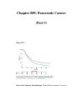
Physical Findings Patients with early disease may not have any significant abnormalities detectable on physical examination. Jaundice may be a presenting feature in some; in these patients a palpable, nontender gallbladder (Courvoisier's sign) may be palpated under the right costal margin. Patients with more advanced disease may have an abdominal mass, hepatomegaly, splenomegaly, or ascites. The left supraclavicular lymph node (Virchow's node) may be involved with tumor, or widespread peritoneal disease may be palpable on rectal examination in the pouch of Douglas. Diagnostic Procedures Imaging Studies (Fig. 89-1) Ultrasound is often used as an initial investigation for patients with jaundice, or with...
Bình luận(0) Đăng nhập để gửi bình luận!
CÓ THỂ BẠN MUỐN DOWNLOAD


Chịu trách nhiệm nội dung:
Nguyễn Công Hà - Giám đốc Công ty TNHH TÀI LIỆU TRỰC TUYẾN VI NA
LIÊN HỆ
Địa chỉ: P402, 54A Nơ Trang Long, Phường 14, Q.Bình Thạnh, TP.HCM
Hotline: 093 303 0098
Email: support@tailieu.vn
Giấy phép Mạng Xã Hội số: 670/GP-BTTTT cấp ngày 30/11/2015 Copyright © 2022-2032 TaiLieu.VN. All rights reserved.

