


Chapter 098. Iron Deficiency and Other Hypoproliferative Anemias (Part 4)
lượt xem 2
download
 Download
Vui lòng tải xuống để xem tài liệu đầy đủ
Download
Vui lòng tải xuống để xem tài liệu đầy đủ
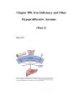
Stages of Iron Deficiency Iron-deficiency anemia is the condition in which there is anemia and clear evidence of iron lack. The progression to iron deficiency can be divided into three stages (Fig. 98-2). The first stage is negative iron balance, in which the demands for (or losses of) iron exceed the body's ability to absorb iron from the diet. This stage results from a number of physiologic mechanisms, including blood loss, pregnancy (in which the demands for red cell production by the fetus outstrip the mother's ability to provide iron), rapid growth spurts in the adolescent, or inadequate dietary...
Bình luận(0) Đăng nhập để gửi bình luận!
CÓ THỂ BẠN MUỐN DOWNLOAD


Chịu trách nhiệm nội dung:
Nguyễn Công Hà - Giám đốc Công ty TNHH TÀI LIỆU TRỰC TUYẾN VI NA
LIÊN HỆ
Địa chỉ: P402, 54A Nơ Trang Long, Phường 14, Q.Bình Thạnh, TP.HCM
Hotline: 093 303 0098
Email: support@tailieu.vn
Giấy phép Mạng Xã Hội số: 670/GP-BTTTT cấp ngày 30/11/2015 Copyright © 2022-2032 TaiLieu.VN. All rights reserved.

