


Summary of Chemistry doctoral thesis: Study on isolation and investigation of antibiotic and cytotoxic activity of secondary compounds from three actynomycetes strains Streptomyces G246, G261, G248 collected in some central East Vietnam sea
lượt xem 5
download
 Download
Vui lòng tải xuống để xem tài liệu đầy đủ
Download
Vui lòng tải xuống để xem tài liệu đầy đủ
Summary of Chemistry doctoral thesis: Study on isolation and investigation of antibiotic and cytotoxic activity of secondary compounds from three actynomycetes strains Streptomyces G246, G261, G248 collected in some central East Vietnam sea
Bình luận(0) Đăng nhập để gửi bình luận!
Nội dung Text: Summary of Chemistry doctoral thesis: Study on isolation and investigation of antibiotic and cytotoxic activity of secondary compounds from three actynomycetes strains Streptomyces G246, G261, G248 collected in some central East Vietnam sea
- MINISTRY OF EDUCATION AND VIETNAM ACADEMY TRAINING OF SCIENCE AND TECHNOLOGY GRADUATE UNIVERSITY OF SCIENCE AND TECHNOLOGY ---------------------------- CAO DUC DANH STUDY ON ISOLATION AND INVESTIGATION OF ANTIBIOTIC AND CYTOTOXIC ACTIVITY OF SECONDARY COMPOUNDS FROM THREE ACTINOMYCETES STRAINS STREPTOMYCES G246, G261, G248 COLLECTED IN SOME CENTRAL EAST VIETNAM SEA Major: Organic Chemistry Code: 9 44 01 14 SUMMARY OF CHEMISTRY DOCTORAL THESIS HA NOI- 2019
- This thesis was completed at: Graduate University Science and Technology - Vietnam Academy of Science and Technology Advisor 1: Prof. Dr. Habil. Pham Van Cuong Advisor 2: Dr. Tran Dang Thach 1st Reviewer: 2nd Reviewer: 3rd Reviewer: The thesis will be defended at Graduate University of Science and Technology - Vietnam Academy of Science and Technology, at hour … date … month … 2019 The thesis can be found in - The library of the Graduate University of Science and Technology, Vietnam Academy of Science and Technology. - The National Library of Vietnam.
- 1 INTRODUCTION 1. The urgency of the thesis The ocean accounts for 70% of the earth's surface, where there is the largest biological diversity on earth. It is home to 34 of the 36 animal and plant sectors on earth with more than 300,000 known species. The marine environment has been known as a rich source of natural compounds, such as a huge pharmaceutical warehouse awaiting exploration and discovery. Characteristics of harsh deep sea habitats are the conditions for forming organic compounds with unique chemical structure characteristics and precious biological activity. The study of secondary active ingredients produced from marine microorganisms in the world has achieved many remarkable achievements. Among them are many secondary compounds with interesting chemical structure and biological activity. At the same time many of these compounds are being tested further for medical applications. Vietnam is located in the Pacific region (with sovereignty with an area of about 1,000,000 km2) with a rich and diverse marine system, with great potential for marine resources. The Government of Vietnam has oriented the development of marine economy, exploitation of natural resources and the study of natural products from the sea. However, the study of secondary compounds from Vietnam's marine microbial source has only just begun, there are very few published studies, although the source of marine diversity of our country is very large. Infectious diseases account for the majority of human and animal diseases. Around the second half of the 19th century, it was discovered that microorganisms are the cause of infectious diseases. Therefore, chemotherapy aims at pathogenic microorganisms that have been developed into the main therapy. In 1928, Alexander Fleming discovered Penicillin - a powerful antibiotic compound and purified by Abraham, Chain, and Florey in a stable form that had a therapeutic effect in 1941. Penicillin antibiotics became famous because It saved many lives in World War II. Since then, antibiotics have become an early miracle pharmaceutical that occupies the world's leading position in the pharmaceutical field, with increasingly new results, with increasing demand and increasing production volume. Currently, there are more antibiotics that are extracted from fungi, bacteria, actinomycetes, which occupy most of them with marine actynomycetes. But more and more microorganisms cause disease resistance to existing antibiotics. Therefore, it is necessary to continue to research, find and discover new antibiotics, active ingredients with tuberculosis and anti-cancer resistance. Therefore, we carried out this thesis with the title: “Study on isolation and investigation of antibiotic and cytotoxic activity of secondary compounds from three actynomycetes strains Streptomyces G246, G261, G248 collected in some central East Vietnam sea”
- 2 2. The aim of the thesis - Study to isolate and determine the structure of secondary compounds from cultivation of 3 strains of microorganisms isolated from central East Viet Nam sea. - Survey of cytotoxic activity and testing antimicrobial activity of isolated substances as a scientific basis for the research and application of these compounds. 3. The main contents of the thesis - Review of previous studies on secondary compounds as well as biological activity from marine microbial strains. - Finding procedures for handling culture fluid to create extracts. Purify these extracts on column chromatography to obtain fractions. - Refining substances in the segments to obtain compounds. - Determine the chemical structure of isolated compounds. - Testing antimicrobial activity to test isolated substances. - Testing anticancer activity in vitro for some cancer cell lines such as KB, MCF-7, Hep-G2, Lu-1 of compounds isolated. CHAPTER 1. OVERVIEW 1.1. The distribution and diversity of marine microorganisms 1.2. Bacterial radiation and the formation of secondary compounds from actinomycetes 1.3. Common marine bacteriophage families 1.4. Secondary compounds with biological activity from marine bacteriophages 1.4.1. Finding in the world 1.4.1.1. The compounds have antibiotic activity 1.4.1.2. The compounds are active against tuberculosis 1.4.1.3. The compounds with cytotoxic activity 1.4.2. Finding in our country CHAPTER 2. MATERIALS AND METHODS 2.1. Materials and research equipment 2.1.1. Material 2.1.3. Device 2.2. Research Methods 2.2.1. Sample collection method 2.2.2. Marine bacteriophage isolation method 2.2.3. Methods for cleaning strains with loop implants 2.2.4. Method to keep the actinomycetes after isolation
- 3 2.2.5. Activation and culture methods 2.2.6. Method of extraction from cultured fluid for biological activity screening 2.2.7. Method of identifying bacteriophages 2.2.8. Mass birth method 2.2.9. Methods of isolation of secondary compounds. 2.2.10. Methods for determining the chemical structure of clean compounds can be extracted 2.2.11. Method of testing antibiotic antimicrobial activity 2.2.12. Test method of cytotoxic activity 2.2.13. Test method for anti-tuberculosis activity. CHAPTER 3. EXPERIMENT AND RESULTS 3.1. Sample collection results 25 samples have been collected from central East Vietnam sea, namely: - In Thanh Hoa sea area, 2 sediment samples were collected. - In Quang Binh sea area, 2 sediment samples, 1 sea form, 2 seaweed samples and 1 coral sample were collected. In the Quang Tri sea area, there were 2 sediment samples, 1 sea pattern, 1 seaweed sample. - In the water of Son Tra peninsula, Da Nang obtained 1 sample of sediment, 2 samples of seaweed, 1 sample of seaweed, 1 sample of thorny skin, 1 sample of bag. - In Cu Lao Cham coastal area, Quang Nam obtained 3 sediment samples, 1 model of thorny skin, 1 soft body sample, 1 sea rabbit model, 1 solid tail pattern. 3.2. Result of isolation of actinomycete strains - From 25 samples collected in the central waters of Vietnam, 32 bacteria strains with different morphology and colors were isolated. - From 12 samples collected in the waters of Thanh Hoa, Quang Binh and Quang Tri, 12 bacteria strains were isolated. - From 13 samples collected in Da Nang and Quang Nam waters, 20 bacteria strains were isolated. 3.3. Results of bioactivity test of actinomycete strains Antiretroviral resistance results: 29/32 active strains (see details in table 3.1). Results of anti-tuberculosis screening were performed at UIC University for Mycobacterium tuberculosis H37Rv strain. The results showed that 21/32 strains showed anti- tuberculosis activity, in which 1 strain showed very good activity with> 90% inhibitory value, 7 strains showed good activity with inhibitory%> 50% (see details in table 3.2).
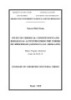
- 4 Coarse residues of cultured extracts of 32 studied strains were tested for cytotoxic activity against human cancer cell line (KB - carcinoma (CCL-17). Toxic cancer of KB epithelial cells (see details in Table 3.3) 3.4. Results of identification of bioactive antibiotic strains 3.4.1. Observe morphological characteristics of studied strains 3.4.2. Multiplication of 16S RNA riboxom gene 3.4.3. Genome sequencing The nucleotide sequence of the 16S ARN riboxom gene section of strains G246, G261, G248 showed that the studied strains belong to genus Streptomyces sp. (see PLIII.1.4) 3.5. The results of high biomass of strains have good biological activity. The strains of G246, G261, G248 have good biological activity, they are fermented in large quantities of 50 liters in suitable conditions to conduct further studies. 3.6. Extraction and chemical structure of secondary compounds from Streptomyces sp. G246 3.6.1. Sample processing, creating a residue 3.6.2. Isolate substances from the extract Figure 3. 1. Diagram of isolation of Streptomyces sp. G246
- 5 3.6.3. Physical parameters and spectral data of compounds isolated from Streptomyces sp. G246 3.7. Extraction and chemical structure of secondary compounds from Streptomyces sp. G261 3.7.1. Sample processing, creating a residue 3.7.2. Isolate from Streptomyces sp. G261 Figure 3. 2. Diagram of isolation of Streptomyces sp. G261 3.7.3 Physical parameters and spectral data of secondary compounds from the Streptomyces sp. G261 3.8. Extraction and chemical structure of secondary compounds from Streptomyces sp. G248 3.8.1. Sample processing, creating a residue 3.8.2. Isolate substances from the extract 3.8.3. Physical parameters and spectral data of secondary compounds from the Streptomyces sp. G248
- 6 Figure 3. 3. Diagram of isolation of Streptomyces sp. G248 3.9. Results of bioactivity test of compounds isolated from actinomycete strains 3.9.1. Performance test results for test microorganisms of some isolated compounds Of the 26 substances isolated, we have tested antimicrobial activity. As a result, there are 13 substances with antimicrobial antibiotic activity, many of which have very strong activity such as G246-2, G248-11, G248-12, G248-9 and many substances with good antifungal activity as G246-6, G261-11, G248-11 (see details in table 3.5). 3.9.2. The results of anti-tuberculosis activity test of some isolated compounds The results of tuberculosis resistance testing were performed at UIC University. Some substances tested for anti-tuberculosis activity with Mycobacterium tuberculosis H37Rv, in which G246-1 exhibited the best activity with MIC value of 6.00 µg / ml, G248-10 showed
- 7 good activity with MIC value = 11.17 µg / ml, while G248-9 showed weak activity with MIC value of 48.02 µg/ ml The remaining substances did not show activity at the study concentration (50 µg/ ml) (see details table 3.6). 3.9.3. Test results of cytotoxic activity of some isolated compounds 14 compounds with tested antimicrobial activity, tuberculosis resistance from three research strains (G246, G261 and G248) were tested for cytotoxic activity with four cancer cell lines in: KB - Schedule cancer tissue (CCL - 17TM); Hep G2 - liver cancer (HB - 8065TM); MCF-7 - breast cancer (HTB - 22TM) and LU-1 - lung cancer (HTB-57TM). The results are detailed in Table 3.5. The result has 5 compounds with cytotoxic activity, including 4 active substances with KB cell line, 5 active compounds with liver cancer cell line Hep-G2, 4 active compounds calculated with Lu-1 cell lines, 4 active compounds with MCF7 cell lines (see details in table 3.7) CHAPTER 4. DISCUSSION OF RESULTS 4.1. Sample collection results A total of 25 samples included: 12 samples in the waters of Thanh Hoa, Quang Binh and Quang Tri; 13 samples in Da Nang and Quang Nam waters. Specifically, there are 10 samples of sediment, 4 samples of seaweed, 4 samples of seaweed, 1 sample of soft corals, 2 samples of echinoderms and 4 other samples. 4.2. Result of isolation of actinomycete strains From 25 samples, 32 isolates with different morphology and colony color were isolated from the collected sample (details are described in Appendix PL III.1.2). 4.3. Results of bioactivity test of strains obtained. - The results of the test of antimicrobial activity with crude scale showed that 29/32 isolated strains are resistant to the strains of test microorganisms. - The results showed that 21/32 strains exhibited anti-tuberculosis activity. - The results showed that 20 strains had toxic activity of KB epithelial cancer cells (IC50
- 8 Identified the actinomycetes strains G246, G261, G248 all belong to genus Streptomyces sp. (See details at PLIII.1.4) 4.5. The results of high biomass of strains have good biological activity. Successful fermentation of 50 liters for strains G246, G261, G248 to continue the next study. 4.6. Chemical structure of secondary compounds from Streptomyces sp. G246 From the culture fluid of the microorganism strain Streptomyces sp. G246, we isolated and determined the chemical structure of 10 compounds, including 2 new compounds G246-1 and G246-2. The structure of the compounds was determined by NMR, MS spectroscopy methods and compared with reference materials, the compounds were identified as (2S, 2 ″ S) - 6-lavandulyl-7-methoxy-5 , 2 ′, 4′-trihydroxylflavanone (G246-1), (2 ″ S) -5′-lavandulyl-4′- methoxy-2,4,2 ′, 6′-tetrahydroxylchalcone (G246-2), cyclo- ( Pro-Gly (G246-3), cyclo- (L-Pro- L-Tyr) (G246-4), cyclo- (D-Pro-L-Tyr) (G246-5), cyclo- (Pro-Ala) ) (G246-6), norharman (G246-7), tryptophan (G246-8), 3-indol carboxylic acid (G246-9), phenyl alanine (G246-10), including 2 new substances G246-1 and G246-2. The absolute configuration of the two new compounds G246-1 and G246-2 is determined based on the method of calculating quantum circular electron chemistry (ECD) based on Gaussian 09 software. Figure 4.28. Chemical structure of compound G246-1 to G246-10 4.6.1. Compound G246-1: (2S,2″S) -6-lavandulyl-7-methoxy-5,2′,4′-trihydroxylflavanone (new compound) Compound 1 was isolated as an amorphous solid, and optically active [a]25D -28(c, 0.8, MeOH). Its positive HR-ESI mass spectrum showed the proton adduct ion [M+H]+ at m/z 13 439.2115 (calcd. for C26H31O6, 439.2121) which, together with C-NMR data, are consistent
- 9 with the molecular formula of C26H30O6. The IR spectrum showed the presence of hydroxyl (3299 cm-1) and carbonyl (1670 cm-1) functionalities. In the 1H-NMR spectrum of 1, the presence of an ABX system at δH 6,35 (1H, d, J = 2.0 Hz, H-3′), 6,37 (1H, dd, J = 2.0, 8.5 Hz, H-5′), 7.31 (1H, d, J = 8.5 Hz, H-6′), and a singlet proton at δH 6.13 was observed in the aromatic region. Additionally, the resonances of protons at dH 5.56, 4.98, 4.54 and 4.59, a methoxy group at dH 3.84, three singlet methyls at δH 13 1.49, 1.58 and 1.65, and a number of aliphatic protons were noted. Analysis of the C-NMR data with the aid of HSQC experiment revealed 26 carbon resonances for G246-1, including one ketone group (δC 193.9), ten sp2 quaternary carbons, five sp2 methines, one sp2 methylene, two sp3 methines, three sp2 methylenes, three methyls and one methoxy group. Beside the aromatic ABX system, two other spin-spin coupling systems were revealed from the COSY spectrum: H-2/CH2-3 (I) and CH2-1”/H-2”/CH2-3”/H-4” (II)). The chemical shifts of carbons at δC 75.5 (C-2), 156.6 (C-20), 159.5 (C-400), 164.9 (C-5), 161.9 (C-7) and 164.9 (C-9) suggested their linkage to oxygen (Table 4.1). In the HMBC spectrum, cross-peaks of C-200 (δC 48.2) with CH3-10” (δH 1.65) and CH2-9” (δH 4.54), and those of C-8” (δC 149.8) with protons of CH2-1” (δH 2.64) indicated the linkage of C-2” of the coupling system II with the isopropenyl group. Furthermore, the HMBC correlations of H-4” (δH 4.98) with C-6” (δC 17.8) and C-7” (δC 25.9), and CH2-3” (δH 2.02) with C-5” (δC 132.0) indicated the lavandulyl group in the structure of G246-1. The presence of the B-ring was confirmed by cross-peaks of H-3’ (δH 6.35) with C-1’ (δC 118.5), C-2’ (δC 156.6), C-4’ (δC 159.5) and C-5’(δC 107.6). Similarly, the A-ring was established by HMBC correlations of H-8 (δH 6.13) with C-6 (δC 109.6), C-7 (δC 161.9), C-9 (δC 164.9) and C-10 (δC 105.7) (Figure 4.1). Figure 4.1. Chemical structure and interactions HMBC (H → C), COSY (H─H) of compound G246-1 and chemical structure of reference compounds. The spectral features of coupling systems I and the carbonyl group deduced from HMBC signals indicated structural similarity of G246-1 to flavanone compounds. The connection of
- 10 the lavandulyl group to the A-ring at C-6 via C-6/C-1” linkage was revealed by HMBC cross- peaks of the protons of CH2-1” with C-5, C-6 and C-7. Table 4. 1. NMR spectral data of compound G246-1 and reference compound C C#,b Ca H (multi, J, Hz) 2 75,3 75,5 5,56 (dd, 2,5; 13,0) 3 42,8 48,2 2,89 (dd, 13,0; 16,5) 2,71 (dd, 2,5; 16,5) 4 198,2 193,9 5 163,0 164,9 6 96,2 109,6 7 165,3 161,9 8 107,8 93,4 6,13 (s) 9 162,1 164,9 10 103,2 105,7 1′ 117,9 118,5 2′ 156,1 156,6 3′ 103,4 103,4 6,35 (d, 2,0) 4′ 159,4 159,5 5′ 107,8 107,6 6,37 (dd, 2,0; 8,5) 6′ 128,6 128,5 7,31 (d, 8,5) 1′′ 27,7 28,2 2,64 (m) 2′′ 47,8 48,2 2,52 (m) 3′′ 31,9 32,4 2,02 (m) 4′′ 124,5 124,8 4,98 (t, 6,5) 5′′ 131,6 132,0 6′′ 17,9 17,8 1,97 (s) 7′′ 25,8 25,9 1,98 (s) 8′′ 149,1 149,8 9′′ 111,2 111,2 4,54 (s) 4,59 (s) 10′′ 19,1 19,2 1,65 (s) OMe 55,9 3,84 (s) a) measured in CD3OD; b) measured in CD3COCD3; δC # data of sophoraflavanone G compound [89]
- 11 Finally, the methoxy group at δH 3.85 was attached to C-7 as indicated by their correlation in the HMBC spectrum. Complete analyses of 2D-NMR spectra established the structure of G246-1 as 6-lavandulyl-7-methoxy-5,2’,4’-trihydroxylflavanone. Despite extensive effort, attempts to produce suitable crystals of 1 and 2 for X-ray diffraction analysis were unsuccessful. Alternatively, the absolute configurations of G246-1 were resolved by comparison of their experimental and calculated electronic circular dichroism (ECD) spectra. The ECD quantum chemical calculations were performed using the Gaussian 09 software. To obtain minimum energy conformers, geometry optimization of each possible isomer of these compounds was conducted. The calculated ECD spectra of compound G246-1 were generated using the time-dependent density functional method at the B3LYP/6-31++G(d,p) level. Since, the CD spectrum of compound G246-1 displayed a positive Cotton effect at 336nm (Δ +2.98 mdeg) and a negative Cotton effect at 292nm (Δ -9.08 mdeg), the S configuration was thus assigned for carbon C-2 of G246-1 [89]. To determine the absolute configuration of C-2” of G246-1, the ECD spectra of the two isomers, 2S,2”S-1 and 2S,2”R-G246-1, were performed in gas phase. The experimental CD spectrum of G246-1 showed excellent agreement with the calculated ECD of 2S,2”S-G246-1 (Figure 4.1). Thus, the S-configuration was suggested for both C-2 and C-2” of compound G246-1. 4.6.2. Compound G246-2: (2″S)-5′-lavandulyl-4′-methoxy-2,4,2′,6′-tetrahydroxylchalcone (new compound) Figure 4.2. Chemical structure and interactions HMBC (H → C), COSY (H─H) of compound G246-2 and chemical structure of reference compounds. Compound G246-2 was isolated as an optically active [a]25D -8.6 (c, 0.25, CH2Cl2). Its HR-ESI-MS showed the proton adduct ion [M+H]+ at m/z 439.2115 (calcd. For C26H31O6, 13 439.2121). A long with the C-NMR data, a molecular formula of C9H8O4 was suggested for G246-2. Comparison of the 1D-NMR spectra with those of G246-1 revealed the same substructures, lavandulyl group, A- and B-ring systems for compound G246-2. The differences were noted for the signals of the CH=CH system [characterized from protons at H 7.82 (d, J =
- 12 16.0 Hz, H-a) and 7.96 (d, J = 16.0 Hz, H-b)] of G246-2 instead of the resonances of the coupling system CH-2/CH2-3 in the structure of G246-1. Table 4. 1. NMR spectral data of compound G246-2 and reference compound C# C#,b C Ca H (multi, J, Hz) 1′ 118,5 1 116,5 2′ 156,6 2 156,9 3′ 103,4 3 106,2 6,37 (br,s) 4′ 159.5 4 159,1 5′ 107,6 5 109,1 6,43 (br d, 8,0) 6′ 128,5 6 131,0 7,39 (d, 8,0) 10 105,7 1′ 106,2 9 164,9 2′ 161,0 8 93,4 3′ 90,9 5,92 (s) 7 161,9 4′ 161,0 6 109,6 5′ 107,4 5 164,9 6′ 165,8 1′′ 28,2 1′′ 29,7 2,70 (dd, 6,0; 14,0) 2,71 (dd, 8,0; 14,0) 2′′ 48,2 2′′ 46,5 2,39 (m) 3′′ 32,4 3′′ 31,9 2,14 (m) 4′′ 124,8 4′′ 122,8 5,11 (t, 6,5) 5′′ 132,0 5′′ 132,9 6′′ 17,8 6′′ 18,0 1,60 (s) 7′′ 25,9 7′′ 25,8 1,69 (s) 8′′ 149,8 8′′ 150,5 9′′ 111,2 9′′ 110,7 4,76 (s)/ 4,79 (s) 10′′ 19,2 10′′ 20,7 1,72 (s) OH-6′ 14,46 (s) OMe 55,9 OMe 55,7 3,85 (s) 3 48,2 α 126,0 7,82 (d, 16,0) 2 75,5 β 137,5 7,96 (d, 16,0) 4 193,9 γ 193,2 a) measured in CDCl3; b) measured in CD3OD; δC# data of G246-1 compound
- 13 This observation suggested a chalcone skeleton for G246-2 which was confirmed by cross-peaks of H-b with C-2 (C 156.9), C-6 (dC 131.0) and C- γ (C 193.2) in the HMBC spectrum of G246-2. The location of the lavandulyl at C-5’ was indicated by the HMBC correlations of CH2-1” (H 2.70 and 2.71) with C-5’ (C 164.9) and C-6’ (C 109.6), and H-2” (H 2.39) with C-6’ (C 109.6) (Figure 4.2). Similarly, HMBC cross-peaks of C-4’ (C 161.0) with the protons of the methoxy at H 3.85 assigned the connection of C-4’ with the methoxy group. Since, a strong coupling constant (J = 16.0 Hz) was noted for H-a and H-b, a trans- configuration was thus assigned for CH-a/CH-b double bond. Complete analyses of the 2D- NMR spectra identified the structure of H246-2 as 5’-lavandulyl-4’-methoxy-2,4,2’,6’- tetrahydroxylchalcone. Despite extensive effort, attempts to produce suitable crystals of G246-2 for X-ray diffraction analysis were unsuccessful. Alternatively, the absolute configurations of G246-2 were resolved by comparison of their experimental and calculated electronic circular dichroism (ECD) spectra. The ECD quantum chemical calculations were performed using the Gaussian 09 software. To obtain minimum energy conformers, geometry optimization of each possible isomer of these compounds was conducted. The calculated ECD spectra of compound G246-2 were generated using the time-dependent density functional method at the B3LYP/6- 31++G(d,p) level. Similarly, the S-configuration was also assigned for compound G246-2 by comparison of its experimental CD spectrum with the calculated ECD spectra (in gas phase) of S- and R-configuration of G246-2. 4.7. Chemical structure of secondary compounds from Streptomyces sp. G261 From the culture fluid of the microorganism strain Streptomyces sp. G261 isolated and determined the chemical structure of 13 compounds: norharman (G261-1), 2,3-butanediol (G261-2), 1H-pyrrole-2-carboxylic acid (G261-3), 2- oxo-2,3-dihydrobenzo [d] oxazole-4- carboxylic acid (G261-4), 3-hydroxy-4-methoxybenzoic acid (G261-5), 2-acetamidobenzamide (G261-6), phenyl alanine (G261-7), cyclo-(Pro-Gly) (G261-8), cyclo- (Pro-Ala) (G261-9), cylo-(Pro-Leu) (G261-10), cyclo-(Pro-Tyr) (G261-11), cyclo-trans-4-OH-(Pro-Phe) (G261- 12), cyclo-(Leu-Tyr) (G261-13), in which 1 substance was first isolated from nature are 2-oxo- 2,3-dihydrobenzo [d] oxazole-4-carboxylic acid (G261-4). Below is a detailed description of the method of determining the structure of compounds G261-4 which is the first compound isolated from nature.
- 14 Figure 4. 2. Chemical structure of compound G261-1 to G261-13 4.7.1. Compound G261-4: 2-oxo-2,3-dihydrobenzo [d] oxazole-4-carboxylic acid Figure 4.32. Chemical structure and interactions on HMBC spectrum of G261-4 ESI-HRMS mass spectra of G261-4 for the molecular mimic ion peak at m/z 178,0152 [M-H]- (theoretical calculation for molecular formula C8H4NO4 m/z 178,0140). 1H-NMR spectra show signal appearance of aromatic ring protons in δH 7.24 (1H, d, J = 8.0 Hz, H-7); 7.23 (1H, dd, J = 8.0; 8.0 Hz, H-6); 7.49 (1H, dd, J = 2.0; 7.0 Hz, H-5). 13C-NMR spectrum in combination with HSQC showed 3 aromatic methine groups at δC 118.1 (C-5); 123.6 (C-7); 126.4 (C-6); and 3 carbon quaternary sp2 at δC 116.7 (C-4); 144,2 (C-3); 146.5 (C-8); In addition, the presence of a cabonyl group in the low-field region associated with nitrogen allergens at δC 149,2 and a carbonyl group at δC 163.4 was also observed. In HMBC spectrum, there is a long-term interaction of proton H-5 (δH 7.49) with C-4 (δC 116.7); C-3 (δC 144.2) and C = O (δC 163.4) interaction of protons H-7 (δH 7.24) with C-8 ((C 146,5), C-6 (δC 123, 6). Combining spectral data and comparing references [102] allowed the determination of G261-4 as 2-oxo-2,3-dihydrobenzo [d] oxazole-4-carboxylic acid. This is the first compound isolated from nature [103].
- 15 4.8. Chemical structure of secondary compounds from Streptomyces sp. G248 From the cultured extract of the Streptomyces sp. G248, by chromatography and spectroscopic methods MS, 1D-NMR, 2D-NMR, isolated and determined the chemical structure of 13 symbol compounds from G248-1 to G248-13 including cyclo-(Pro-Leu) (G248- 1), cyclo-(Pro-Phe) (G248-2), norharman (G248-3), cyclo-(Pro-Tyr) (G248-4), cyclo-(Pro-Gly) (G248-5), cyclo-(Pro-Trp) (G248-6), Adenine (G248-7), 2-(4-hydroxyphenyl) acetic acid (G248-8), (2S,2″S)-6-lavandulyl-7,4′-dimethoxy-5,2′-dihydroxylflavanone (G248-9), (2S) -6- prenyl-4′-methoxy-5,7-dihydroxylflavanone G248-10, (2S,2″S)-6-lavandulyl-5,7,2′,4′- tetrahydroxylflavanone(G248-11), (2″S) -5′-lavandulyl-2′-methoxy-2,4,4′,6′- tetrahydroxylchalcone (G248-12), (2S,2″S)-6-lavandulyl-7-methoxy-5,2′,4′- trihydroxylflavanone (G248-13). Including 3 new compounds are G248-9, G248-11, G248-12 of flavonoids compound. Figure 4.81. Chemical structure of compound G248-1 to G248-13 4.8.1. Compound G248-9: (2S,2″S)-6-lavandulyl-7,4′-dimethoxy-5,2′-dihydroxylflavanone Figure 4. 3. Chemical structure and interactions HMBC (H → C), COSY (H─H) of G248-9 compound and chemical reference structure.
- 16 Compound G248-9 was isolated as an amorphous solid, and optically active [α]25D = - 5 (c 0.176, MeOH). Its positive HR-ESI mass spectrum showed the proton adduct ion [M+H]+ at m/z 453.2270 (calcd for C27H33O6, 453.2277) which, together with 13C NMR data, is consistent with the molecular formula of C27H31O6. The IR spectrum indicated the presence of hydroxyl groups at 3366 cm-1, and a carbonyl functionality at 1696 cm-1. In the 1H-NMR spectrum, the presence of an ABX system at δH 6.47 (d, J = 2.5 Hz, H-3’), 6.47 (dd, J = 2.0, 8.5 Hz, H-5’) and 7.38 (d, J = 8.5 Hz, H-6’), and a singlet proton at δH 6.12 (s, H-8) was observed at the aromatic region. Additionally, the resonances of protons at H 5.55, 4.96, 4.52 and 4.59, two methoxy groups at H 3.82 and 3.83, three singlet methyls at H 1.48, 1.57 and 1.64, and a number of aliphatic protons were noted. Analysis of 13C-NMR spectrum with the aid of HSQC experiment revealed 27 carbon resonances for G248-9, including one ketone group (C 193.6), three methyls, one sp2 methylene, three sp2 methylenes, five sp2 methines, two sp3 methines, two methoxy groups and ten sp2 quaternary carbons (Table 1). Beside the aromatic ABX system, two other spin-spin coupling systems were revealed from the COSY spectrum: H- 2/CH2-3 (I), and CH2-1’’/H-2’’/CH2-3’’/H-4’’ (II). The chemical shifts of carbons at C 75.0 (C-2), 160.1 (C-2’), 159.0 (C-4’’), 164.7 (C-5), 161.9 (C-7) and 165.4 (C-9) suggested their linkage to oxygen. In the HMBC spectrum, cross-peaks of C-2’’ with CH3-10’’ and CH2-9’’, and those of C-8’’ with protons of CH2-1’’ indicated the linkage of C-2’’ of the coupling system II with the isopropenyl group. Additionally, HMBC correlations of H-4’’ with C-6’’ and C-7’’, and CH2-3’’ with C-5’’ indicated the lavandulyl group in the structure of G248-9. The presence of the A-ring was confirmed by cross-peaks of H-8 with C-6, C-7, C-9 and C-10. Similarly, the B-ring was established by HMBC correlations of H-3’ with C-1’, C-2’, C-4’ and C-5’. The spectral features of coupling systems I and the carbonyl group deduced from HMBC signals indicated structural similarity of G248-9 to flavanone compounds. Furthermore, the connection of the lavandulyl group to the A-ring at C-6 via C-6/C-1’’ linkage was revealed by HMBC cross-peaks of the protons of CH2-1’’ with C-5, C-6 and C-7. Finally, two methoxy group at H 3.82 and 3.83 was attached to C-7 and C-4’ as indicated by their correlation in the HMBC spectrum (Figure 2). Complete analyses of 2D-NMR spectra established the planar structure of G248-9 as 6-lavandulyl-7,4’-dimethoxy-5,2’-dihydroxylflavanone. The G248-9 CD spectrum gives a positive Cotton effect at 336 nm (Δ +2.7) and a negative Cotton effect at 292 nm (Δ - 8.5), allowing for absolute configuration at C- 2 is 2S [89]. The absolute configuration at the C-2 ″ position of G248-9 is determined based on the method of calculating the quantum electron quantum chemistry (ECD - Electronic Circular Dichroism) based on the Gaussian 09 software. The theory for 2S,2″S- and 2S,2″R- allows the
- 17 determination of 2S, 2″S configurations of G248-9 compound in accordance with the calculation model (Figure 4. 66). Table 4.3. NMR spectral data of compounds G248-9 and compounds refer to G246-1 C C#,a Ca H (multi, J, Hz) 2 75,5 75,0 5,55 (dd, 2,5; 13,0) 3 48,2 45,4 2,82 (dd, 6,0; 17,0) 2,90 (dd, 13,0; 17,0) 4 193,9 198,8 5 164,9 164,7 6 109,6 109,6 7 161,9 161,9 8 93,4 93,6 6,13 (s) 9 164,9 165,2 10 105,7 105,6 1′ 118,5 119,7 2′ 156,6 160,0 3′ 103,4 99,8 6,49 (d, 2,0) 4′ 159,5 159,0 5′ 107,6 108,1 6,47 (dd, 2,0; 8,0) 6′ 128,5 128,5 7,39 (d, 8,5) 1′′ 28,2 28,2 2,61 (m) 2′′ 48,2 48,2 2,50 (m) 3′′ 32,4 32,4 2,03 m 4′′ 124,8 124,8 4,97 (t, 6,5) 5′′ 132,0 132,0 6′′ 17,8 17,8 1,48 (s) 7′′ 25,9 25,8 1,58 (s) 8′′ 149,8 149,8 9′′ 111,2 111,2 4,53 (m)/ 4,58 (m) 10′′ 19,2 19,1 1,65 (s) OMe 55,9 55,9 3,83 (s) OMe 55,9 3,82 (s) a) measured in CD3OD; δC# data of G246-1 compound
- 18 From the analysis on HR-ESI-MS, IR, 1D and 2D NMR spectra, CD and ECD spectrum has been established, the structure of G248-9 is (2S,2″S)-6-lavandulyl-7,4′ -dimethoxy-5.2′- dihydroxylflavanone. Finding on the Scifinder database allows to conclude that this is a new compound. 4.8.2. Compound G248-12: (2″S)-5′-lavandulyl-2′-methoxy-2,4,4′,6′-tetrahydroxylchalcone Figure 4. 4. Chemical structure and interactions HMBC (H → C), COSY (H─H) of G248-12 compound and chemical reference structure. Compound G248-12 was isolated as an amorphous solid, with negative optical rotation [α]25D -1.8 (c 0.54, CH2Cl2) . It’s positive HR-ESI-MS showed the proton adduct ion [M+H]+ at 13 m/z 439.2117 (calcd for C26H31O6, 439.2121). Considering the C-NMR data, a molecular formula of C26H30O6 was suggested for G248-11. Comparison of the 1D NMR spectra with those of G248-9 revealed the same substructures, l A- and B-ring systems, avandulyl group for compound G248-12. The differences were noted for the signals of the CH=CH system instead of the resonances of the coupling system CH-2/CH2-3. This observation suggested a chalcone skeleton for G248-12 which was confirmed by cross-peaks of H- with C-2, C-6 and C-γ in the HMBC spectrum of G248-12. The location of the lavandulyl at C-5’ was indicated by the HMBC correlations of H-1’’ with C-5’ (δC 108.9) and C-6’ (δC 166.6), and H-2″ with C-6’ (δC 166.6) (Figure 2). Similarly, HMBC cross-peaks of C-2′ (δC 162.3) with the protons of the methoxy at H 3.91 assigned the connection of C-2’ with the methoxy group. Complete analyses of 2D-NMR spectra established the structure of G248-12 as 5’-lavandulyl-2′- methoxy-2,4,2’,6’-tetrahydroxylchalcone. This compound has the same planar structure of kuraridin, which was previously isolated from Albizzia julibrissin [6]. However, the two compounds have different rotational degrees, so in terms of stereoscopic structure of the compound G248-12 is different from kuraridin (with polarity [α]25D −25.5 (c 0.1, MeOH). Absolutely at position C-2″ of G248-12 is determined based on the method of calculating quantum electron quantum chemistry (ECD - Electronic Circular Dichroism) based on software Gaussian 09. Theoretical calculation for 2 isomers 2″S- and 2″R- allow to determine the configuration 2″S of the compound G248-12 in accordance with the calculation model (Figure
CÓ THỂ BẠN MUỐN DOWNLOAD

-

Summary of chemistry doctoral thesis: Study on synthesis, characteristics, and adsorption properties of toxic organic substances in the water environment of mesoporous carbon materials
 28 p |
  43
|
43
|  7
7
-

Summary of chemistry doctoral thesis: Study on the fabrication of magnetic fluids based on superparamagnetic iron oxide nanoparticles (SPIONs) applied to magentic resonance imaging (MRI) application
28 p | 47
| 5
-

Summary of chemistry doctoral thesis: Study on chemical constituents and biological activities of Tacca vietnmensis and Tacca chantrierispecies growing in Vietnam
27 p | 50
| 5
-

Summary of Chemistry Doctoral thesis: Study on chemical constituents and cytotoxic activities of Glochidion Glomerulatum and Glochidion Hirsutum growing in study on chemical constituents and cytotoxic activities of glochidion glomerulatum and glochidion hirsutum growing in Vietnam
27 p | 54
| 5
-

Summary of the Doctoral Thesis in Philosophy of Education: Develop the competence of teaching chemical experiment for pedagogical students through the module “The practical experiment about teaching chemistry method in high school
27 p | 49
| 5
-

Summary of Organic Chemistry Doctoral Thesis: Study on fabrication nano Platinum modified glassy carbon electrode for application to analyze lead, cadmium in the water environment
28 p | 32
| 4
-

Summary of Chemistry Doctoral Thesis: Study on antimicrobial secondary metabolites isolated from selected marine-derived actinobacteria strains belong to Streptomyces genus
26 p | 31
| 4
-

Summary of Chemistry doctoral thesis: Study on chemical constituents and biological activities of several Antidesma species growing in Vietnam
27 p | 33
| 4
-

Summary of Chemical doctoral thesis: Study on fabrication nano Platinum modified glassy carbon electrode for application to analyze lead, cadmium in the water environment
28 p | 35
| 4
-

Summary of Chemistry doctoral thesis: Study on chemical constituents and biological activities of Vitex limonifolia Wall. Ex C.B.Clarke and Vitex trifolia L.
27 p | 35
| 4
-

Summary of Chemistry Doctoral thesis: Study on chemical constituents and biological activities from Culcita Novaeguineae Müller & Troschel, 1842 and Pentaceraster Gracilis (Lutken, 1871) in Viet Nam
28 p | 28
| 4
-
Summary of chemistry doctoral thesis: Study on chemical constituents and inhibitory activities of enzymes α-glucosidase and α amylase of Gymnema sylvestre (RETZ.) R.BR. EX SM. and Gymnema latifolium wall. EX wight
27 p | 22
| 4
-

Summary of Chemistry doctoral thesis: The accumulation, elimination and effect of heavy metals (As, Cd, Pb) on cortisol levels in Oreochromis sp.
27 p | 38
| 3
-
Summary of Chemical doctoral thesis: Study on enhancement of technical characteristics for some composite rubbers with nano additive
27 p | 32
| 3
-
Summary Of Chemistry Doctoral Thesis: Study on chemical constituents and biological activities from the tubers of Ophiopogon Japonicus (L.F.) KER-GAWL
27 p | 45
| 3
-

Summary of chemistry doctoral thesis: Study on chemical constituents and biological activities from the leaves of Excoecaria agallocha L. and Excoecaria cochinchinensis Lour.
27 p | 34
| 3
-

Summary of Biology Doctoral thesis: Study on chemistry and biological activity of some compounds from marine fungi isolated in central Vietnam
27 p | 44
| 2


Chịu trách nhiệm nội dung:
Nguyễn Công Hà - Giám đốc Công ty TNHH TÀI LIỆU TRỰC TUYẾN VI NA
LIÊN HỆ
Địa chỉ: P402, 54A Nơ Trang Long, Phường 14, Q.Bình Thạnh, TP.HCM
Hotline: 093 303 0098
Email: support@tailieu.vn
Giấy phép Mạng Xã Hội số: 670/GP-BTTTT cấp ngày 30/11/2015 Copyright © 2022-2032 TaiLieu.VN. All rights reserved.

