


báo cáo hóa học:" Highly efficient transduction of human plasmacytoid dendritic cells without phenotypic and functional maturation"
lượt xem 5
download
 Download
Vui lòng tải xuống để xem tài liệu đầy đủ
Download
Vui lòng tải xuống để xem tài liệu đầy đủ
Tuyển tập các báo cáo nghiên cứu về hóa học được đăng trên tạp chí sinh học quốc tế đề tài : Highly efficient transduction of human plasmacytoid dendritic cells without phenotypic and functional maturation
Bình luận(0) Đăng nhập để gửi bình luận!
Nội dung Text: báo cáo hóa học:" Highly efficient transduction of human plasmacytoid dendritic cells without phenotypic and functional maturation"
- Journal of Translational Medicine BioMed Central Open Access Research Highly efficient transduction of human plasmacytoid dendritic cells without phenotypic and functional maturation Philippe Veron1,2, Sylvie Boutin1, Samia Martin1, Laurence Chaperot3, Joel Plumas3, Jean Davoust1,4 and Carole Masurier*1 Address: 1Laboratoire d'Immunologie, GENETHON, CNRS UMR 8115, 91002 EVRY Cedex, France, 2GENOSAFE SA, 91002 EVRY Cedex, France, 3Service EFS Rhône-Alpes, La Tronche, F-38701 Inserm, U823, Immunobiologie et Immunothérapie des cancers, La Tronche, F-38706, Univ Joseph Fourier, Grenoble, F-38041 France and 4INSERM U580, Hôpital Necker-Enfants-Malades, Université Paris Descartes, Faculté de Médecine René Descartes, 75015 Paris, France Email: Philippe Veron - veron@genethon.fr; Sylvie Boutin - boutin@genethon.fr; Samia Martin - martin@genethon.fr; Laurence Chaperot - Laurence.Chaperot@efs.sante.fr; Joel Plumas - joel.plumas@wanadoo.fr; Jean Davoust - jean.davoust@necker.fr; Carole Masurier* - masurier@genethon.fr * Corresponding author Published: 27 January 2009 Received: 5 September 2008 Accepted: 27 January 2009 Journal of Translational Medicine 2009, 7:10 doi:10.1186/1479-5876-7-10 This article is available from: http://www.translational-medicine.com/content/7/1/10 © 2009 Veron et al; licensee BioMed Central Ltd. This is an Open Access article distributed under the terms of the Creative Commons Attribution License (http://creativecommons.org/licenses/by/2.0), which permits unrestricted use, distribution, and reproduction in any medium, provided the original work is properly cited. Abstract Background: Gene modified dendritic cells (DC) are able to modulate DC functions and induce therapeutic immunity or tolerance in an antigen-specific manner. Among the different DC subsets, plasmacytoid DC (pDC) are well known for their ability to recognize and respond to a variety of viruses by secreting high levels of type I interferon. Methods: We analyzed here, the transduction efficiency of a pDC cell line, GEN2.2, and of pDC derived from CD34+ progenitors, using lentiviral vectors (LV) pseudotyped with different envelope glycoproteins such as the vesicular stomatitis virus envelope (VSVG), the gibbon ape leukaemia virus envelope (GaLV) or the feline endogenous virus envelope (RD114). At the same time, we evaluated transgene expression (E-GFP reporter gene) under the control of different promoters. Results: We found that efficient gene transfer into pDC can be achieved with VSVG-pseudotyped lentiviral vectors (LV) under the control of phoshoglycerate kinase (PGK) and elongation factor-1 (EF1α) promoters (28% to 90% of E-GFP+ cells, respectively) in the absence of phenotypic and functional maturation. Surprisingly, promoters (desmin or synthetic C5–12) described as muscle- specific and which drive gene expression in single strand AAV vectors in gene therapy protocols were very highly active in pDC using VSVG-LV. Conclusion: Taken together, our results indicate that LV vectors can serve to design pDC-based vaccines in humans, and they are also useful in vitro to evaluate the immunogenicity of the vector preparations, and the specificity and safety of given promoters used in gene therapy protocols. two subsets of DC are known in the blood, myeloid DC Background Dendritic cells (DC) are antigen-presenting cells (APC) (also known as interstitial or dermal DC), and plasmacy- with a role in controlling the balance between immunity toid DC (pDC) and Langerhans cells (LC) in the tissues and immunological tolerance [1,2]. In humans, at least [3]. Plasmacytoid DC also called "natural interferon pro- Page 1 of 12 (page number not for citation purposes)
- Journal of Translational Medicine 2009, 7:10 http://www.translational-medicine.com/content/7/1/10 ducing cells" (NIPC), represent 0.2–0.8% of peripheral Recombinant AAV is unique among viral vectors that are blood cells and have also been found in the spleen, bone being developed for gene therapy applications in that the marrow, tonsils, lymph nodes, foetal liver and thymus wild-type virus counterpart has never been shown to cause [2,4-6]. Plasmacytoid DC are well known for their ability human disease. So far, transduction efficiencies of DC to recognize and respond to a variety of viruses [6]. They subsets have been shown to be low and variable [36,38]. recognize viral genomic nucleic acids of dsDNA viruses [7- 10] and ssRNA viruses [11-13] via Toll-like receptor 9 In this study, we compared the transduction efficiency (TLR9) and TLR7, respectively in the acidified endosomes into a human pDC cell line and in CD34-pDC, with i) LV without becoming infected themselves. Plasmacytoid DC pseudotyped with different envelopes encoding E-GFP. In are characterized by their high secretion levels of type I this context, we also tested different promoters: two pro- interferon in response to viruses [14,15], which not only moters with high activity in hematopoietic cells (PGK and have direct inhibitory effects on viral replication, but also EF1a) and two promoters described as muscle-specific can promote the function of natural killer cells, B cells, T [39-41] (C5–12 and desmin) in order to evaluate the pro- cells and myeloid DC [16]. Human pDC do not express moter leak in pDC, ii) rAAV of different serotypes. We lineage specific markers, but are characterized by the found that efficient gene transfer into pDC can be expression of HLA-DR, CD4, CD123, BDCA2 and BDCA4 achieved mainly with VSVG-pseudotyped LV under the [3]. These scarce cells can be generated from CD34+ pro- control of PGK and EF1 promoters. Surprisingly, promot- genitor cells [17]. Recently, a pDC cell line called GEN2.2 ers described as muscle-specific were also highly active in established from leukemic pDC was described as sharing pDC. Gene transfer into pDC could be of high importance most of the phenotypic and functional features of normal for the design of new DC-based vaccines, or for induction pDC [18] and so represents a good model for study of the of peripheral tolerance for dedicated therapeutic applica- physiology of their normal counterpart [19]. tions. Over the classical antigen loading methods usually con- Methods sidered, such as peptide or protein loading, gene modified Culture of pDC line DC offer potential advantages: 1) they ensure long-lasting Gen2.2 is a pDC cell line derived from a leukaemia expression of the antigen and production of an entire patient. Tumor cells were characterized as pDC like [18]. array of epitopes presented by the autologous HLA mole- Briefly, they grow on a murine fibroblast feeder cell line cules, 2) antigens are delivered to both endogenous MHC MS5 in RPMI, 10% FCS complemented with 1% L- class I and class II antigen presentation pathways [2,20]. glutamine, non essential amino acids, gentamycin and Lentiviral vectors (LV) pseudotyped with the vesicular sto- 0.2% sodium pyruvate. matitis virus envelope glycoprotein (VSVG) are efficient gene delivery vectors for dividing and non-dividing cells Lentiviral vector constructions and production and were shown to be applicable to many cell types The VSV-G pseudotyped LV vectors were produced in 293 including human conventional DC and LC [21-26]. T cells by transient transfection of three plasmids, the Transduced DC and LC retained their immature pheno- transfer vector (pRRL-SIN-PPT-hPGK-GFP-WPRE, pRRL- type, were able to respond to maturation signals, and SIN-PPT-hEF1-GFP-WPRE, pRRL-SIN-PPT-C512-GFP- maintain immunostimulatory potential in both autolo- WPRE, or pRRL-SIN-PPT-desmin-GFP-WPRE or pRRL- gous and allogeneic settings [22,26,27]. To our knowl- SIN-PPT-C512-MART1-WPRE the packaging construct pCMVΔR8.74 and the vesicular stomatitis virus envelope- edge, the transduction capacity of LV into pDC has not yet been evaluated. LV can be pseudotyped with a variety of expressing construct pMD.G. High-titer stocks were pre- envelope glycoproteins [28,29] such as the gibbon ape pared by ultracentrifugation as described [42]. Also, leukaemia virus envelope (GaLV) or the feline endog- GALV-pseudotyped LV vectors or RD114-pseudotyped LV enous virus envelope (RD114) which have been reported vectors were produced in 293 T cells by transient transfec- to be efficient in the transduction of hematopoietic cells tion of the transfer vector pRRL-SIN-PPT-hPGK-GFP- [30-32]. The elongation factor-1α (EF1α) and WPRE, the packaging construct pCMVΔR8.74 and either phoshoglycerate kinase (PGK) promoters were shown to the gibbon ape leukaemia virus chimeric envelope plas- have an activity in a human CD34+ cell and in cultured mid (pBA-GaLV-ampho) or feline leukaemia virus type C cord blood cells and transgene-expressing myeloid DC chimeric envelope plasmid (pBA-RD114-ampho). Vector were obtained from them [23,26,33,34]. supernatants were also concentrated by ultracentrifuga- tion. Expression titers were determined by flow cytometry One of the alternate vectors used to transduce monocytes (FACSCalibur, Becton Dickinson, Mountain View, CA), or DC was the recombinant adeno-associated virus on C2C12 cells for LV constructs with desmin and C5–12 (rAAV) with a genome conventionally packaged as single- promotors, and on HCT116 cells for the other constructs. Titers were 7.7 × 10⁷ to 7.9 × 10⁹ transducing units/ml. stranded molecules (ss) [35-37], characterized by its abil- ity to transduce both dividing and non-dividing cells. Page 2 of 12 (page number not for citation purposes)
- Journal of Translational Medicine 2009, 7:10 http://www.translational-medicine.com/content/7/1/10 24 hours. All cells were cultured in a humidified incubator AAV vector construction and production Pseudotyped AAV vectors were generated by packaging at 37°C and 5% CO2. AAV2-based recombinant genomes in AAV1, AAV2 or AAV5 capsids. All the vectors used in the study were pro- Transduction of GEN2.2 duced using the three-plasmid transfection protocol as GEN2.2 were transduced by lentiviral vectors at multiplic- described elsewhere [43]. Briefly, HEK293 cells were tri- ity of infection (MOI) of 18 TU/ml or adeno-associated vector at 9 × 10³ to 25 × 103 viral genome (Vg)/cell. Trans- transfected with the adenovirus helper plasmid pXX6 [44], a pAAV packaging plasmid expressing the rep and cap ductions were carried out just after thawing at a fixed con- centration of 2–5 × 10⁶ of cells per 200–500 μl of genes (pACG2.1 for AAV2, pLT-RC02 for AAV1 and pLT- RC03 for AAV5) and the relevant pAAV2 vector plasmid. medium. After 3 hours at 37°C, cells were placed in com- ssAAV vectors were produced with conventional pGG2 plete medium and analysed by flow cytometry between AAV2 vector plasmid expressing E-GFP under the tran- day 5 and day 60. scriptional control of the cytomegalovirus immediate early (CMV IE) promoter associated with a SV40 polyA Transduction of CD34-pDC signal. Recombinant vectors were purified by double- Semi-adherent and non-adherent cells in culture were har- CsCl2 ultracentrifugation followed by dialysis against ster- vested at day 6 and transduced by LV-VSVG-PGK at an MOI of 18 TU/ml and at a fixed concentration of 1 × 10⁶ ile phosphate-buffered saline (PBS). Physical particles were quantified by real time PCR and vector titers are cells/ml, in RPMI 1640. After 3 hours at 37°C, cells were expressed as viral genomes per ml (vg/ml). replaced in the same complete medium then cultured for 5–6 additional days. Cell line The OP9 stroma cell line coding for human delta 1 (OP9- Transduction of monocytes Del1) was kindly provided by A. Galy (Genethon, Evry, After thawing, monocytes were transduced by LV-VSVG- France) and maintained as previously described [17]. PGK at an MOI of 18 TU/ml and at a fixed concentration of 1 × 10⁶ cells/ml respectively, in RPMI 1640. After 3 Culture of peripheral blood monocytes and CD34+ hours at 37°C, cells were cultured in complete medium as described above to generate Mo-DC. progenitors Monocytes were generated from normal volonteers' monocytes after elutriation of peripheral blood according ELISA Human interferon-α levels were determined using specific to the french EFS procedures (Pr Jacky Bernard, Reims, France). This method yielded purified (92.2% +/- 5.1) ELISA kit (R&D Systems, Minneapolis, MN). Lower limit CD14+CD45+ cells as assessed by flow cytometry. Briefly, of detection was 10 pg/ml. cryopreserved monocytes were cultured in 6-well plates, at a density of 1 × 10⁶ cells/ml in RPMI 1640 (Invitrogen Life Mixed leukocyte reaction (MLR) technology, Auckland, USA) supplemented with 10% of Enriched naïve CD45RA+ T-cells were recovered after elu- FCS (Hyclone, Logan, Utah, USA) and 1% L-glutamin triation of monocytes. This method yielded purified (Invitrogen). Monocytes were differentiated in cDC (Mo- (83.6% +/- 7.3) CD45RA+cells as assessed by flow cytom- DC) in presence of 50 ng/ml of recombinant human (rh) etry. CD45RA+T cells were labelled with carboxyfluores- GM-CSF (Novartis, Bâle, Switzerland), and 15 ng/ml of cein diacetate succinimidyl ester (CFSE) at a final concentration of 0.5 μM, for 20 min at 37°C before being rhIL-4 (Tebu-bio, le Perray, France). Maturation was induced in some experiments by addition of LPS (7 μg/ml extensively washed. E-GFP negative and positive GEN2.2 Sigma-aldrich, St.Louis, MO, USA) at day 8, for 24 hours. were sorted on a MoFlow cytometer (Dako, Glostrup, Denmark). For the mixed leukocyte reaction, CpG pDC were generated from cord blood CD34+ cells (CD34- matured allogeneic pDC were extensively washed and cul- pDC) following protocols previously described by Olivier tured in 96-well U-bottom plates at different cell numbers et al [17]. 2 × 10⁴ CD34+ progenitors were added onto with 1 × 10⁵ CFSE labelled CD45RA++ T-cells. On day 4, OP9-Del1 cells seeded one day before, in 24-well plates at cells were harvested, washed, labelled for T specificity 3 × 10⁴ cells/well. Cells were cultured in RPMI 1640 (Inv- with anti-CD3 antibody and analysed by flow cytometry. itrogen) supplemented with 10% FCS (Hyclone), 1% L- The percentage of dividing T-cells was linearly correlated glutamine and 1% Penicillin/Streptomycin (Gibco) in the with the decrease in CFSE fluorescence. presence of recombinant human Fms-like tyrosine kinase- Activation of a MART-1 CD8+ T cell clone by transduced 3-Ligand (FLT3-L; 5 ng/ml) and rIL-7 (5 ng/ml; R&D Sys- tems, Minneapolis, MN). Maturation of CD34-pDC was DC subpopulations Matured HLA-A2+ DC subpopulations were obtained after induced in some experiments by addition of CpG oligode- oxynucleotide type A (ODN 2216 at 2 μM) at day 10, for transduction of cells by LV coding for a MART-1 peptide Page 3 of 12 (page number not for citation purposes)
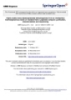
- Journal of Translational Medicine 2009, 7:10 http://www.translational-medicine.com/content/7/1/10 Figure 1 Transduction efficiencies of GEN2.2 Transduction efficiencies of GEN2.2. The pDC cell line, GEN2.2, was non-transduced (NT) or transduced with E-GFP encoding vectors then analysed 5 days posttransduction. GEN2.2 were gated in forward/side scatter, then analyzed for the expression E-GFP by flow cytometry. (A) GEN2.2 were transduced by LV with a PGK promoter pseudotyped with either VSVG and RD114 envelopes at a MOI of 18 or with the GaLV envelope at a MOI of 9. (B) GEN2.2 were transduced with VSVG pseudotyped-LV with a PGK, EF1, desmin or C5–12 promoter, at a MOI of 18. (C) GEN2.2 were transduced by rAAV of serotype 1, 2 or 5 with a CMV promoter, with the number of viral genomes/cell indicated. Results are expressed as mean percentage of cell +/- SD over the number of independent experiments indicated. using a PGK promoter. Non-transduced matured HLA- Flow cytometric analysis A2+ GEN2.2, CD34-pDC and Mo-DC and transduced The pDC phenotype was assessed using three color immu- GEN2.2 cells, CD34-pDC and Mo-DC were co-cultured in nostaining with biotinylated, phycoerythrin (PE)-, Cy- 96-well U-bottom plates at different ratios with 1 × 10⁵ Chrome (CyC)-and allophycocyanin (APC) -conjugated cells/well of a specific MART-1 CD8+ T-cell clone HLA-A2 monoclonal anti-CD40 (5C3), anti-CD80 (L307.4), restricted (LT12) and labelled with CFSE as described ear- CD83 (HB15e), anti-CD86 (FUN-1), anti-HLA-DR lier for the MLR. On day 5, cells were harvested, washed, (G46.6) antibodies (purchased from Becton Dickinson, labelled with an anti-CD8 antibody and analysed by flow Mountain View, CA, Pharmingen product, San Diego, CA) cytometry. The percentage of dividing T-cells was linearly and anti-BDCA2 (AC-144), anti-BDCA4 (AD5-17F6) and correlated with the loss in CFSE fluorescence. anti-CD123 (AC145) (from Miltenyi Biotech). Data were Page 4 of 12 (page number not for citation purposes)
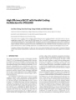
- Journal of Translational Medicine 2009, 7:10 http://www.translational-medicine.com/content/7/1/10 Figure 2 Transduction efficiencies of CD34-pDC Transduction efficiencies of CD34-pDC. CD34-pDC were non-transduced (NT) or transduced with E-GFP encoding vec- tors at day 6 after the induction of differentiation, then cultured for 6 additional days. pDC were gated in forward/side scatter, then analyzed for the expression of E-GFP by flow cytometry. (A) CD34+ progenitors were transduced by LV with a PGK pro- moter pseudotyped with either VSVG and RD114 envelopes at a MOI of 18 or with the GaLV envelope at a MOI of 9. (B) CD34+ progenitors were transduced with VSVG pseudotyped-LV with a PGK, EF1, desmin or C5–12 promoter, at a MOI of 18. (C) CD34+ progenitors were transduced by rAAV of serotype 1, 2 or 5 with a CMV promoter, with the number of viral genomes/cell indicated. Results are expressed as mean percentage of cell +/- SD over the number of independent experiments indicated. acquired using a FACSCalibur flow cytometer (Becton using LV pseudotyped with different envelopes from Dickinson) and data analysis was performed using the VSVG, GaLV or RD114 viruses. E-GFP expression can be CellQuest program (Becton Dickinson). easily and accurately monitored by FACS analysis. Prelim- inary experiments performed with LV encoding E-GFP under the control of the ubiquitous PGK promoter with Statistical analyses Results were presented as the mean +/- standard devia- different MOI (5–50), at a fixed cell density, showed that tion. Student's t-test for paired data was use to determine maximum transduction levels were reached at a MOI of significant differences between the two groups. A p- 18 for VSVG-LV and RD114-LV and at a MOI of 9 for value
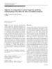
- Journal of Translational Medicine 2009, 7:10 http://www.translational-medicine.com/content/7/1/10 Figure 3 Immunophenotype of transduced pDC Immunophenotype of transduced pDC. Comparative phenotypes of transduced and untransduced GEN2.2 in absence of maturation agent, at day 5. Overlay histograms show the expression of CD123 or HLA-DR for untransduced (thin line), total transduced (thick line) and E-GFP+ gated (green line) GEN2.2, versus isotype-matched controls (dotted line). (A) GEN2.2 transduced by LV with a PGK promoter pseudotyped with either VSVG, RD114 or GaLV envelopes. (B) GEN2.2 were trans- duced with VSVG pseudotyped-LV with a PGK, EF1, desmin or C5–12 promoter (C) GEN2.2 were transduced by rAAV of serotype 1, 2 or 5 with a CMV promoter. The results are representative of at least 4 experiments. ever, it was difficult in our hands to obtain high enough titers to reach a MOI of 18 using similar transduction con- ditions without cellular toxicity. So, at a two-fold lower MOI, a single exposure of GEN2.2 to GaLV-LV led to only 13.3% +/- 5.5% of E-GFP positive cells (figure 1A). Similar results were obtained on human CD34-pDC transduced at day 6 (figure 2A) and monitored 6 days posttransduc- tion. Long-term expression of the transgene for GEN2.2 was maintained in all cases until at least day 60, as checked by flow cytometry (data not shown). In a second step, we then selected the VSVG-LV pseudo- type at MOI of 18 to transduce the pDC cell line, and eval- uated the expression of GFP under the control of different promoters such as the ubiquitous PGK promoter, the hematopoietic cell-specific EF1 promoter, the muscle-spe- cific desmin and the synthetic C512 promoters. The per- centage of E-GFP+ cells obtained was very high with both EF1 (79% +/- 15.3%) and C5–12 (94% +/- 2.8%) promot- ers which are 2.6 to 3 more efficient than the PGK pro- moter for transducing GEN2.2 (figure 1B). Surprisingly, a second muscle-specific promoter, desmin, was also highly Figure 4 Transduction of GEN2.2 does not induce maturation efficient in pDC, since 47.7% +/- 11.1% of cells were E- Transduction of GEN2.2 does not induce maturation. GFP+ (figure 1B). Similar results were obtained on human Comparative phenotype of transduced and non transduced CD34-pDC transduced at day 6 (figure 2B) and moni- GEN2.2. Overlay histograms show the expression of rele- tored 6 days posttransduction. Altogether, these results vant antigens for untransduced (thin line) and transduced show that VSVG-pseudotyped LV encoding the E-GFP as (thick line) with LV-VSVG/PGK or rAAV2/2, versus isotype- transgene under the control of the EF1 or C5–12 promot- matched controls (dotted line). The results are representa- ers are very efficient for transduction of pDC. tive of at least 3 experiments. Page 6 of 12 (page number not for citation purposes)
- Journal of Translational Medicine 2009, 7:10 http://www.translational-medicine.com/content/7/1/10 Figure 5 CpG induced maturation of transduced pDC CpG induced maturation of transduced pDC. Comparative phenotype of transduced GEN2.2, 6 days posttransduction, in the absence and presence of CpG for 24 hours. Overlay histograms show the expression of relevant antigens for transduced GEN2.2 cultured without CpG (thin line), with CpG (thick line) and with CpG and gated on E-GFP+ (green line) versus isotype- matched controls (dotted line). (A, B, C) Transduced GEN2.2 vectors are the same ones as those described in figure 2. Values indicated are MFI of the transduced populations cultured without CpG versus transduced populations cultured with CpG. The results are representative of at least 4 experiments. In a similar protocol, we used AAV vectors of different transduced cells ranging from around 3% to less than 1% of E-GFP+ cells (figure 1C and 2C). These results indicate serotypes (rAAV2/1, rAAV2/2 and rAAV2/5) to transduce the pDC cell line and CD34-pDC and compared their effi- that pDC are not susceptible to transduction by single- cacy. We previously showed that single-stranded rAAV2/1 strand AAV vectors of serotype 1, 2 or 5. and rAAV2/2 were very poorly efficient in transducing human pDC generated in vitro from CD34+ progenitor Immunophenotypical analysis of transduced pDC cells [38]. We evaluated here, whether cells fully differen- The GEN2.2 cell line was previously characterized by its tiated into pDC could be transduced by rAAV of serotypes phenotype as a pDC cell line. These cells have been shown 1 and 2, but also of serotype 5. Preliminary experiments to express the human leukocyte antigen-DR (HLA-DR), performed with different amounts of viral particles (5 × the IL3-receptor (CD123) and the CD4 [18]. Moreover, as 103 to 5 × 10⁴ vg/cell), at a fixed cell density, showed that a hallmark of pDC, these cells are BDCA2 and BDCA4 maximum transduction levels were reached with 2.5 × 10⁴ (type II C lectin)-positive and CD11c- and CD1a-negative. vg/cell for rAAV2/1 and rAAV2/2 and with 9 × 10³ vg/cell Trypan blue exclusion and cell counting of LV and rAAV for rAAV2/5, with no cellular toxicity (data not shown). transduced GEN2.2 at the end of the culture period indi- GEN2.2 and CD34-pDC were monitored for CD123, cated that transduction had no deleterious effect on cell HLA-DR and E-GFP expression, but only at day 5 to 6 viability compared to control cells (data not shown). We posttransduction, since pDC are dividing cells and AAV explored in detail the immunophenotype of these trans- vectors are mainly episomal. A single exposure of pDC to duced and control GEN2.2 by flow cytometry. We showed rAAV2/1, rAAV2/2 or rAAV2/5 led to very low levels of that whatever lentiviral or rAAV vectors used to trans- Page 7 of 12 (page number not for citation purposes)
- Journal of Translational Medicine 2009, 7:10 http://www.translational-medicine.com/content/7/1/10 T-cell stimulatory capacity of non-transduced and transduced GEN2.2 in mixed lymphocyte alloreactions Figure 6 T-cell stimulatory capacity of non-transduced and transduced GEN2.2 in mixed lymphocyte alloreactions. Day 5 transduced GEN2.2 were matured in CpG for 24 hours, before cell sorting on an E-GFP expression basis. (A-E) Total non- transduced (NT), E-GFP- and E-GFP+cell sorted GEN2.2 transduced by the same LV as those described in figure 3 were incu- bated with allogeneic T cells stained with CFSE. (F) Total non-transduced (NT) and rAAV2/1, rAAV2/2 or rAAV2/5 transduced unsorted GEN2.2 were incubated with allogeneic T-cells stained with CFSE. After 4 days of co-culture, percentages of CD3+ dividing T cells measured by flow cytometry were linearly correlated with the loss of CFSE fluorescence. Dot plots inserted in graphs show one representative CFSE profile at the ratio 3/1 for GFP+ cells. The data are shown as the means of 3 independent experiments. duced the GEN2.2 cells, no significant modification of the cate that the LV transduction does not alter the phenotype CD123 and HLA-DR expression (figure 3) of the CD4, of pDC or their capacity to mature. BDCA2 and BDCA4 (data not shown) or of the costimu- latory molecules and maturation marker CD80, CD86, Functional properties of transduced pDC CD40 and CD83 as illustrated figure 4, with two vectors, We evaluated the ability of different transduced GEN2.2 was observed, compared to control cells. Comparative to stimulate allogeneic T cells in an allogeneic mixed lym- phenotypic analysis of unactivated and CpG-activated phocyte reaction (MLR). GEN2.2 transduced with the dif- transduced GEN2.2 revealed a normal upregulation of the ferent E-GFP encoding vectors were matured with CpG for co-stimulatory molecule CD86, demonstrating that the 24 hours, then sorted by flow cytometry on the basis of E- maturation capacity of transduced subpopulations was GFP expression. Non-transduced, E-GFP negative and unaltered (Figure 5). Similarly, the phenotype of human positive sorted GEN2.2 were used for stimulation of allo- transduced CD34-pDC was not modified compare to geneic T-cells labelled with CFSE. Both negative and posi- non-transduced cells (data not shown). Our results indi- tive E-GFP GEN2.2 populations displayed similar Page 8 of 12 (page number not for citation purposes)
- Journal of Translational Medicine 2009, 7:10 http://www.translational-medicine.com/content/7/1/10 with evidence of functional differences in their ability to regulate the T-cell responses, to produce antiviral type I IFN and to cross-present exogenous antigens to CD8+ T cells [47]. We previously showed that VSVG-pseudotyped HIV-1 vectors are good candidates for efficient transduc- tion of monocyte- and CD34+-derived LC, without induc- ing phenotypic and functional maturation [26]. More recently, we also showed that self-complementary duplex strands but not single strands rAAV2/1 and 2 were also very efficient in transducing major DC subsets generated in vitro, including CD34+-derived pDC [38]. In this study, we extended LV transduction to pDC, using IFN-α production by pDC Figure 7 different pseudotyped HIV-1 vectors encoding E-GFP IFN-α production by pDC. GEN2.2 and day 6 CD34-pDC under the control of different promoters and showed that were non transduced (NT) or transduced by LV-VSVG at an VSVG-pseudotyped LV encoding E-GFP under the control MOI of 18 (LV-VSVG), then 6 days later, the IFNα produc- of EF1 or C512 promoters are the most efficient combina- tion was measured in cell culture supernatants before or tions, leading to transduction of 60% to 90% of the pDC after maturation in CpG, for 24 hours. The data are shown cell line, GEN2.2 [18] and CD34-pDC. Of note, we as the means of 3 independent experiments. showed that transduction did not alter alloreactive pres- entation properties of pDC. Furthermore, pDC trans- allostimulatory capacity compared to non-transduced duced with LV expressing a MART-1 peptide was as efficient as Mo-DC for activation of a specific CD8+ T cell GEN2.2, whatever vector used (figure 6 and data not shown). In response to these viruses, pDC are known to clone. Altogether, these results show that antigen-loading secrete high levels of type I IFN [14,15]. Of note, IFN-α of pDC through ex-vivo LV transduction may represent a was not detected in cell supernatant of any transduced relevant immunotherapy approach for particular clinical GEN2.2 cultures when checked between 24 hours and 10 applications. Indeed, compared with antigen loading pro- days following contact with the different viral particles. tocols using whole tumor cell lysates or recombinant Nevertheless, GEN2.2 and CD34-pDC were always able to tumor-associated antigen peptides, LV transduction offers secrete IFN-α upon stimulation by the CpG motif via the the advantage of direct antigen processing from cytosolic toll-like receptor signalling pathway, as illustrated figure 7 proteins and of long lasting antigen expression. for pDC transduced with a LV pseudotyped with VSVG coding for E-GFP under the control of the PGK promoter. Previous publications [30-32] reported efficient transduc- Moreover, we evaluated the capacity of the HLA-A0201 tion levels of hematopoietic cells with LV pseudotyped expressing pDC to activate a CD8+ T cell clone after trans- with GaLV or RD114 envelopes. Here, the highest pDC duction with a LV coding for the MART-1 peptide under transduction levels were obtained with the VSVG enve- the control of the PGK promoter. The transduced GEN2.2 lope, which was also previously shown to efficiently trans- obtained were efficient in activating a specific CD8+ T-cell duce human hematopoietic progenitor and leukaemia clone (Figure 8A). Results were confirmed on CD34-pDC cells [26,48,49] as well as fully differentiated human transduced with the same LV expressing a MART-1 peptide monocyte-derived DC [50,51], with a long lasting expres- sion. The EF1α promoter was shown to have a stronger (Figure 8B). Interestingly these transduced CD34-pDC activity than the PGK promoter in a human CD34+ cell were as efficient as Mo-DC for activation of a specific CD8+ T cell clone (Figure 8B). line [33] and in cultured cord blood cells [33,34] and allowed to obtain transgene-expressing myeloid DC [23]. Altogether, these results indicate that the functional prop- Here, we showed that after a single exposure to VSVG- erties of pDC were not altered by LV or rAAV transduction. pseudotyped LV, the percentage of E-GFP expressing pDC Furthermore, LV-transduced pDC were able to activate a was 2.6 fold higher when the expression was driven by the CD8+ T-cell clone. EF1 compared to the PGK promoter. The average copy number of the vector in transduced pDC under both con- ditions was similar (3–4 copies per cell), as determined by Discussion The attractiveness of dendritic cells as a target for genetic real-time quantitative PCR (data not shown). This indi- manipulation is a consequence of their ability to initiate cates that the integration levels are similar with both con- and orchestrate primary immune responses, including structions but that, as previously described, the promoter tolerogenic responses [1,45,46]. At least two circulating activity is different. We also evaluated two other promot- subsets of DC have been described: myeloid DC and pDC ers described to be muscle restricted [39-41], the desmin Page 9 of 12 (page number not for citation purposes)
- Journal of Translational Medicine 2009, 7:10 http://www.translational-medicine.com/content/7/1/10 CD8+ T 8 ell clone activation by LV transduced pDC Figure c CD8+ T cell clone activation by LV transduced pDC. In vitro antigen presentation capacities of LV transduced HLA-A2 pDC cells and Mo-DC. Cells were transduced with LV encoding the MART-1 peptide under the control of the PGK promoter. (A) Mature non-transduced (NT) and transduced (VSVG-PGK-MART-1) GEN2.2 or (B) CD34-pDC and Mo-DC were co-cul- tured with the HLA-A2 restricted CD8+ T-cell clone specific for the MART-1 peptide (LT12) stained with CFSE. After 5 days of co-culture, percentages of CD8+ dividing T-cells measured by flow cytometry were linearly correlated with the loss of CFSE fluorescence. The data in panel A are shown as the mean of triplicate and represent one out of 3 independent experiments whereas the data in panel B were performed once. and synthetic C512 promoters which have been shown in ducing major DC subsets [38] might elicit high immune gene therapy studies to specifically target muscles and to responses against the transgene. drive gene expression in a context of ss rAAV vectors [41]. As in our previous report [38], we showed here that even Conclusion with an ubiquitous promoter like CMV, only a very low DC transduction with LV preparations can serve as vaccine transduction efficiency could be reached with ss rAAV in vehicles in human through efficient transduction levels the different DC subsets. So, in order to investigate the and are also useful in vitro to evaluate the immunogenicity potential leak of these promoters in human DC subsets, of the vector preparations and the specificity and safety of we constructed and produced LV vectors carrying the two promoters used in gene therapy protocols. different cassettes. Surprisingly, we showed that the per- centages of E-GFP expressing pDC with desmin and C512 Competing interests promoters were very high and equivalent to those The authors declare that they have no competing interests. obtained with PGK and EF1 promoters, respectively. The average copy number in pDC for desmin and C512 pro- Authors' contributions moters were 4 and 1 copies per cell, respectively, showing VP contributed to the experimental design, data acquisi- that the C512 promoters was at least as efficient as an tion and analysis, and drafting of the manuscript. BS con- ubiquitous promoter (data not shown). In contrast to the tributed to the data acquisition and analysis. MS designed desmin promoter, the C512 promoter was also active in lentiviral vector constructions. CL provided the Gen2.2 monocyte-derived DC and LC (around 10% of E-GFP+ cell line. PJ provided the Gen2.2 cell line and critically cells) and in a human colorectal carcinoma (HCT116) revised the manuscript. DJ gave the final approval of the (data not shown). Nevertheless, transgene expression version to be published. MC conceived of the study, par- with these cassettes in ss AAV vectors was not detectable ticipated in its design and coordination and drafted the (data not shown). Taken together, these data suggest that manuscript. All authors read and approved the final man- the use of desmin or C5–12 promoters in ss rAAV, for clin- uscript. ical gene therapy protocols, will not induce transgene expression in DC subsets. Nevertheless, the use of these Acknowledgements promoters in sc rAAV, which are highly efficient for trans- VP is supported by a CIFRE convention from Association Nationale de la Recherche Technique, France. This work was supported by the Association Page 10 of 12 (page number not for citation purposes)
- Journal of Translational Medicine 2009, 7:10 http://www.translational-medicine.com/content/7/1/10 Française contre les Myopathies (AFM), CNRS and an ATIGE grant to JD 20. Jaraquemada D, Marti M, Long EO: An endogenous processing pathway in vaccinia virus-infected cells for presentation of from Genopole Evry, France and by INCa – Canceropole 2004–05. We cytoplasmic antigens to class II-restricted T cells. J Exp Med wish to thank Anne Galy for providing the OP9-Del1 cell line, Isabelle Lam- 1990, 172:947-954. bert for providing viral vectors and Florence Faure for giving us the HLA- 21. Chinnasamy N, Chinnasamy D, Toso JF, Lapointe R, Candotti F, Mor- A0201 restricted MART-1 specific CD8+ T-cell clone. We thank Laurent gan RA, Hwu P: Efficient gene transfer to human peripheral blood monocyte-derived dendritic cells using human immu- Poujades for real-time quantitative PCR. We thank Susan Cure for the crit- nodeficiency virus type 1-based lentiviral vectors. Hum Gene ical reading of the manuscript. Ther 2000, 11:1901-1909. 22. Schroers R, Sinha I, Segall H, Schmidt-Wolf IG, Rooney CM, Brenner References MK, Sutton RE, Chen SY: Transduction of human PBMC- derived dendritic cells and macrophages by an HIV-1-based 1. Banchereau J, Steinman RM: Dendritic cells and the control of lentiviral vector system. Mol Ther 2000, 1:171-179. immunity. Nature 1998, 392:245-252. 23. Salmon P, Arrighi JF, Piguet V, Chapuis B, Zubler RH, Trono D, Kin- 2. Banchereau J, Briere F, Caux C, Davoust J, Lebecque S, Liu YJ, Pulen- dler V: Transduction of CD34+ cells with lentiviral vectors dran B, Palucka K: Immunobiology of dendritic cells. Annu Rev enables the production of large quantities of transgene- Immunol 2000, 18:767-811. expressing immature and mature dendritic cells. J Gene Med 3. Pulendran B: Modulating vaccine responses with dendritic cells 2001, 3:311-320. and Toll-like receptors. Immunol Rev 2004, 199:227-250. 24. Rouas R, Uch R, Cleuter Y, Jordier F, Bagnis C, Mannoni P, Lewalle P, 4. Blom B, Ho S, Antonenko S, Liu YJ: Generation of interferon Martiat P, Broeke A Van den: Lentiviral-mediated gene delivery alpha-producing predendritic cell (Pre-DC)2 from human in human monocyte-derived dendritic cells: optimized CD34(+) hematopoietic stem cells. J Exp Med 2000, design and procedures for highly efficient transduction com- 192:1785-1796. patible with clinical constraints. Cancer Gene Ther 2002, 5. Shortman K, Liu YJ: Mouse and human dendritic cell subtypes. 9:715-724. Nat Rev Immunol 2002, 2:151-161. 25. Breckpot K, Dullaers M, Bonehill A, van Meirvenne S, Heirman C, de 6. Colonna M, Trinchieri G, Liu YJ: Plasmacytoid dendritic cells in Greef C, Bruggen P van der, Thielemans K: Lentivirally transduced immunity. Nat Immunol 2004, 5:1219-1226. dendritic cells as a tool for cancer immunotherapy. J Gene 7. Lund J, Sato A, Akira S, Medzhitov R, Iwasaki A: Toll-like receptor Med 2003, 5:654-667. 9-mediated recognition of Herpes simplex virus-2 by plasma- 26. Veron P, Boutin S, Bernard J, Danos O, Davoust J, Masurier C: Effi- cytoid dendritic cells. J Exp Med 2003, 198:513-520. cient transduction of monocyte- and CD34(+)- derived Lang- 8. Hochrein H, Schlatter B, O'Keeffe M, Wagner C, Schmitz F, Schie- erhans cells with lentiviral vectors in the absence of mann M, Bauer S, Suter M, Wagner H: Herpes simplex virus type- phenotypic and functional maturation. J Gene Med 2006, 1 induces IFN-alpha production via Toll-like receptor 9- 8:951-961. dependent and -independent pathways. Proc Natl Acad Sci USA 27. Dyall J, Latouche JB, Schnell S, Sadelain M: Lentivirus-transduced 2004, 101:11416-11421. human monocyte-derived dendritic cells efficiently stimu- 9. Krug A, Luker GD, Barchet W, Leib DA, Akira S, Colonna M: Herpes late antigen-specific cytotoxic T lymphocytes. Blood 2001, simplex virus type 1 activates murine natural interferon-pro- 97:114-121. ducing cells through toll-like receptor 9. Blood 2004, 28. Reiser J: Production and concentration of pseudotyped HIV- 103:1433-1437. 1-based gene transfer vectors. Gene Ther 2000, 7:910-913. 10. Tabeta K, Georgel P, Janssen E, Du X, Hoebe K, Crozat K, Mudd S, 29. Stitz J, Buchholz CJ, Engelstadter M, Uckert W, Bloemer U, Schmitt I, Shamel L, Sovath S, Goode J, et al.: Toll-like receptors 9 and 3 as Cichutek K: Lentiviral vectors pseudotyped with envelope essential components of innate immune defense against glycoproteins derived from gibbon ape leukemia virus and mouse cytomegalovirus infection. Proc Natl Acad Sci USA 2004, murine leukemia virus 10A1. Virology 2000, 273:16-20. 101:3516-3521. 30. Hanawa H, Kelly PF, Nathwani AC, Persons DA, Vandergriff JA, Har- 11. Diebold SS, Kaisho T, Hemmi H, Akira S, Reis e Sousa C: Innate grove P, Vanin EF, Nienhuis AW: Comparison of various enve- antiviral responses by means of TLR7-mediated recognition lope proteins for their ability to pseudotype lentiviral of single-stranded RNA. Science 2004, 303:1529-1531. vectors and transduce primitive hematopoietic cells from 12. Heil F, Hemmi H, Hochrein H, Ampenberger F, Kirschning C, Akira human blood. Mol Ther 2002, 5:242-251. S, Lipford G, Wagner H, Bauer S: Species-specific recognition of 31. Loo JC van der, Liu BL, Goldman AI, Buckley SM, Chrudimsky KS: single-stranded RNA via toll-like receptor 7 and 8. Science Optimization of gene transfer into primitive human hemat- 2004, 303:1526-1529. opoietic cells of granulocyte-colony stimulating factor-mobi- 13. Lund JM, Alexopoulou L, Sato A, Karow M, Adams NC, Gale NW, lized peripheral blood using low-dose cytokines and Iwasaki A, Flavell RA: Recognition of single-stranded RNA comparison of a gibbon ape leukemia virus versus an RD114- viruses by Toll-like receptor 7. Proc Natl Acad Sci USA 2004, pseudotyped retroviral vector. Hum Gene Ther 2002, 101:5598-5603. 13:1317-1330. 14. Cella M, Jarrossay D, Facchetti F, Alebardi O, Nakajima H, Lanzavec- 32. Relander T, Johansson M, Olsson K, Ikeda Y, Takeuchi Y, Collins M, chia A, Colonna M: Plasmacytoid monocytes migrate to Richter J: Gene transfer to repopulating human CD34+ cells inflamed lymph nodes and produce large amounts of type I using amphotropic-, GALV-, or RD114-pseudotyped HIV-1- interferon. Nat Med 1999, 5:919-923. based vectors from stable producer cells. Mol Ther 2005, 15. Siegal FP, Kadowaki N, Shodell M, Fitzgerald-Bocarsly PA, Shah K, Ho 11:452-459. S, Antonenko S, Liu YJ: The nature of the principal type 1 inter- 33. Ramezani A, Hawley TS, Hawley RG: Lentiviral vectors for feron-producing cells in human blood. Science 1999, enhanced gene expression in human hematopoietic cells. 284:1835-1837. Mol Ther 2000, 2:458-469. 16. Liu YJ: IPC: professional type 1 interferon-producing cells and 34. Salmon P, Kindler V, Ducrey O, Chapuis B, Zubler RH, Trono D: plasmacytoid dendritic cell precursors. Annu Rev Immunol 2005, High-level transgene expression in human hematopoietic 23:275-306. progenitors and differentiated blood lineages after transduc- 17. Olivier A, Lauret E, Gonin P, Galy A: The Notch ligand delta-1 is tion with improved lentiviral vectors. Blood 2000, a hematopoietic development cofactor for plasmacytoid 96:3392-3398. dendritic cells. Blood 2006, 107:2694-2701. 35. Liu Y, Chiriva-Internati M, Grizzi F, Salati E, Roman JJ, Lim S, Her- 18. Chaperot L, Blum A, Manches O, Lui G, Angel J, Molens JP, Plumas J: monat PL: Rapid induction of cytotoxic T-cell response against Virus or TLR agonists induce TRAIL-mediated cytotoxic cervical cancer cells by human papillomavirus type 16 E6 activity of plasmacytoid dendritic cells. J Immunol 2006, antigen gene delivery into human dendritic cells by an 176:248-255. adeno-associated virus vector. Cancer Gene Ther 2001, 19. Angel J, Chaperot L, Molens JP, Mezin P, Amacker M, Zurbriggen R, 8:948-957. Grichine A, Plumas J: Virosome-mediated delivery of tumor 36. Ponnazhagan S, Mahendra G, Curiel DT, Shaw DR: Adeno-associ- antigen to plasmacytoid dendritic cells. Vaccine 2007, ated virus type 2-mediated transduction of human mono- 25:3913-3921. Page 11 of 12 (page number not for citation purposes)
- Journal of Translational Medicine 2009, 7:10 http://www.translational-medicine.com/content/7/1/10 cyte-derived dendritic cells: implications for ex vivo immunotherapy. J Virol 2001, 75:9493-9501. 37. Chiriva-Internati M, Liu Y, Salati E, Zhou W, Wang Z, Grizzi F, Roman JJ, Lim SH, Hermonat PL: Efficient generation of cytotoxic T lymphocytes against cervical cancer cells by adeno-associ- ated virus/human papillomavirus type 16 E7 antigen gene transduction into dendritic cells. Eur J Immunol 2002, 32:30-38. 38. Veron P, Allo V, Riviere C, Bernard J, Douar AM, Masurier C: Major subsets of human dendritic cells are efficiently transduced using self-complementary adeno-associated viral vectors 1 and 2. J Virol 2007, 81:5385-5394. 39. Li X, Eastman EM, Schwartz RJ, Draghia-Akli R: Synthetic muscle promoters: activities exceeding naturally occurring regula- tory sequences. Nat Biotechnol 1999, 17:241-245. 40. Gonin P, Arandel L, Van Wittenberghe L, Marais T, Perez N, Danos O: Femoral intra-arterial injection: a tool to deliver and assess recombinant AAV constructs in rodents whole hind limb. J Gene Med 2005, 7:782-791. 41. Bartoli M, Roudaut C, Martin S, Fougerousse F, Suel L, Poupiot J, Gic- quel E, Noulet F, Danos O, Richard I: Safety and efficacy of AAV- mediated calpain 3 gene transfer in a mouse model of limb- girdle muscular dystrophy type 2A. Mol Ther 2006, 13:250-259. 42. Follenzi A, Ailles LE, Bakovic S, Geuna M, Naldini L: Gene transfer by lentiviral vectors is limited by nuclear translocation and rescued by HIV-1 pol sequences. Nat Genet 2000, 25:217-222. 43. Riviere C, Danos O, Douar AM: Long-term expression and repeated administration of AAV type 1, 2 and 5 vectors in skeletal muscle of immunocompetent adult mice. Gene Ther 2006, 13:1300-1308. 44. Xiao X, Li J, Samulski RJ: Production of high-titer recombinant adeno-associated virus vectors in the absence of helper ade- novirus. J Virol 1998, 72:2224-2232. 45. Steinman RM: The dendritic cell system and its role in immu- nogenicity. Annu Rev Immunol 1991, 9:271-296. 46. Steinman RM, Hawiger D, Nussenzweig MC: Tolerogenic den- dritic cells. Annu Rev Immunol 2003, 21:685-711. 47. Pulendran B: Variegation of the immune response with den- dritic cells and pathogen recognition receptors. J Immunol 2005, 174:2457-2465. 48. Case SS, Price MA, Jordan CT, Yu XJ, Wang L, Bauer G, Haas DL, Xu D, Stripecke R, Naldini L, et al.: Stable transduction of quiescent CD34(+)CD38(-) human hematopoietic cells by HIV-1-based lentiviral vectors. Proc Natl Acad Sci USA 1999, 96:2988-2993. 49. Stripecke R, Cardoso AA, Pepper KA, Skelton DC, Yu XJ, Mascaren- has L, Weinberg KI, Nadler LM, Kohn DB: Lentiviral vectors for efficient delivery of CD80 and granulocyte-macrophage- col- ony-stimulating factor in human acute lymphoblastic leuke- mia and acute myeloid leukemia cells to induce antileukemic immune responses. Blood 2000, 96:1317-1326. 50. Lizee G, Gonzales MI, Topalian SL: Lentivirus vector-mediated expression of tumor-associated epitopes by human antigen presenting cells. Hum Gene Ther 2004, 15:393-404. 51. Dullaers M, Thielemans K: From pathogen to medicine: HIV-1- derived lentiviral vectors as vehicles for dendritic cell based cancer immunotherapy. J Gene Med 2006, 8:3-17. Publish with Bio Med Central and every scientist can read your work free of charge "BioMed Central will be the most significant development for disseminating the results of biomedical researc h in our lifetime." Sir Paul Nurse, Cancer Research UK Your research papers will be: available free of charge to the entire biomedical community peer reviewed and published immediately upon acceptance cited in PubMed and archived on PubMed Central yours — you keep the copyright BioMedcentral Submit your manuscript here: http://www.biomedcentral.com/info/publishing_adv.asp Page 12 of 12 (page number not for citation purposes)
CÓ THỂ BẠN MUỐN DOWNLOAD

-

báo cáo hóa học:" High dose concentration administration of ascorbic acid inhibits tumor growth in BALB/C mice implanted with sarcoma 180 cancer cells via the restriction of angiogenesis"
 9 p |
9 p |  80
|
80
|  7
7
-

báo cáo hóa học:" Anti-angiogenic effect of high doses of ascorbic acid"
10 p | 67
| 6
-
Báo cáo hóa học: " High P–T Nano-Mechanics of Polycrystalline Nickel Yusheng Zhao Æ T. D. Shen Æ Jianzhong Zhang"
16 p | 42
| 6
-
báo cáo hóa học:" Highly selective fluorescent chemosensor for Zn2+ derived from inorganic-organic hybrid magnetic core/shell Fe3O4@SiO2 nanoparticles"
24 p | 55
| 6
-
Báo cáo hóa học: "High Capacity Downlink Transmission with MIMO Interference Subspace Rejection in Multicellular CDMA Networks"
20 p | 42
| 6
-

Báo cáo hóa học: " High-Speed Turbo-TCM-Coded Orthogonal Frequency-Division Multiplexing Ultra-Wideband Systems"
12 p | 42
| 5
-

Báo cáo hóa học: " High expression of transcriptional coactivator p300 correlates with aggressive features and poor prognosis of hepatocellular carcinoma"
11 p | 52
| 5
-

báo cáo hóa học:" High correlation of the proteome patterns in bone marrow and peripheral blood blast cells in patients with acute myeloid leukemia"
8 p | 63
| 5
-
Báo cáo hóa học: " High-yield Synthesis of Multiwalled Carbon Nanotube by Mechanothermal Method"
7 p | 46
| 5
-

Báo cáo hóa học: "High activity of sequential low dose chemo-modulating Temozolomide in combination with Fotemustine in metastatic melanoma. A feasibility study"
8 p | 85
| 5
-

Báo cáo hóa học: " Research Article New Structured Illumination Technique for the Inspection of High-Reflective Surfaces: Application for the Detection of Structural Defects"
14 p | 70
| 4
-
Báo cáo hóa học: " Highly stable meso-diaminopimelate dehydrogenase from an Ureibacillus thermosphaericus strain A1 isolated from a Japanese compost: purification, characterization and sequencing"
37 p | 49
| 4
-

Báo cáo hóa học: " Research Article High-Speed Smart Camera with High Resolution"
16 p | 33
| 4
-
Báo cáo hóa học: "High Efficiency EBCOT with Parallel Coding Architecture for JPEG2000"
14 p | 44
| 4
-

Báo cáo hóa học: " High-Performance Wireless via the Merger of CI Chip-Shaped DS-CDMA and Oscillating-Beam Smart Antenna Arrays"
8 p | 49
| 4
-

Báo cáo hóa học: " Research Article A High-End Real-Time Digital Film Processing Reconfigurable Platform"
15 p | 46
| 3
-
Báo cáo hóa học: " High-rate low-temperature dc pulsed magnetron sputtering of photocatalytic TiO2 films: the effect of repetition frequency"
7 p | 43
| 3


Chịu trách nhiệm nội dung:
Nguyễn Công Hà - Giám đốc Công ty TNHH TÀI LIỆU TRỰC TUYẾN VI NA
LIÊN HỆ
Địa chỉ: P402, 54A Nơ Trang Long, Phường 14, Q.Bình Thạnh, TP.HCM
Hotline: 093 303 0098
Email: support@tailieu.vn
Giấy phép Mạng Xã Hội số: 670/GP-BTTTT cấp ngày 30/11/2015 Copyright © 2022-2032 TaiLieu.VN. All rights reserved.

