


Báo cáo hóa học: " Impact of g-chain cytokines on EBV-specific T cell cultures"
lượt xem 8
download
 Download
Vui lòng tải xuống để xem tài liệu đầy đủ
Download
Vui lòng tải xuống để xem tài liệu đầy đủ
Tuyển tập báo cáo các nghiên cứu khoa học quốc tế ngành hóa học dành cho các bạn yêu hóa học tham khảo đề tài: Impact of g-chain cytokines on EBV-specific T cell cultures
Bình luận(0) Đăng nhập để gửi bình luận!
Nội dung Text: Báo cáo hóa học: " Impact of g-chain cytokines on EBV-specific T cell cultures"
- Merlo et al. Journal of Translational Medicine 2010, 8:121 http://www.translational-medicine.com/content/8/1/121 RESEARCH Open Access Impact of g-chain cytokines on EBV-specific T cell cultures Anna Merlo1†, Riccardo Turrini1†, Cristina Trento2, Paola Zanovello1,3, Riccardo Dolcetti4*, Antonio Rosato1,3* Abstract Background: Recent preclinical adoptive immunotherapy studies in murine models prompt to employ “proper” rather than “as many as possible” antigen-specific T cells to gain better therapeutic results. Ideally, “proper” T cells are poorly differentiated in vitro, but retain the capacity to fully differentiate into effector cells in vivo, where they can undergo long-term survival and strong proliferation. Such requirements can be achieved by modifying culture conditions, namely using less “differentiating” cytokines than IL-2. Methods: To evaluate this issue in human T cell cultures, we exploited a well characterized and clinical-grade protocol finalized at generating EBV-specific CTL for adoptive immunotherapy. In particular, we studied the impact of IL-7, IL-15 and IL-21 compared to IL-2 on different aspects of T cell functionality, namely growth kinetics, differentiation/activation marker expression, cytokine production, and short-term and long-term cytotoxicity. Results: Results disclosed that the culture modifications we introduced in the standard protocol did not improve activity nor induce substantial changes in differentiation marker expression of EBV-specific CTL. Conclusions: Our data indicated that the addition of g-chain cytokines other than IL-2 for the generation of EBV- specific T cell cultures did not produce the improvements expected on the basis of recent published literature. This fact was likely due to the intrinsic differences between murine and human models and highlights the need to design ad hoc protocols rather than simply modify the cytokines added in culture. Background recirculation and in vivo expansion capability. These fea- tures are highlighted by a well-defined “marker expres- Infusion of antigen-specific T cells proved to be safe and sion signature ” , namely CD27 low/neg , CD28 low/neg , effective against both virus infections (e.g., CMV [1]) CD62Llow/neg, CCR7low/neg, and CD57high. Thus, the new and cancer, in particular melanoma and EBV-driven malignancies [2]. The vast majority of current protocols trend in adoptive cell therapy (ACT) focuses on the rely on the infusion of a high number of effector cells infusion of a more limited number of cells, but with the “proper” phenotype and functional characteristics, which that require long-term in vi tro cultures, in particular when dealing with Tumor Infiltrating Lymphocytes can promote prolonged in vivo persistence and expan- (TIL) or clonal cultures. Consequently, this aspect sion, and induction of immunological memory to pro- implies labor-intensive and cost-ineffective procedures vide protection against possible relapses. The and, furthermore, has a potential negative impact on the potentiality to expand and persist in the host also relies on the possibility for the infused cells to find an “immu- characteristics of cells infused. Indeed, as advanced by nological space” to colonize. This is “naturally” accom- Gattinoni and colleagues [3,4], long-term T cell cultures move toward a differentiate d phenotype characterized plished in Post Transplant Lymphoproliferative Disease by a high cytotoxic potential, but also a poor (PTLD) after Haemopoietic Stem Cell Transplantation (HSCT), in which patients are immunocompromised * Correspondence: rdolcetti@cro.it; antonio.rosato@unipd.it due to the immunosuppressive regimens; in patients † Contributed equally with other tumors, it has been achieved by chemother- 1 University of Padova, Dept. of Oncology and Surgical Sciences, Via apy and irradiation [5] or by immunodepleting (anti- Gattamelata 64, 35128 Padova, Italy 4 CRO, Centro Riferimento Oncologico IRCCS, Via F. Gallini 2, 33081 Aviano, CD45) antibodies [6]. In these conditions, infused T Italy cells have a favourable environment with fewer Full list of author information is available at the end of the article © 2010 Merlo et al; licensee BioMed Central Ltd. This is an Open Access article distributed under the terms of the Creative Commons Attribution License (http://creativecommons.org/licenses/by/2.0), which permits unrestricted use, distribution, and reproduction in any medium, provided the original work is properly cited.
- Merlo et al. Journal of Translational Medicine 2010, 8:121 Page 2 of 8 http://www.translational-medicine.com/content/8/1/121 competitors for and elevated availability of homeostatic LCL were maintained in RPMI 1640 (Euroclone, Pero, cytokines (IL-7 and IL-15), and possibly less numerous Milan, Italy) supplemented with 10% heat-inactivated T regulatory (Treg) populations. type AB Human Serum (HS, Lonza BioWhittaker; Basel, Although much attention has been paid to shorten the Switzerland), 1 mM Na Pyruvate, 10 mM Hepes Buffer, generation protocols in the clinical settings, a stringent 2 mM Ultraglutamine (all from Lonza BioWhittaker), correlation between phenotype (and so differentiation) 1% Antibiotic/antimycotic (Gibco, Invitrogen Corpora- and outcome has been shown mainly in mouse models tion), hereafter referred to as HS complete medium. thus far [4,7,8], with few notable exceptions [9]. In this Generation of EBV-specific CD4+ and CD8+ T-cell lines context, several reports have described the impact of dif- ferent g-chain cytokines on the differentiation status and EBV-specific T cells were established as previously functional properties of T-cell cultures in vitro and, described [10], with modifications. Briefly, PBMC were more importantly, in vivo. Overall, they suggested that co-cultivated with irradiated (40 Gy) autologous LCL at certain g-chain cytokines, in particular IL-15 and IL-21, a ratio of 40:1 in 24-well plates (Corning Incorporated; are superior to the commonly used IL-2 in maintaining Corning, NY) in HS complete medium. PBMC were seeded at a concentration of 2 × 10⁶ cells/ml and main- a less differentiated phenotype of cultured T cells, thus possibly resulting in a better therapeutic activity. In this tained at 37°C in a 6.5% CO2 humidified atmosphere. regard, the eradication of large established melanomas On day 10 and weekly thereafter, CTL were re-stimu- (approximately 50 mm 2 tumor area) was achieved by lated with irradiated LCL at a 4:1 ratio. Recombinant the infusion of as little as 5 × 10⁵ IL-21 cultured T cells IL-2 (35 I.U./ml, Proleukin, Chiron Corporation; Emery- [7]. ville, CA) or IL-7 (10 ng/ml; Peprotech; Rocky Hill, NJ) To explore this critical issue in human T cell cultures, or IL-15 (10 ng/ml; Peprotech) or IL-21 (10 ng/ml; we took advantage of a well established and clinical- eBioscience; San Diego, CA) were added on day 14 and graded protocol aimed at generating EBV-specific CTL replenished every 2 days. On day 14, before cytokine addition, CD4+ T cells were immunomagnetically sorted for ACT. We slightly modified the protocol by adding using the CD4+ T cell Isolation Kit II (Miltenyi Biotec; to the cultures IL-7, IL-15 or IL-21 instead of IL-2. Bergisch Gladbach, Germany), and both CD8+ and CD4 Moreover, we separated and maintained in parallel cul- tures CD4+ and CD8+ T cells to better discriminate the + T cells were cultured in parallel. At each subsequent re-stimulation with LCL, CD4+ T cells were adjusted to impact of the different cytokines on the two subsets. 1.5 × 10⁶ cells/ml and CD8+ T cells to 2 × 10⁶ cells/ml. We therefore compared the proliferative potential, phe- notype, cytokine production, and cytotoxic activity of effector cells obtained in different culture conditions. Cytotoxicity assays Cytotoxic activity of CD4+ and CD8+ T cells was deter- On the whole, addition of different cytokines did not mined in a standard 4-h 51Cr release assay, as previously produce any clear improvement or substantial differ- ences between T cell lines. Therefore, to obtain more reported [11]. Autologous LCL were used as target cells, active T cells for therapy, we infer that several other while K562 cell line served as indicator of NK-like activ- conditions need to be optimized other than the use of ity. All tests were carried out with an excess of unmarked ("cold”) K562 (5:1 ratio between “cold” and different cytokines, namely ad hoc protocols able to “hot” target). Where indicated, CD4+ T cells were pre- appropriately balance the effector cell expansion and the treated for 2 h at 37°C with either 20 μM brefeldin A timing of culture. (BFA, Sigma-Aldrich; St. Louis, MO) or 100 nM conca- Methods namycin A (CMA, Sigma-Aldrich) and assayed in the presence of the drugs. To assess calcium-dependence of Lymphoblastoid cell lines (LCL) EBV-transformed lymphoblastoid cells were generated cytolytic activity, 4 mM EGTA (Sigma-Aldrich) was from peripheral blood mononuclear cells of HLA-typed added to the assay. For antibody blocking experiments, T cells were pre-incubated with 10 μg/ml of anti-FasLi- healthy donors using culture supernatant from the EBV- producing marmoset cell line B95.8 (American Type gand mAb (clone NOK-1; BioLegend; San Diego, CA). Culture Collection). Signed informed consent was obtained from the donors and the research protocol was Flow cytometry approved by the institutional ethical review board of the Surface markers were determined by staining with Istituto Oncologico Veneto, in accordance with the ethi- FITC- or PE-conjugated antibodies and the respective cal standards of Helsinki Declaration. isotypes. CTL lines were stained with antibodies to Cyclosporin A (CsA, Sandoz Pharmaceuticals AG; CD3, CD16, CD56 (BD-Pharmingen; San Diego, CA), Cham, Switzerland) was initially added to the cultures CD4 and CD8 (BD Biosciences; San Diego, CA), CCR7 to inhibit T cell growth (final concentration, 700 ng/ml). (eBioscience), CD27, CD28, CD57, CD62L and CD127
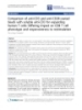
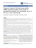
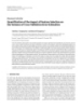
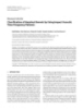
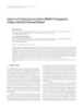
- Merlo et al. Journal of Translational Medicine 2010, 8:121 Page 3 of 8 http://www.translational-medicine.com/content/8/1/121 (IL7Ra; BioLegend). Cells (2 × 10⁵) were washed with + + CD4 CD8 phosphate-buffered saline (PBS; Sigma-Aldrich) and re- 50 120 IL2 Total CTL count (x106) suspended in 50 μl of staining solution (PBS, 3% FBS IL7 100 IL15 40 and 0,1% NaN3) containing an optimal concentration of IL21 80 30 antibody. After a 20-minute incubation in ice, cells were 60 20 washed again and analyzed using a FacsCalibur (BD) 40 10 flow cytometer. Flow cytometry data were analyzed with 20 0 FlowJo software (Tree Star, Inc.; Ashland, OR). 0 4 4 1 2 3 1 2 3 Stimulation number Figure 1 Growth kinetics of CD4+ and CD8+ T cell lines. The ELISA test extrapolated mean total cell counts of CD4+ (left) and CD8+ (right) Cytokine ELISA tests were performed using Human T cell lines cultured with IL-2, IL-7, IL-15 and IL-21 before each re- TNF a Screening Set and Human IFN g Screening Set stimulation with LCL is represented. Figure shows mean values from (Thermo Scientific, Rockford, IL), according to the man- at least two independent experiments. ufacturer’s instructions. Briefly, 2 × 10⁵ effector cells and 2 × 10⁵ autologous LCL were seeded in 96-well round- after 3 to 7 re-stimulations (Figure 1 and data not bottom plates. Positive controls were represented by shown). IL-15 produced a similar trend in CD4 + and effector T cells incubated with PMA-ionomycin (40 ng/ CD8+ T cell growth and proved to be superior to other ml and 4 μg/ml, respectively; Sigma-Aldrich). Baseline tested cytokines in inducing the expansion of both sub- cytokine production was determined in supernatants populations, while IL-7 supported the expansion of CD4+ from unstimulated T cells, or LCL only. Cytokine secre- T cells only, albeit at different degrees of magnitude tion was measured after 5h-incubation. for different donors. In deep contrast, IL-21 alone allowed survival but did not sustain the expansion of Outgrowth assay either subsets of T cells, in line with previously reported Outgrowth assay was carried out as previously described data [14-16]. [12]. Briefly, target LCL were seeded as replicates in U- bottom 96-well plates at doubling dilution, starting from 104 cells/well to 78 cells/well. T cells were added to half Assessment of phenotype of the replicates at 104 cells/well in HS complete med- The use of different cytokines in culture could impact on differentiation, trafficking and functional properties ium without IL-2. Plates were then incubated at 37°C in of T cells, characteristics that have a counterpart on 6.5% CO2 and re-feeded weekly by replacing half of the specific surface marker expression [3]. We therefore medium. LCL outgrowth was scored after 4 weeks by analyzed the expression of CD27, CD28, CD57, CD62L, visual examination with an inverted microscope. Results IL7Ra, and CCR7 at different time points during cul- are expressed as the minimum number of LCL required ture. We performed flow cytometry analysis at day 0 for successful outgrowth in 50% of replicate wells. just before seeding, at day 14 before immunomagnetic separation and cytokine addition, and after 1 month of Results culture. At 2 months, phenotype of CD4+ T cells only Analysis of in vitro growth kinetics could be evaluated, since CD8+ T lymphocytes did not To dissect the impact of different g-chain cytokines on proliferate so long. The phenotype of IL-21 T cells human T cell in vitro expansion, we took advantage of a could not be determined due to the low number of lym- well defined protocol aimed at generating EBV-specific phocytes obtained in these cultures. As shown in Figure T cells cultures [10,13]. First, we evaluated the prolifera- 2, overall we found more pronounced differences in the tive potential of CTL lines cultured with IL-15, IL-7 or phenotypic profile of CD8+ and CD4+ T cells prior to IL-21 in comparison to IL-2. Briefly, we seeded PBMC the addition of the various cytokines than after their from healthy donors with autologous LCL without cyto- supplement to cultures. Indeed, immediately after ex kine addition for the selection phase. Two weeks later, vivo collection, nearly all CD4+ T cells expressed CD27, the expansion phase was started by supplying different CD28, CD62L, IL7Ra, in comparison to only about 50% cytokines to purified CD8+ and CD4+ T cells, to assess of CD8 + T cells. Conversely, CD8 + T cells tended to their proliferative response. As expected, we found that both CD8+ and CD4+ T cells grew vigorously when cul- acquire CD27 and CD28 expression in culture, differ- ently from what observed by Vanhoutte et al. [17], while tured with IL-2, although with differential magnitudes. IL7Ra and CD62L were poorly represented in this sub- In particular, CD4+ T cells grew for a longer time (more set respect to the CD4+ T cell counterpart. These latter than 14 weeks) in comparison to CD8 + T cells, which cells, on the contrary, partly lost the CD27 expression disclosed an initial phase of logarithmic growth followed during culture. The expression of CCR7, which by a progressive reduction of their active proliferation
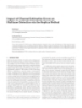
- Merlo et al. Journal of Translational Medicine 2010, 8:121 Page 4 of 8 http://www.translational-medicine.com/content/8/1/121 + + CD4 CD8 100 100 % expression 80 80 day 0 60 60 40 40 20 20 0 0 IL7Ralfa CD27 CD28 CD62L IL7Ralfa CD27 CD28 CD62L 100 100 80 80 % expression 60 60 day 14 40 40 20 20 0 0 IL7Ralfa CD27 CD28 CD62L IL7Ralfa CD27 CD28 CD62L IL2 IL2 100 100 IL15 IL15 IL7 IL7 80 80 % expression 60 60 1 month 40 40 20 20 0 0 IL7Ralfa CD27 CD28 CD62L IL7Ralfa CD27 CD28 CD62L IL2 100 IL15 IL7 80 % expression 60 2 months 40 20 0 IL7Ralfa CD27 CD28 CD62L Figure 2 Expression of maturation/differentiation markers. Figure shows marker expression by CD4+ and CD8+ T cells at day 0, 14 (before separation and cytokine supply), 1 month and, for CD4+ T cells only, 2 months. Figure shows mean +/- SD of 3 replicate cultures from 2 donors.
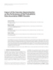
- Merlo et al. Journal of Translational Medicine 2010, 8:121 Page 5 of 8 http://www.translational-medicine.com/content/8/1/121 a ppeared initially quite variable between CD4 + and in response to LCL stimulation in comparison to IL-7 CD8+ T cells, was lost by all T cell lines from the third cultures, but comparable levels in response to PMA- week of culture and thereafter (data not shown); on the ionomycin (Figure 3). Cytokine production by IL-21 T other hand, CD44 was expressed at high intensity in cells could not be assessed due to the low number of nearly all T cells for the entire period of culture (data lymphocytes obtained in cultures. not shown). CD57 expression was quite different between CD4+ and CD8+ T cells (4.35 +/- 3.44% versus Analysis of in vitro functional activity 22.99 +/- 5.15% immediately after ex vivo collection, In vitro functional activity was assessed both in short- respectively); in fact, it was rapidly up-regulated and term and long-term assays. Standard cytotoxicity tests then lost by CD4 + T cells, while retained by CD8 + were performed with T cell lines at 21 days of culture. T cells (data not shown). Finally, after 1 month of cul- At this time point (third restimulation, see Figure 1), we could test all the cell lines obtained but IL-21 CD4+ T ture the phenotypic profile tended to stabilize and did not further modify substantially at least for the CD4+ cells. Although NK cell presence was negligible (< 1%), T cell subset, the only one that could be tested. nevertheless all tests were carried out in the presence or absence of an excess of “ cold ” K562 to eliminate any possible influence of NK-like activity. As shown in Evaluation of cytokine production Next, we investigated the production of cytokines by Figure 4a, the addition of different cytokines did not modify the lytic activity of either CD8+ or CD4+ T cells. cultures in response to different stimuli, such as autolo- gous LCL and PMA-ionomycin, to verify whether the Notably, in contrast with recently published data [7], IL-21-cultured CD8+ T cells showed a strong lytic activ- conditions tested have an impact on cytokine produc- tion. In particular, we studied the production of Th1 ity similar to that of cognate IL-2 cultures. To assess the cytokines, namely IFN g and TNF a , which play an mechanisms involved in lytic activity we focused on CD4 + T cells, as no clear preferential use of granule important role in anti-tumor immunity [18,19]. We found that IL-2, IL-7, and IL-15 CD8+ T cell cultures exocytosis or apoptosis induction is described for this produced comparable amounts of IFN g and TNF a in subset. By using compounds that selectively inhibit per- response to both stimuli (Figure 3). Moreover, while IL- forin-based or Fas/FasL-based pathway, we found that 2, IL-7, and IL-15 CD4+ T cells did not display relevant all CD4+ T cells obtained, irrespectively of culture con- differences in the amount of TNFa secreted, IL-2 and ditions, killed their targets through the cytotoxic granule IL-15 CD4+ T cells produced a higher amount of IFNg content release (Figure 4b). These findings are in line with our previous observations [13] and the vast major- ity of data related to EBV-specific cultures [20]. Once again, cytokines used in cultures did not modify func- CD4+ CD8+ tional activity. 4000 4000 Although commonly used to evaluate functionality of TNFa production (pg/ml) IL2 IL2 IL7 IL7 effector T cells, the cytotoxic activity does not always IL15 IL15 3000 3000 correlate with in vivo efficacy, as recently demon- strated not only in mouse models [4] but also in clini- 2000 2000 cal trials [21]. After adoptive transfer, a clue characteristic is the capacity of effector cells to per- 1000 1000 form sequential killings before exhaustion. As this 0 0 issue can not be adequately addressed in a short-term us us PMA PMA LCL LCL test, we performed outgrowth assays that evaluate the IL2 1400 1400 IL2 IFNg production (pg/ml) IL15 IL7 ability of a fixed input of T cells to inhibit long-term IL15 1200 1200 growth of different numbers of target cells, without the 1000 1000 addition of cytokines. This experimental design closely 800 800 resembles in vivo adoptive transfer protocols, which 600 600 are based on a single infusion of effector T cells with- 400 400 out exogenous cytokine supply [13,22]. In both cases, 200 200 0 0 T cells do not likely survive longer than a few days, us us PMA PMA L CL LCL when they can display their killing potential. Thus, the Figure 3 Th1 cytokine production. Figure shows TNFa and IFNg extent of target elimination could be predictive of the production by CD4+ (left panel) and CD8+ (right panel) T cells in response to stimulation with autologous LCL or PMA-ionomycin, or outcome: even few surviving tumor cells can ultimately unstimulated (us), as assessed by ELISA test. Figure shows mean ± lead to a successful microculture outgrowth or to the SD of 3 replicate cultures from 2 donors. death of the engrafted animals. Due to the low number
- Merlo et al. Journal of Translational Medicine 2010, 8:121 Page 6 of 8 http://www.translational-medicine.com/content/8/1/121 CD8+ CD4+ CD4+ a) b) LCL LCL LCL + CMA K562 LCL + EGTA 80 80 80 LCL + BFA LCL + anti FasL 60 60 60 IL2 40 40 40 20 20 20 0 0 0 1 3 6 12 25 50 1 3 6 12 25 50 1 3 6 12 25 50 80 80 80 60 60 60 IL7 40 40 40 20 20 20 0 0 0 1 3 6 12 25 50 1 3 6 12 25 50 1 3 6 12 25 50 Specific lysis (%) 80 80 80 60 60 60 IL15 40 40 40 20 20 20 0 0 0 1 3 6 12 25 50 1 3 6 12 25 50 1 3 6 12 25 50 Specific lysis (%) Effector:target ratio 80 60 IL21 40 20 0 1 3 6 12 25 50 Effector:target ratio Figure 4 Lytic activity of EBV-specific CD8+ and CD4+ T cells. A) Cytotoxic activity was tested by standard 4 h 51Cr-release assay in the presence of “cold” K562 at a 5:1 ratio of “cold": “hot” target. B) Lytic mechanisms involved in cytotoxicity. CD4+ T cell line cytotoxicity was evaluated in the presence of CMA and EGTA that block perforin-based pathway, and BFA and anti-FasL mAb that interfere with Fas/FasL-based pathway. Figure shows mean values from 3 independent experiments carried out for each donor cell line. o f cells required (as few as 0.32 × 10 6 cells for each Discussion test), in this case we could test every cell line obtained. Recent advances in immunotherapeutic approaches have In line with our previous results (data not shown), IL- highlighted the importance of infusing antigen-specific 2-cultured CD8+ T cells disclosed a superior ability to T cells that have ideally a poorly differentiated pheno- inhibit long-term growth of target cells in comparison type and are characterized by a strong proliferative to their CD4+ T cell counterpart; a similar trend was potential upon in vivo transfer. These conditions have observed for CD8+ T lymphocytes cultured in IL-7 or been partially met by acting on recipient patients with IL-15. Instead, the reverse was true for CD8 + T cells lymphodepleting strategies or by proposing the shorten- supplied with IL-21. Finally, striking was the finding ing of T cell in vitro expansion protocols with the use that IL-15 CD4 + T cells, despite a vigorous in vitro of “less differentiating” cytokines. With regard to this cytotoxic activity in short-term assay, did not exert any latter issue, we exploited a protocol successfully used in inhibitory potential (Figure 5). immunotherapeutic approaches for EBV-related
- Merlo et al. Journal of Translational Medicine 2010, 8:121 Page 7 of 8 http://www.translational-medicine.com/content/8/1/121 belonging to the memory compartment and therefore the obtainment of less differentiated cells is expected to >10000 be difficult. The long and likely confounding selection 10000 phase could be bypassed by performing faster (e.g., over- 5000 night) peptide mix stimulation followed by immunomag- LCL input netic isolation of cytokine-producing T cells, as recently 2500 proposed [25], or by introducing the wanted antigen 1250 specificity through CAR- or transgenic TCR-coding vec- tor transduction [26,27]. In these cases, the alternatively 625 chosen cytokines could be added in a less precondi- 313 tioned milieu, thus driving a less pronounced differen- 157 tiation of responding T lymphocytes, or, in the case of CAR or TCR transfer, of the whole population of trans- 78 + + + + + + + + CD4 CD8 CD4 CD8 CD4 CD8 CD4 CD8 duced peripheral T cells. IL2 IL7 IL15 IL21 Overall, although the use of g-chain cytokines other Figure 5 Inhibition of LCL outgrowth by EBV-specific CTL than IL-2 did not produce any substantial in vitro cultured with different cytokines. Results are expressed as the improvement, a realistic and clear-cut description of the minimum LCL number required for successful outgrowth at day 28 activity of a determined T cell population should be of culture (black circles). These values are compared with the derived by in vivo studies. In this regard, however, we corresponding results for outgrowth of LCL seeded without effector T cells (dotted line). Figure shows mean values from 3 independent could not produce definitive results since we only had experiments performed for each donor cell line. the possibility to test those cultures that reach a suffi- cient number for infusion. Moreover, the PTLD-SCID mouse model suffers from different intrinsic biases that malignancies to compare the impact of different g-chain might have frustrated the purpose of our study. In fact, we have evidence that human T cells survive no longer cytokines on phenotype and functionality of cultured T than 24 hr after in vivo transfer [13], even when this fol- cells, as suggested by recent studies [4,7,8]. We analyzed lows irradiation or cyclophosphamide treatment of reci- purified CD8+ and CD4+ T cells to avoid potential pient mice. Moreover, this poor survival was verified not influence of one population on the other one; indeed, despite a trend toward a “natural” expansion of CD8+T only for EBV-specific T cells, but also for less differen- tiated, CAR-transduced antigen-specific T cells (data not cells, the percentage of CD4+T cells in cultures turns shown). In addition, due to the intrinsic differences out to be quite various among different donors and dif- between mouse and human adhesion molecules and ferent preparations from the same donor. Our choice receptors, it is hard to evaluate the lymph node homing furthermore took into account the increasing attention paid on the CD4 + T cells as actual effector cells in and recirculation capacity that have a fundamental role in the more physiological model described by Gattinoni immunotherapeutic approaches [23,24]. et al. [4], which envisages the transfer of mouse T cells Intriguingly, the results presented herein are pro- into a syngeneic murine microenvironment. In such foundly different from those of recently published stu- experimental context, moreover, the concomitant vacci- dies. Previous reports, in fact, mainly rely on murine T nation strategies make the lymph node homing proper- cells derived from mice expressing transgenic TCR spe- ties even more relevant, as they dramatically contribute cific for the antigen of interest. All T cells have there- to the improvement of the final outcome [4]. Thus, it is fore the desired specificity and hence they only need to left to be verified in a human context the impact of dif- be activated in vitro, bypassing a potentially long selec- ferent lymphoid homing marker expression on the out- tion phase. Conversely, this phase was absolutely come of adoptive transfer strategies. required by our protocol, and covered the first 14 days of culture. Moreover, our protocol envisages the addi- Conclusions tion of cytokine only after this phase. During this gap, As a whole, our results indicate the need to design ad EBV-specific T cells that are present in PBMC of sero- hoc protocols to appreciate the impact of g-chain cyto- positive donors respond to the viral antigens presented kines other than IL-2 on the functionality of CTL for by LCL, very likely producing IL-2 that in turn can adoptive cell therapy. influence the culture. In this regard, IL-21 has been reported to be capable of reverting the IL-2-induced dif- ferentiation [7], but no information is available for IL-7 Acknowledgements and IL-15. In addition, it must be noted that in vitro This study was partly supported by grants from the Italian Ministry of Health expansion selectively involved EBV-specific precursors (Progetto oncologico di medicina molecolare: i tumori femminili; Progetto
- Merlo et al. Journal of Translational Medicine 2010, 8:121 Page 8 of 8 http://www.translational-medicine.com/content/8/1/121 strategico: Farmaci cellulari, vaccini e bioterapie innovative dei tumori; 12. Long HM, Haigh TA, Gudgeon NH, Leen AM, Tsang CW, Brooks J, Landais E, Alleanza Contro il Cancro, ACC-4), the European Community (FP6 VITAL, Houssaint E, Lee SP, Rickinson AB, Taylor GS: CD4+ T-cell responses to Contract no. 037874) and the Italian Association for Cancer Research (AIRC). Epstein-Barr virus (EBV) latent-cycle antigens and the recognition of EBV-transformed lymphoblastoid cell lines. J Virol 2005, 79:4896-4907. Author details 13. Merlo A, Turrini R, Bobisse S, Zamarchi R, Alaggio R, Dolcetti R, Mautner J, 1 University of Padova, Dept. of Oncology and Surgical Sciences, Via Zanovello P, Amadori A, Rosato A: Virus-Specific Cytotoxic CD4+ T Cells Gattamelata 64, 35128 Padova, Italy. 2Department of Haematology, Imperial for the Treatment of EBV-Related Tumors. J Immunol 2010, 184:5895-902. College, Du Cane Road, London, UK. 3Istituto Oncologico Veneto IRCCS, Via 14. Zeng R, Spolski R, Casas E, Zhu W, Levy DE, Leonard WJ: The molecular Gattamelata 64, 35128 Padova, Italy. 4CRO, Centro Riferimento Oncologico basis of IL-21-mediated proliferation. Blood 2007, 109:4135-4142. IRCCS, Via F. Gallini 2, 33081 Aviano, Italy. 15. Kaka AS, Shaffer DR, Hartmaier R, Leen AM, Lu A, Bear A, Rooney CM, Foster AE: Genetic modification of T cells with IL-21 enhances antigen Authors’ contributions presentation and generation of central memory tumor-specific cytotoxic AM analyzed and interpreted data and wrote the manuscript. RT performed T-lymphocytes. J Immunother 2009, 32:726-736. 16. Kinter AL, Godbout EJ, McNally JP, Sereti I, Roby GA, O’Shea MA, Fauci AS: flow cytometry analysis and wrote the manuscript. CT carried out experimental work. PZ and RD critically revised the manuscript. AR The common gamma-chain cytokines IL-2, IL-7, IL-15, and IL-21 induce conceived the study, and participated in its design and coordination. All the expression of programmed death-1 and its ligands. J Immunol 2008, authors read and approved the final manuscript. 181:6738-6746. 17. Vanhoutte VJ, McAulay KA, McCarrell E, Turner M, Crawford DH, Haque T: Competing interests Cytolytic mechanisms and T-cell receptor Vbeta usage by ex vivo The authors declare that they have no competing interests. generated Epstein-Barr virus-specific cytotoxic T lymphocytes. Immunology 2009, 127:577-586. Received: 17 August 2010 Accepted: 22 November 2010 18. Tannenbaum CS, Hamilton TA: Immune-inflammatory mechanisms in Published: 22 November 2010 IFNgamma-mediated anti-tumor activity. Semin Cancer Biol 2000, 10:113-123. 19. Knutson KL, Disis ML: Tumor antigen-specific T helper cells in cancer References immunity and immunotherapy. Cancer Immunol Immunother 2005, 1. Peggs KS: Adoptive T cell immunotherapy for cytomegalovirus. Expert 54:721-728. Opin Biol Ther 2009, 9:725-736. 20. Sun Q, Burton RL, Lucas KG: Cytokine production and cytolytic 2. Rosenberg SA, Restifo NP, Yang JC, Morgan RA, Dudley ME: Adoptive cell mechanism of CD4(+) cytotoxic T lymphocytes in ex vivo expanded transfer: a clinical path to effective cancer immunotherapy. Nat Rev therapeutic Epstein-Barr virus-specific T-cell cultures. Blood 2002, Cancer 2008, 8:299-308. 99:3302-3309. 3. Gattinoni L, Powell DJ Jr, Rosenberg SA, Restifo NP: Adoptive 21. Haque T, Wilkie GM, Jones MM, Higgins CD, Urquhart G, Wingate P, immunotherapy for cancer: building on success. Nat Rev Immunol 2006, Burns D, McAulay K, Turner M, Bellamy C, Amlot PL, Kelly D, MacGilchrist A, 6:383-393. Gandhi MK, Swerdlow AJ, Crawford DH: Allogeneic cytotoxic T-cell 4. Gattinoni L, Klebanoff CA, Palmer DC, Wrzesinski C, Kerstann K, Yu Z, therapy for EBV-positive posttransplantation lymphoproliferative disease: Finkelstein SE, Theoret MR, Rosenberg SA, Restifo NP: Acquisition of full results of a phase 2 multicenter clinical trial. Blood 2007, 110:1123-1131. effector function in vitro paradoxically impairs the in vivo antitumor 22. Lacerda JF, Ladanyi M, Louie DC, Fernandez JM, Papadopoulos EB, efficacy of adoptively transferred CD8+ T cells. J Clin Invest 2005, O’Reilly RJ: Human Epstein-Barr virus (EBV)-specific cytotoxic T 115:1616-1626. lymphocytes home preferentially to and induce selective regressions of 5. Dudley ME, Yang JC, Sherry R, Hughes MS, Royal R, Kammula U, Robbins PF, autologous EBV-induced B cell lymphoproliferations in xenografted C.B- Huang J, Citrin DE, Leitman SF, Wunderlich J, Restifo NP, Thomasian A, 17 scid/scid mice. J Exp Med 1996, 183:1215-1228. Downey SG, Smith FO, Klapper J, Morton K, Laurencot C, White DE, 23. Quezada SA, Simpson TR, Peggs KS, Merghoub T, Vider J, Fan X, Blasberg R, Rosenberg SA: Adoptive cell therapy for patients with metastatic Yagita H, Muranski P, Antony PA, Restifo NP, Allison JP: Tumor-reactive melanoma: evaluation of intensive myeloablative chemoradiation CD4+ T cells develop cytotoxic activity and eradicate large established preparative regimens. J Clin Oncol 2008, 26:5233-5239. melanoma after transfer into lymphopenic hosts. J Exp Med 2010, 6. Louis CU, Straathof K, Bollard CM, Gerken C, Huls MH, Gresik MV, Wu MF, 207:637-650. Weiss HL, Gee AP, Brenner MK, Rooney CM, Heslop HE, Gottschalk S: 24. Xie Y, Akpinarli A, Maris C, Hipkiss EL, Lane M, Kwon EK, Muranski P, Enhancing the in vivo expansion of adoptively transferred EBV-specific Restifo NP, Antony PA: Naive tumor-specific CD4+ T cells differentiated in CTL with lymphodepleting CD45 monoclonal antibodies in NPC patients. vivo eradicate established melanoma. J Exp Med 2010, 207:651-667. Blood 2009, 113:2442-2450. 25. Moosmann A, Bigalke I, Tischer J, Schirrmann L, Kasten J, Tippmer S, 7. Hinrichs CS, Spolski R, Paulos CM, Gattinoni L, Kerstann KW, Palmer DC, Leeping M, Prevalsek D, Jaeger G, Ledderose G, Mautner J, Klebanoff CA, Rosenberg SA, Leonard WJ, Restifo NP: IL-2 and IL-21 confer Hammerschmidt W, Schendel DJ, Kolb HJ: Effective and long-term control opposing differentiation programs to CD8+ T cells for adoptive of EBV PTLD after transfer of peptide-selected T cells. Blood 2010, immunotherapy. Blood 2008, 111:5326-5333. 115:2960-70. 8. Hinrichs CS, Borman ZA, Cassard L, Gattinoni L, Spolski R, Yu Z, Sanchez- 26. Vera J, Savoldo B, Vigouroux S, Biagi E, Pule M, Rossig C, Wu J, Heslop HE, Perez L, Muranski P, Kern SJ, Logun C, Palmer DC, Ji Y, Reger RN, Rooney CM, Brenner MK, Dotti G: T lymphocytes redirected against the Leonard WJ, Danner RL, Rosenberg SA, Restifo NP: Adoptively transferred kappa light chain of human immunoglobulin efficiently kill mature B effector cells derived from naive rather than central memory CD8+ T lymphocyte-derived malignant cells. Blood 2006, 108:3890-3897. cells mediate superior antitumor immunity. Proc Natl Acad Sci USA 2009, 27. Bobisse S, Rondina M, Merlo A, Tisato V, Mandruzzato S, Amendola M, 106:17469-17474. Naldini L, Willemsen RA, Debets R, Zanovello P, Rosato A: Reprogramming 9. Kaneko S, Mastaglio S, Bondanza A, Ponzoni M, Sanvito F, Aldrighetti L, T lymphocytes for melanoma adoptive immunotherapy by T-cell Radrizzani M, La Seta-Catamancio S, Provasi E, Mondino A, Nagasawa T, receptor gene transfer with lentiviral vectors. Cancer Res 2009, Fleischhauer K, Russo V, Traversari C, Ciceri F, Bordignon C, Bonini C: IL-7 69:9385-9394. and IL-15 allow the generation of suicide gene-modified alloreactive self-renewing central memory human T lymphocytes. Blood 2009, doi:10.1186/1479-5876-8-121 113:1006-1015. Cite this article as: Merlo et al.: Impact of g-chain cytokines on EBV- 10. Rooney CM, Smith CA, Ng CY, Loftin S, Li C, Krance RA, Brenner MK, specific T cell cultures. Journal of Translational Medicine 2010 8:121. Heslop HE: Use of gene-modified virus-specific T lymphocytes to control Epstein-Barr-virus-related lymphoproliferation. Lancet 1995, 345:9-13. 11. Rosato A, Milan G, Collavo D, Zanovello P: DNA-based vaccination against tumors expressing the P1A antigen. Methods 1999, 19:187-190.
CÓ THỂ BẠN MUỐN DOWNLOAD

-

Báo cáo hóa học: " Impact of Video Coding on Delay and Jitter in 3G Wireless Video Multicast Services"
 7 p |
7 p |  35
|
35
|  7
7
-
Báo cáo hóa học: " Impact of AFM-induced nano-pits in a-Si:H films on silicon crystal growth"
6 p | 58
| 7
-

Báo cáo sinh học: " Impact of changes in diet on the availability of land, energy demand and greenhouse gas emissions of agriculture"
27 p | 52
| 7
-

Báo cáo hóa học: " Prognostic impact of ZAP-70 expression in chronic lymphocytic leukemia: mean fluorescence intensity T/B ratio versus percentage of positive cells"
11 p | 61
| 6
-
Báo cáo hóa học: "Comparison of anti-CD3 and anti-CD28-coated beads with soluble anti-CD3 for expanding human T cells: Differing impact on CD8 T cell phenotype and responsiveness to restimulation"
15 p | 51
| 5
-
Báo cáo hóa học: "Prognostic impact of clinical course-specific mRNA expression profiles in the serum of perioperative patients with esophageal cancer in the ICU: a case control study"
11 p | 67
| 5
-
Báo cáo hóa học: " Impact of the Gaussian Approximation on the Performance of the Probabilistic Data Association MIMO Decoder"
5 p | 44
| 5
-

Báo cáo hóa học: " Research Article Impact of Radio Link Unreliability on the Connectivity of Wireless Sensor Networks"
16 p | 52
| 5
-
Báo cáo hóa học: " The Impact of Channel Estimation Errors and Co-antenna Interference on the Performance of a Coded MIMO System"
18 p | 48
| 4
-
Báo cáo hóa học: " Impact and Mitigation of Multiantenna Analog Front-End Mismatch in Transmit Maximum Ratio Combining"
14 p | 32
| 4
-
Báo cáo hóa học: " Research Article Quantification of the Impact of Feature Selection on the Variance of Cross-Validation Error Estimation"
11 p | 60
| 4
-
Báo cáo hóa học: " Research Article Classification of Hazelnut Kernels by Using Impact Acoustic Time-Frequency Patterns"
11 p | 35
| 4
-

Báo cáo hóa học: " Research Article Assessing Task Migration Impact on Embedded Soft Real-Time Streaming Multimedia "
15 p | 50
| 4
-

Báo cáo hóa học: " Research Article Assessing Task Migration Impact on Embedded Soft Real-Time Streaming Multimedia Applications"
15 p | 46
| 4
-

Báo cáo hóa học: "Prognostic Impact of MiR-155 in Non-Small Cell Lung Cancer Evaluated by in Situ Hybridization"
9 p | 63
| 4
-
Báo cáo hóa học: "Impact of Clustering in Indoor MIMO Propagation Using a Hybrid Channel Model"
14 p | 39
| 4
-
Báo cáo hóa học: " Impact of Channel Estimation Errors on Multiuser Detection via the Replica Method"
12 p | 45
| 3


Chịu trách nhiệm nội dung:
Nguyễn Công Hà - Giám đốc Công ty TNHH TÀI LIỆU TRỰC TUYẾN VI NA
LIÊN HỆ
Địa chỉ: P402, 54A Nơ Trang Long, Phường 14, Q.Bình Thạnh, TP.HCM
Hotline: 093 303 0098
Email: support@tailieu.vn
Giấy phép Mạng Xã Hội số: 670/GP-BTTTT cấp ngày 30/11/2015 Copyright © 2022-2032 TaiLieu.VN. All rights reserved.

