


Báo cáo y học: "Inactivation of HIV-1 in breast milk by treatment with the alkyl sulfate microbicide sodium dodecyl sulfate (SDS)"
lượt xem 2
download
 Download
Vui lòng tải xuống để xem tài liệu đầy đủ
Download
Vui lòng tải xuống để xem tài liệu đầy đủ
Tuyển tập các báo cáo nghiên cứu về y học được đăng trên tạp chí y học quốc tế cung cấp cho các bạn kiến thức về ngành y đề tài: "Inactivation of HIV-1 in breast milk by treatment with the alkyl sulfate microbicide sodium dodecyl sulfate (SDS)...
Bình luận(0) Đăng nhập để gửi bình luận!
Nội dung Text: Báo cáo y học: "Inactivation of HIV-1 in breast milk by treatment with the alkyl sulfate microbicide sodium dodecyl sulfate (SDS)"
- Retrovirology BioMed Central Open Access Research Inactivation of HIV-1 in breast milk by treatment with the alkyl sulfate microbicide sodium dodecyl sulfate (SDS) Sandra Urdaneta*1,8, Brian Wigdahl2, Elizabeth B Neely1,3, Cheston M Berlin Jr4,5, Cara-Lynne Schengrund6, Hung-Mo Lin7 and Mary K Howett1,8 Address: 1Department of Microbiology and Immunology, Penn State College of Medicine, Hershey, Pennsylvania 17033 USA, 2Department of Microbiology and Immunology, Institute for Molecular Medicine and Infectious Diseases, Drexel University, College of Medicine, Philadelphia, Pennsylvania 19104 USA, 3Department of Neural and Behavioral Sciences, Penn State College of Medicine, Hershey, Pennsylvania 17033 USA, 4Department of Pediatrics, Penn State College of Medicine, Hershey, Pennsylvania 17033 USA, 5Department of Pharmacology, Penn State College of Medicine, Hershey, Pennsylvania 17033 USA, 6Department of Biochemistry, Penn State College of Medicine, Hershey, Pennsylvania 17033 USA, 7Department of Health Evaluation Sciences, Penn State College of Medicine, Hershey, Pennsylvania 17033 USA and 8Department of Bioscience and Biotechnology, Drexel University, College of Medicine, Philadelphia, Pennsylvania 19104 USA Email: Sandra Urdaneta* - sandra.urdaneta@drexel.edu; Brian Wigdahl - Brian.Wigdhal@DrexelMed.edu; Elizabeth B Neely - eneely@psu.edu; Cheston M Berlin - cmb6@drexel.edu; Cara-Lynne Schengrund - cxs8@psu.edu; Hung-Mo Lin - hlin@psu.edu; Mary K Howett - mkh28@drexel.edu * Corresponding author Published: 29 April 2005 Received: 14 February 2005 Accepted: 29 April 2005 Retrovirology 2005, 2:28 doi:10.1186/1742-4690-2-28 This article is available from: http://www.retrovirology.com/content/2/1/28 © 2005 Urdaneta et al; licensee BioMed Central Ltd. This is an Open Access article distributed under the terms of the Creative Commons Attribution License (http://creativecommons.org/licenses/by/2.0), which permits unrestricted use, distribution, and reproduction in any medium, provided the original work is properly cited. Abstract Background: Reducing transmission of HIV-1 through breast milk is needed to help decrease the burden of pediatric HIV/AIDS in society. We have previously reported that alkyl sulfates (i.e., sodium dodecyl sulfate, SDS) are microbicidal against HIV-1 at low concentrations, are biodegradable, have little/no toxicity and are inexpensive. Therefore, they may be used for treatment of HIV-1 infected breast milk. In this report, human milk was artificially infected by adding to it HIV-1 (cell-free or cell-associated) and treated with ≤1% SDS (≤10 mg/ml). Microbicidal treatment was at 37°C or room temperature for 10 min. SDS removal was performed with a commercially available resin. Infectivity of HIV-1 and HIV-1 load in breast milk were determined after treatment. Results: SDS (≥0.1%) was virucidal against cell-free and cell-associated HIV-1 in breast milk. SDS could be substantially removed from breast milk, without recovery of viral infectivity. Viral load in artificially infected milk was reduced to undetectable levels after treatment with 0.1% SDS. SDS was virucidal against HIV-1 in human milk and could be removed from breast milk if necessary. Milk was not infectious after SDS removal. Conclusion: The proposed treatment concentrations are within reported safe limits for ingestion of SDS by children of 1 g/kg/day. Therefore, use of alkyl sulfate microbicides, such as SDS, to treat HIV1-infected breast milk may be a novel alternative to help prevent/reduce transmission of HIV- 1 through breastfeeding. Page 1 of 10 (page number not for citation purposes)
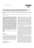
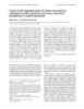
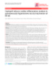
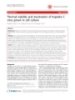
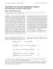
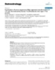
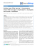
- Retrovirology 2005, 2:28 http://www.retrovirology.com/content/2/1/28 (i.e., SDS) of breast milk infected with HIV-1 has been Background As proven in developed countries, MTCT of HIV-1 is pre- examined. We hypothesize that treatment of expressed ventable with highly active antiretroviral therapy com- breast milk with this microbicide will effectively inactivate bined with total avoidance of breastfeeding. The most HIV-1 in breast milk. Efficiency of viral inactivation in widely promoted mode of replacement feeding is the use breast milk is hereon reported. The effects of microbicidal of infant formula. However, thus far, it has not been treatment on breast milk components have also been applicable in resource-constrained countries, the epi- studied (i.e., gross protein content, immunoglobulins, center of the HIV/AIDS epidemic. In this setting, lack of lipids and energy content, cellular fraction, electrolytes) clean water, absence of financial resources to purchase and no significant changes were observed[10,11]. The formula, and cultural stigma represent stumbling blocks results of the biochemical analysis of breast milk treated for a generalized implementation of this prevention plan. with SDS will be published elsewhere. Alternatives to reduce, if not prevent, the risk of transmis- sion of HIV-1 through breast milk are in demand to act in Results synergy with antiretroviral regimens that prevent peripar- Virucidal activity of SDS against HIV-1 in breast milk tum transmission of HIV-1. Here we introduce the novel The virucidal activity of SDS against cell-free HIV-1 in concept of using microbicides to treat HIV-1 infected breast milk was assessed by adding high titer HIV-1 IIIB to breast milk to prevent MTCT of HIV-1. breast milk obtained from apparently healthy donors of unknown HIV serostatus. Within 1 min of incubation of The alkyl sulfate family of microbicides are agents with breast milk containing cell-free HIV-1 with 0.1% SDS, both surfactant and protein denaturant properties. The HIV-1 infectivity was decreased to uninfected control lev- prototypic alkyl sulfate, sodium dodecyl sulfate (SDS, els (Figure 1A). The minimum concentration of 0.05% C12H26O4SNa, CAS No. 151-21-3), is an anionic sur- was required to observe inactivation of HIV-1 (Figure 1B). factant and detergent. SDS is a common ingredient used Infectivity of cell-associated HIV-1 (i.e., HIV-1-infected in the cosmetic and personal care products industry (e.g., Sup-T1 cells) was abolished with treatment with 0.1% toothpastes, shampoos, bubble baths, dishwashing for- SDS. This inactivation was due to induced lysis of Sup-T1 mulations, moisturizing lotions, baby wipes, etc.), and in cells at this concentration (data not shown). Cell-associ- the laboratory environment as a denaturing agent in gel ated HIV-1 was partially susceptible to 0.01% SDS (Figure electrophoresis and other protein solubilization tech- 2A). Nonetheless, even when cell lysis is absent is not an niques[1,2]. SDS is listed in the Generally Recognized As issue low SDS concentrations abolished cell-associated Safe (GRAS) list of chemicals of the United States Food HIV-1 infectivity. With 0.01% SDS, maximum inactiva- and Drug Administration (FDA)[3]. Also, the United tion of infectious cell-associated HIV-1 was achieved Nations Environment Programme (UNEP) has classified within 7 min of treatment (Figure 2B). Using branched SDS as "readily biodegradable" and, after extensive toxico- DNA technology to determine HIV-1 load in spiked breast milk samples treated with ≥1% SDS, it was determined logical analysis, UNEP concluded that "sodium dodecyl sulfate is of no concern with respect to human health"[2]. that viral RNA titers were reduced to undetectable levels According to this report, the Estimated Human Exposure (Figure 3). (EHE) level of SDS on a daily basis is 0.158 mg/kg/day and 0.034 mg/kg/day, in children (15 kg of weight) and Removal of SDS from breast milk babies (5 kg) respectively. This includes exposure by Despite the overall benign nature of SDS, the possibility means of body lotions and oral intake by means of con- of removing SDS from breast milk in case it was deemed taminated water or food and toothpaste. The maximum necessary or desirable prior to feeding was still examined. safe ingested dose for children is estimated to be up to 1.0 Several methods were assessed with respect to their effi- g/kg/day[4]. ciency of removing SDS from the breast milk preparations (i.e. potassium salts, Microcon® YM-10 [Amicon®, Inc.], SDS 300-Detergent-Out® [Geno Technology, Inc.]). Of We have previously reported that SDS and related com- these, the SDS-300 Detergent-Out® Medi kit was as effi- pounds inactivate sexually transmitted viruses including HIV-1, herpes simplex virus type 2 (HSV-2) and human cient as potassium salts[12] with respect to the removal of papillomaviruses [5-9]. SDS can inactivate cell-free mac- the surfactant from breast milk (data not shown). The rophage-tropic (i.e., CCR5 receptor-using), T-cell tropic mechanism of action of this resin is proprietary informa- (i.e., CXCR4 receptor-using) or dual receptor tropic HIV-1 tion. However, >90% of the SDS initially present was (i.e., strain 89.6) with concentrations as low as removed from all samples, with a remaining concentra- 0.025%[5,6]. There is an urgent need to develop safer tion of SDS of 0.1% or less, as determined with reagents methods to provide infants of HIV-1-infected women the provided in the kit (Figure 4). Differences among treat- benefits of human milk without the risk of the disease. To ment groups were not statistically significant (p > 0.05). If this end, the possible use of treatment with alkyl sulfates removal of the microbicide would be necessary or Page 2 of 10 (page number not for citation purposes)
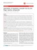
- Retrovirology 2005, 2:28 http://www.retrovirology.com/content/2/1/28 Media Breast milk 1.4 2.5 1.2 2.0 Media: β -gal expression by P4-R5 cells (RLU/s) Milk: β -gal expression by P4-R5 cells (RLU/s) 1.0 1.5 0.8 0.6 1.0 0.4 0.5 ` ` 0.2 0.0 0.0 -0.2 -0.5 Untreated 1 min 3 min 5 min 7 min 10 min 15 min Before SDS-removal After SDS-removal 10,000,000 p=0.0002 p=0.0056 1,000,000 RLU/s 100,000 10,000 1,000 S S S S ilk ilk ilk ilk ilk ilk ia ia SD SD SD SD ed ed m m m M m m M m % 1% % 1% in in in in in 05 05 in S S S IV S 0. 0. SD SD SD SD 0. 0. IV H + H + 1% % 1% % IV IV 05 05 H 0. 0. H 0. 0. + + IV IV H H Figure 1 Irreversible inactivation of cell-free HIV-1 in breast milk treated with SDS Irreversible inactivation of cell-free HIV-1 in breast milk treated with SDS. A. Breast milk from a healthy donor was artificially infected with cell-free HIV-1 IIIB and treated with 0.1% SDS for up to15 min at 37°C prior to plating on P4-R5 MAGI indicator cells (see methods section for details). Two days later, β-gal expression was measured in relative luminescent units per second (RLU/s) in triplicate samples. Results shown are representative of three experiments. B. Infectivity of cell-free HIV- 1 in breast milk treated with SDS (0.05% and 0.1%) was assessed before and after removal of SDS with SDS-300 Detergent- Out™ (see methods section for details). Results are representative of two experiments, each with triplicate samples. Page 3 of 10 (page number not for citation purposes)
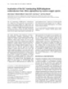
- Retrovirology 2005, 2:28 http://www.retrovirology.com/content/2/1/28 Media Breast Milk 1.2 β-gal expression by P4-R5 cells (RLU/s) 1.0 0.8 0.6 0.4 0.2 ` 0.0 Untreated 0.01% SDS 0.1% SDS Treatment of Sup-T1 cells 0.7 β -gal expression by P4-R5 cells 0.6 0.5 0.4 0.3 0.2 0.1 ` 0.0 ed in in nd in in in in m m m m m m at ou re 10 15 1 3 5 7 gr nt ck U Ba Length of treatment Figure 2 Inactivation of cell-associated HIV-1 in breast milk with SDS Inactivation of cell-associated HIV-1 in breast milk with SDS. A. Supt-T1 cells infected with HIV-1 IIIB were mixed into breast milk from a healthy donor and treated with 1% or 0.1% SDS for 10 min at 37°C prior to plating on P4-R5 MAGI indicator cells (see methods section for details). Two days later, β-gal expression was measured in relative luminescent units per second (RLU/s) in triplicate samples. Levels of β-gal expression by P4-R5 cells correlates with infectivity of cell-associated HIV-1 (i.e., infected Sup-T1 cells). Results are representative of four experiments. B. Representative results of the time-course of inactivation of cell-associated HIV-1. Sup-T1 cells in media infected with HIV-1 IIIB were treated for up to 15 min with 0.01% SDS and assayed for infectivity using P4-R5 indicator cells. Samples were assayed in triplicate. Page 4 of 10 (page number not for citation purposes)
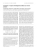
- Retrovirology 2005, 2:28 http://www.retrovirology.com/content/2/1/28 Experiment 1 Experiment 2 600,000 500,000 400,000 RNA copies/ml 300,000 200,000 100,000 0 HIV IIIB HIV + 0.01% SDS HIV + 0.5% SDS HIV + 1% SDS Figure 3 Reduction of HIV-1 RNA levels in breast milk treated with SDS Reduction of HIV-1 RNA levels in breast milk treated with SDS. Cell-free HIV-1 IIIB was added to breast milk and treated with ≤1% SDS for 10 min prior to viral load determination using branched DNA technology. Shown are results of 2 independent experiments. Assay sensitivity range: 75–500,000 RNA copies/ml. desirable prior to feeding the mother's milk, it is relevant text of HIV-1 infection [13-15]. Here we report that, with to determine the potential reversal of the antiviral effect concentrations as low as 0.1% SDS (1 mg/ml), we can after removal of SDS. To this end, the effect of SDS inactivate in vitro high titers of HIV-1 added to breast milk. removal with Detergent-Out™ on infectivity of HIV-1 was This is evidenced by the irreversible loss of infectivity of also assessed. HIV-1 infectivity was not recovered either cell-free and cell-associated HIV-1, and by significant after removal of SDS (Figure 1B). Passage of virus solu- decrease in HIV-1 RNA titers. At treatment concentrations tions through the resin itself decreased infectivity by 40% of 0.1% SDS, Sup-T1 cells were lysed contributing to the – 60% (Figure 1B). Paired t-test of HIV-1-infected media lack of infectivity observed. This result is congruent with and milk samples before and after being passed through our previously reported findings[16]. However, T cells, as the column showed this difference to be statistically sig- well as macrophages, in colostrum were conserved after nificant (Media p = 0.0056, Milk p = 0.0002). treatment with this concentration (data not shown). This discrepancy is possibly due to differences in membrane lipid and protein composition among these cell popula- Discussion We have previously shown that SDS, has broad-spectrum tions[17]. At this time, we do not understand why the effi- microbicidal activity, including anti-HIV-1 activity with ciency of treatment with 0.01% SDS in inactivating cell- concentrations as low as 0.025% [5-9]. The positive associated HIV-1 in breast milk is lower at 10 and 15 min impact of feeding mother's own milk on infant health and of treatment. However, this should not be confused with survival are well known and promoted, even in the con- increased infectivity because infectivity at these time Page 5 of 10 (page number not for citation purposes)
- Retrovirology 2005, 2:28 http://www.retrovirology.com/content/2/1/28 Bovine Serum Albumin Whole Bovine Milk Human Milk 0.16 p=0.3202 n=5 n=8 Final SDS concentration remaining in solution (%) + SD 0.14 0.12 0.1 0.08 0.06 p=0.4940 p=0.5794 n=10 0.04 n=10 n=8 n=8 0.02 n=2 n=5 n=4 0 No SDS 0.1% SDS 1% SDS Initial SDS concentration Efficiency of SDS removal from breast milk, whole bovine milk and bovine serum albumin Figure 4 Efficiency of SDS removal from breast milk, whole bovine milk and bovine serum albumin. Mixtures of human milk, cow's milk or bovine serum albumin (BSA) containing SDS (0.1%-1%) were subject to SDS removal with SDS-300 Deter- gent-Out®, as per manufacturer's instructions. SDS remaining in solution was quantified spectrophotometrically with the rea- gents included in the SDS-300 Detergent-Out® kit. points was still significantly reduced relative to the results and their interpretation should not be affected. untreated milk sample (Figure 2B). When comparing media with breast milk, we are compar- ing the overall efficacy of SDS in each milieu, and we can Adequate methods of milk storage were put in place to observe that efficacy is comparable. minimize the effects of freeze-thaw cycles on milk compo- nents[18,19]. Surprisingly, P4-R5 cells exposed to infected The decrease in HIV-1 RNA titers after microbicidal treat- breast milk had higher expression of β-gal than those ment (Figure 3) has also been observed by other research- exposed to infected media (Figures 1A and 2A), and the ers using microbicidal compounds (e.g., Nonoxynol-9) in opposite would have been expected considering the anti- cervico-vaginal fluids, and may be due to exposure of the HIV-1 properties inherent to breast milk. However, viral RNA to RNases in the milk after dissolution of the because the results are expressed in relative luminescent viral envelope (Deborah J. Anderson, Ph.D., personal units per seconds (RLU/s), any change in β-gal expression communication 12/19/03). If deemed necessary or desir- is relative to its matched controlled. Any interference in able, a commercially available resin resuspended in water the milk control would be the same across all milk sam- that can remove SDS from milk has been identified. The ples in that experiment because the milk from the same effects of SDS-removal with this method on human milk donor was used for all test samples in a single experiment. nutrients are data presented in a separate manuscript to be In addition, we did not pool donors' milk. Therefore, the published elsewhere, where we report conservation of Page 6 of 10 (page number not for citation purposes)
- Retrovirology 2005, 2:28 http://www.retrovirology.com/content/2/1/28 total milk protein species, conservation of milk milk can sit at room temperature for up to 6–8 hours and immunoglobulins (number and function), and conserva- still be considered bacteriologically safe[18,34], and SDS tion of milk's energy value[10,11]. also has microbicidal activity at room temperature (~23°C) (data not shown). Limitations of our proposed To date, we have only tested this method on very small method may be the need for bottle-feeding in settings volumes (up to 1 ml) using a column device to filter the where cup feeding may be the norm, and milk expression SDS out of milk. On a greater scale, we envision a model may represent a two-fold stumbling block for a wide in which breast milk could be expressed manually or spread use of this method because: (1) of the time it may mechanically (depending on the living conditions of the require to express milk, and (2) of the added cost of the nursing mother) into a recipient container or bottle con- final device if a mechanical milk pumping device would taining SDS. Due to the fast acting effect of SDS against be required. An economic assessment of this milk treat- HIV-1 and other pathogens, milk decontamination would ment option has not yet been performed. Feasibility of occur as warm milk gets expressed into the container. The this preventative option also needs to be determined broad-spectrum action of SDS could also clear milk of because we, as others, face one of the worst aspects of this other pathogens (e.g., secondary bacterial contamination) epidemic: stigma of not breastfeeding. that could potentially contaminate it during expression and handling. If removal of SDS prior to feeding would be Conclusion required, a filtering device comprised by the ion-exchange Here we have introduced the novel concept of using resin could be located within the nipple manifold in such microbicides (e.g., SDS) to treat HIV-1 infected breast a way that milk would be filtered through the resin as it is milk to prevent MTCT of HIV-1. Characteristics of an ideal suctioned out of the bottle. If an infant (assuming 5 kg of microbicide for treatment of breast milk include: (1) weight) ingests about 700 ml of breast milk a day[18], at efficacy at low doses; (2) low level of toxicity; (3) broad- a treatment concentration of 0.1% this would represent spectrum microbicidal activity; (4) tasteless and odorless; an intake of SDS 0.7 g. If 90% of SDS is removed through 5) practical to use; and (6) conservation of milk's nutri- filtration of treated milk, the final SDS concentration tional and immunoprotective functions. SDS meets most ingested at the end of the day would be 0.07 g; or 0.7 g if of these requirements. However, we still need to deter- milk is instead treated with 1% SDS. Because the toxico- mine the effects of SDS treatment on milk's physical prop- logical properties of SDS have been broadly studied in erties (e.g., taste, smell). We anticipate SDS will have animals and humans without toxic effects even at enor- similar efficacy to that here reported in naturally HIV-1 mous doses (e.g., 258 g in 38 days to an adult infected milk. It remains to be determined, though, human)[2,20-23], the need for removal of SDS still whether conservation of milk cells (infected and non- requires further assessment. The metabolism and degrada- infected) with elimination of cell-free HIV-1 is sufficient tion pathway of SDS and other alkyl sulfates has also been to significantly decrease transmission. It is possible that elucidated in Pseudomonas, rats, dogs and humans [24- this may be a simple way to prevent milk-borne transmis- 26]. Sulfatase is known to remove the sulfate, and the car- sion of HIV-1, while allowing HIV-1-infected mothers to bon chain is then metabolized as a fatty acid. We are cur- continue providing the nutritional and immunological rently in the process of identifying other candidate benefits of breast milk to their children. microbicides for potential use to decontaminate breast milk with respect to HIV-1 (unpublished observations). Methods Use of edible compounds that can inactivate HIV-1 in Human milk breast milk would circumvent the issue of removing the Breast milk was obtained, from anonymous healthy microbicide prior to feeding treated milk[10,27-30]. donors, of unknown HIV serostatus, and regardless of age or parity. The subjects who donated milk were either Among the advantages of microbicidal treatment of mothers of children followed in our Outpatient Clinic or expressed HIV-1-infected milk are that it is rapid, discreet nurses that work in our Pediatric Outpatient Clinic. The (i.e., can be performed in private, minutes to hours before study was explained to them, and they signed the consent feeding), of low cost, and able to preserve breast milk's form. The milk samples used were all mature milk (>2 nutritional and protective functions. In light of the sus- weeks postpartum) unless otherwise stated. Aliquots of ceptibility of HIV-1 to heat[31,32], other research groups unpooled milk were stored at -70°C in polypropylene have looked into the use of heat treatment of milk to inac- tubes, and thawed as needed. Because milk samples were tivate HIV-1 [33-38]. However, heat can be detrimental to not pooled, at least two different donors were used for important breast milk constituents[39]. In addition, lack each experiment to control for outcomes that could be of a readily available source of heat in some areas prevents due to individual differences of each donor. This study practical application of this option[40]. Refrigeration of was performed under approval of the Institutional Review expressed milk would not be a sine qua non requirement as Page 7 of 10 (page number not for citation purposes)
- Retrovirology 2005, 2:28 http://www.retrovirology.com/content/2/1/28 Board of the M. S. Hershey Medical Center (Protocol# Landau) were seeded overnight in 12-well plates. Concen- 0628EP). trated HIV-1 IIIB (5 ml; Advanced Biotechnologies, Inc.; Titer: 107.67 TCID50/ml) was treated with SDS (≤0.1% diluted in media or breast milk) for 10 min at 37°C. Microbicidal treatment with sodium dodecyl sulfate (SDS) Stock solutions of 10% (100 mg/ml) SDS (Bio-Rad Labo- Media was then added to each reaction tube (1:100 dilu- ratories) were prepared in sterile water and kept at room tion) and plated in triplicate. After 2 h incubation at temperature for up to two weeks. Volume/volume dilu- 37°C, cells were washed and fresh media (2 ml) was added to each well. β-gal expression was measured 46 h tions in media or breast milk were prepared fresh to obtain concentrations of ≤1%. Treatment of human milk later using a chemiluminescent reporter gene assay system was for 10 min at 37°C with final SDS concentrations of (Galacto-Star™ System, Applied Biosystems). All samples 1%, 0.5% or 0.1%. After treatment, SDS was removed were tested in triplicate. with SDS-300 Detergent-Out™ Medi (Geno Technology, Inc.) as described below. In all experiments untreated, Inactivation of cell-associated HIV-1 was achieved by treating infected Sup-T1 cells (CD4+ human T cells) with uninfected samples were used as controls. SDS (≤1%) for 10 min at 37°C prior to overlaying on P4- R5 cells. In brief, 3 × 10⁶ Sup-T1 cells were infected with a Removal of SDS and SDS Detection SDS removal was accomplished by centrifugation of 1 ml 1:10,000 dilution of stock HIV-1 IIIB. Infected cells were of each sample through ion exchange matrix columns subject to centrifugation, resuspended in fresh media, and incubated in the presence or absence of SDS (≤0.1%, 10 (SDS-300 Detergent-Out™ Medi [Geno Technology, Inc.], Extract Clean™ IC-Ba and Extract Clean™ IC-OH [Alltech min at 37°C), three days later. Infected Sup-T1 cells (1 × 10⁶; incubated in the presence or absence of SDS) were co- Associates, Inc.]). Reagents provided in the SDS-300 Detergent Out kit were used to colorimetrically quantify incubated with indicator P4-R5 cells (1:100 dilution of SDS remaining in solution after removal, in addition to an the inactivation mixture). After 2h, P4-R5 cells were assay using chloroform and methylene blue as previously washed and fed with new media. Chemiluminescent expression of β-gal was measured 46 h later. Inactivation described[41]. Results were compared to a standard curve of SDS in deionized water. Standard curves of SDS diluted of cell-associated HIV-1 in the breast milk was performed in water were compared to breast milk and whole bovine in a similar manner, except that infected Sup-T1 cells were milk. At concentrations ≤0.1% SDS, there was no signifi- resuspended in breast milk instead of media. All samples cant difference between absorbance measured in milk were tested in triplicate. samples (human or bovine) or water samples using the SDS-300 Detegent Out™ reagents (data not shown). The All chemiluminescent data was collected with a Fluoro- sckan® Ascent FL from Thermolab® Systems, except for chloroform-methylene blue assay has the advantage that milk (bovine or human) does not interfere with the data in figure 1B, which was collected with a Zylux Corpo- ration® FB15 luminometer. We have determined that the absorbance of the sample at any SDS concentration in the standards (≤2%) and, therefore, was used for the later final concentrations of SDS to which P4-R5 cells are experiments. Optical density of the samples was measured exposed to in these assays are not toxic[6]. using a visible light spectrophotometer (Spectronic 20®, Bausch & Lomb®). HIV-1 RNA load assay In 10 µl reactions, HIV-1 IIIB (1 µl of virus stock previ- ously diluted 1:100 in media) was added to breast milk or HIV-1 inactivation in vitro Inactivation of infectious cell-free HIV-1 in human milk media, and treated with 1%, 0.5% or 0.1% SDS at 37°C. After 10 min, treatment was blocked by adding 990 µl of was studied by a rapid in vitro system that quantifies remaining viral infectivity after microbicidal treatment. cold media. Samples were then immediately processed in This system, designated MAGI (Multinuclear Activation of the Clinical Laboratories of the M. S. Hershey Medical Galactosidase Indicator) assay[42], is based on the use of Center for viral load determination using the branched DNA (bDNA) VERSANT® HIV-1 RNA 3.0 Assay (Bayer indicator P4-R5 MAGI cells. These cells are HeLa cells (immortalized cervical cancer cell line) stably expressing Corporation, Inc.). This in vitro assay is clinically used to the HIV-1 receptor (CD4) and co-receptors (CXCR4 and directly quantify HIV-1 RNA in plasma of HIV-1-infected CCR5) on the surface, and stably transformed with β- individuals. galactosidase (β-gal) under the control of the HIV-1 long terminal repeat (LTR). Thus, as a result of HIV-1 Tat acti- Statistical Analysis vation of the LTR, cells infected with HIV should express Where indicated, samples were tested in duplicate or trip- β-gal. P4-R5 MAGI cells (8 × 10⁴; obtained through the licates. All experiments were repeated two to four times to AIDS Research and Reference Reagent Program, Division ensure reproducibility of results. All results are presented of AIDS, NIAID, NIH: P4-R5 MAGI from Dr. Nathaniel here in the form of averages ± standard deviations or as Page 8 of 10 (page number not for citation purposes)
- Retrovirology 2005, 2:28 http://www.retrovirology.com/content/2/1/28 representative results, as applicable to each case. Paired t- the manuscript. BW contributed to the design of the study test was used to compare samples before and after and in the interpretation of the data. EBN participated in removal of SDS. ANOVA was used to compare treatment the acquisition of data. CMB obtained the IRB approval groups. for this study and coordinated the collection of breast milk samples. CLS participated in the study design, and supervised some of the technical work. HML contributed List of Abbreviations SDS – Sodium Dodecyl Sulfate with the statistical analysis of the data. MKH conceived the study, supervised the technical work, and contributed HIV-1 – Human Immunodeficiency Virus type 1 to the analysis and interpretation of the data. All authors critically revised the manuscript for intellectual content. AIDS – Acquired Immune Deficiency Syndrome All authors approved of the final version of the manu- script to be published. MTCT – Mother-to-Child Transmission Acknowledgements GRAS – Generally Recognized As Safe The author(s) declare that they have no competing interests. References FDA – Food and Drug Administration 1. Tanford C: Protein Denaturation. In Advances in protein chemistry Volume 23. Edited by: Anfinsen CB NMLEJTRFM. New York, Aca- UNEP – United Nations Environment Programme demic Press; 1968:122-127; 211-222. 2. United Nations Environment Programme (UNEP): SIDS initial assessment report. Sodium dodecyl sulfate (CAS No. 151- EHE – Estimated Human Exposure 21-3). In Screening Information Data Sheet (SIDS) for High Volume Chem- icals Volume 4, Part 2 Volume 4. Edited by: UNEP/OECD/UN/IRPTC . Geneva, United Nations; 1997:1-39. HSV-2 – Herpes Simplex Virus type 2 3. U.S. Food and Drug Administration (FDA): EAFUS: A Food Addi- tive Database. Volume 2003. , Center for Food Safety & Applied MAGI – Multinuclear Activation of Galactosidase Nutrition. Office of Food Additive Safety; 2002. 4. Dreisbach RH, Robertson WO: Miscellaneous Chemicals. In Indicator Handbook of poisoning: Prevention, diagnosis & treatment 12th edition. Norwalk, Appleton & Lange; 1987:286-291. 5. Howett MK, Neely EB, Christensen ND, Wigdahl B, Krebs FC, Mala- LTR – Long Terminal Repeat mud D, Patrick SD, Pickel MD, Welsh PA, Reed CA, Ward MG, Budg- eon LR, Kreider JW: A broad-spectrum microbicide with bDNA – Branched DNA virucidal activity against sexually transmitted viruses. Antimi- crob Agents Chemother 1999, 43:314-321. 6. Krebs FC, Miller SR, Malamud D, Howett MK, Wigdahl B: Inactiva- Competing interests tion of human immunodeficiency virus type 1 by nonoxynol- 9, C31G, or an alkyl sulfate, sodium dodecyl sulfate. Antiviral The funding sources, NIH/NIAID No. PO1 AI37829 Res 1999, 43:157-173. (MKH), NRSA Fellowship NIH/NICHD No. F32 7. Krebs FC, Miller SR, Catalone BJ, Welsh PA, Malamud D, Howett MK, HD41346 (SU), and Lancaster First United Methodist Wigdahl B: Sodium dodecyl sulfate and C31G as microbicidal alternatives to nonoxynol 9: comparative sensitivity of pri- Church Scholarship Fund (SU), had no role in the study mary human vaginal keratinocytes. Antimicrob Agents Chemother design; in the collection, analysis and interpretation of the 2000, 44:1954-1960. data; in the writing of the report; or in the decision to sub- 8. Howett MK, Welsh PA, Budgeon LR, Ward MG, Neely EB, Patrick SD, Weisz J, Kreider JW: Transformation of human vaginal mit the paper for publication. MKH is inventor in and part xenografts by human papillomavirus type 11: Prevention of owner of the U.S. Patent No. 20030129588 that protects infection with a microbicide from the alkyl sulfate chemical family. Pathogenesis 2000, 1:265-276. the intellectual property surrounding the use of sodium 9. Howett MK, Wigdahl B, Malamud D, Christensen ND, Wyrick PB, dodecyl sulfate and related alkyl sulfate compounds as Krebs FC, Catalone BJ: Alkyl sulfates: a new family of broad microbicidal agents. MKH also serves as President of Ren- spectrum microbicides (Abstract No. ThOrC663). Edited by: Editore M. Monduzzi Editore; 2000:707-712. aissance Scientific, LLC, a virtual biotechnology company 10. Urdaneta SL, Neely EB, Berlin CMJ, Wigdahl B, Schengrund CL, Lin founded for the purpose of developing licenses related to HM, Howett MK: Microbicidal compounds for treatment of HIV-1 infected breast milk to prevent postnatal transmission this patent and other patents. To date, MKH has not of HIV-1 (Abstract No. 553): ; October 4-8, Mexico City, received any remuneration in conjunction with alkyl sul- Mexico. ; 2002. fate-related patents. All other authors have no actual or 11. Urdaneta SL, Berlin CM, Wigdahl B, Howett MK: Prevention of mother-to-child transmission of HIV-1 by treatment of HIV- potential, neither personal nor financial conflict of inter- 1-infected breast milk with alkyl sulfate microbicides est that may inappropriately bias their work and/or state- (Abstract No. TuPeF5420): ; July 7-12, 2002, Barcelona, ments here presented. Spain. ; 2002:Monduzzi Editore S.p.A.-Medimond Inc., pp.135-139. 12. Takeda K, Wada A, Sato Y, Hamada S: Removal of Dodecyl Sul- fate Ions Bound to Bovine Serum Albumin and Chymot- Authors' contributions rypsinogen from the Peteins. Effects of Reduction of Disulfide Bridges and Cleavage of Peptide Bonds on the SU contributed to the design of the study, acquisition of Removal Extent. Journal of Colloidal Interface Science 1991, data, analysis and interpretation of the data, and drafted 147:51-56. Page 9 of 10 (page number not for citation purposes)
- Retrovirology 2005, 2:28 http://www.retrovirology.com/content/2/1/28 13. Effect of breastfeeding on infant and child mortality due to 33. Orloff SL, Wallingford JC, McDougal JS: Inactivation of human infectious diseases in less developed countries: a pooled anal- immunodeficiency virus type 1 in human milk: Effects of ysis. WHO Collaborative Study Team on the Role of Breast- intrinsic factors in human milk and pasteurization. J Hum Lact feeding on the Prevention of Infant Mortality. Lancet 2000, 1993, 9:13-17. 355:451-455. 34. Chantry CJ, Morrison P, Panchula J, Rivera C, Hillyer G, Zorrilla C, 14. Coutsoudis A, Pillay K, Spooner E, Kuhn L, Coovadia HM: Influence Diaz C: Effects of lipolysis or heat treatment on HIV-1 provi- of infant-feeding patterns on early mother-to-child transmis- rus in breast milk. JAIDS 2000, 24:325-329. sion of HIV-1 in Durban, South Africa: a prospective cohort 35. Jeffery BS, Mercer KG: Pretoria Pasteurisation: A potential study. South African Vitamin A Study Group. Lancet 1999, method for the reduction of postnatal mother to child trans- 354:471-476. mission of the human immunodeficiency virus. Journal of Trop- 15. Coutsoudis A, Pillay K, Kuhn L, Spooner E, Tsai WY, Coovadia HM: ical Pediatrics 2000, 46:219-223. Method of feeding and transmission of HIV-1 from mothers 36. Jeffery BS, Webber L, Mokhondo KR, Erasmus D: Determination of to children by 15 months of age: prospective cohort study the effectiveness of inactivation of human immunodeficiency from Durban, South Africa. AIDS 2001, 15:379-387. virus by Pretoria pasteurization. J Trop Pediatr 2001, 47:345-349. 16. Krebs FC, Miller SR, Catalone BJ, Fichorova R, Anderson D, Malamud 37. Boisen F, Jørgensen AF: Pasteurization of HIV contaminated D, Howett MK, Wigdahl B: Comparative in vitro sensitivities of breast milk (Abstract No. LbPp122): ; July 7-12, 2000, Dur- human immune cell lines, vaginal and cervical epithelial cell ban, South Africa. ; 2000. lines, and primary cells to candidate microbicides nonoxynol 38. Israel-Ballard K, Donovan R, Enge B, Gesner M, Scott M, Sheppard H, 9, C31G, and sodium dodecyl sulfate. Antimicrob Agents Sage A, Abrams B, Chantry C: A novel approach for evaluating Chemother 2002, 46:2292-2298. pasteurization methods to inactivate HIV in breast milk, 17. le Maire M, Champeil P, Moller JV: Interaction of membrane pro- Poster #899: February 8-11 2004. 2004. teins and lipids with solubilizing detergents. Biochim Biophys 39. Ford JE, Law BA, Marshall VM, Reiter B: Influence of the heat Acta 2000, 1508:86-111. treatment of human milk on some of its protective 18. Lawrence RA, Lawrence RM: Breastfeeding. A guide for the constituents. J Pediatr 1977, 90:29-35. medical profession. 5th edition. St. Louis, Mosby; 1999. 40. Papathakis PC, Rollins NC: Adequacy of feeding recommenda- 19. Clinical Protocol #8: Human milk storage information for tions for infants of HIV-infected mothers: assessment of home use for healthy full term infants. In ABM Protocols (http:// nutrient content, cost and preparation time of breastmilk wwwbfmedorg/protoindxhtml) , Academy of Breastfeeding Medicine replacements in the South African context (Abstract No. (ABM); 2004. MoOrF1030).: ; Barcelona, Spain. Prous Science, S.A.; 2002. 20. Kirsner B, Wolff RA: The effect of sodium alkyl sulfate on the 41. Arand M, Friedberg T, Oesch F: Colorimetric quantitation of peptic activity of the gastric contents and on the healing of trace amounts of sodium lauryl sulfate in the presence of gastric ulcer in man. Gastroenterology 1944, 2:93-101. nucleic acids and proteins. Anal Biochem 1992, 207:73-75. 21. Walker AI, Brown VK, Ferrigan LW, Pickering RG, Williams DA: 42. Kimpton J, Emerman M: Detection of replication-competent Toxicity of sodium lauryl sulphate, sodium lauryl ethoxysul- and pseudotyped human immunodeficiency virus with a sen- phate and corresponding surfactants derived from synthetic sitive cell line on the basis of activation of an integrated beta- alcohols. Food Cosmet Toxicol 1967, 5:763-769. galactosidase gene. J Virol 1992, 66:2232-2239. 22. Gloxhuber C: Toxicological properties of surfactants. Arch Toxicol 1974, 32:245-270. 23. Gloxhuber C, Kunstler K: Anionic Surfactants: biochemistry, toxicology, dermatology. In Surfactant Science Series Volume 43. 2nd edition. New York, Marceld Dekker, Inc.; 1992. 24. Thomas OR, White GF: Metabolic pathway for the biodegrada- tion of sodium dodecyl sulfate by Pseudomonas sp. C12B. Bio- technol Appl Biochem 1989, 11:318-327. 25. Yao G: Dodecyl Sulfate Degradation Pathway Map (http:// www.labmed.umn.edu/umbbd.dds/dds_map.html). . 26. Denner WH, Olavesen AH, Powell GM, Dodgson KS: The metab- olism of potassium dodecyl [35-S]sulphate in the rat. Biochem J 1969, 111:43-51. 27. Kristmundsdottir T, Arnadottir SG, Bergsson G, Thormar H: Devel- opment and evaluation of microbicidal hydrogels containing monoglyceride as the active ingredient. J Pharm Sci 1999, 88:1011-1015. 28. Thormar H, Bergsson G, Gunnarsson E, Georgsson G, Witvrouw M, Steingrimsson O, De Clercq E, Kristmundsdottir T: Hydrogels con- taining monocaprin have potent microbicidal activities against sexually transmitted viruses and bacteria in vitro. Sex Transm Infect 1999, 75:181-185. 29. Neyts J, Kristmundsdottir T, De Clercq E, Thormar H: Hydrogels containing monocaprin prevent intravaginal and intracuta- neous infections with HSV-2 in mice: impact on the search Publish with Bio Med Central and every for vaginal microbicides. J Med Virol 2000, 61:107-110. scientist can read your work free of charge 30. Maguire RA, Bergman N, Phillips DM: Comparison of microbi- cides for efficacy in protecting mice against vaginal challenge "BioMed Central will be the most significant development for with herpes simplex virus type 2, cytotoxicity, antibacterial disseminating the results of biomedical researc h in our lifetime." properties, and sperm immobilization. Sex Transm Dis 2001, Sir Paul Nurse, Cancer Research UK 28:259-265. 31. Pennypacker C, Perelson AS, Nys N, Nelson G, Sessler DI: Local- Your research papers will be: ized or systemic in vivo heat inactivation of human immun- available free of charge to the entire biomedical community odeficiency virus (HIV): a mathematical analysis. J Acquir Immune Defic Syndr Hum Retrovirol 1995, 8:321-329. peer reviewed and published immediately upon acceptance 32. McDougal JS, Martin LS, Cort SP, Mozen M, Heldebrant CM, Evatt BL: cited in PubMed and archived on PubMed Central Thermal inactivation of the acquired immunodeficiency syn- drome virus, human T lymphotropic virus-III/lymphadenop- yours — you keep the copyright athy-associated virus, with special reference to BioMedcentral antihemophilic factor. J Clin Invest 1985, 76:875-877. Submit your manuscript here: http://www.biomedcentral.com/info/publishing_adv.asp Page 10 of 10 (page number not for citation purposes)
CÓ THỂ BẠN MUỐN DOWNLOAD

-
Báo cáo khoa học: "Protective efficacy of commercial inactivated Newcastle disease virus vaccines in chickens against a recent Korean epizootic strain"
 6 p |
6 p |  51
|
51
|  9
9
-

Báo cáo khoa học: "Efficacy of live B1 or Ulster 2C Newcastle disease vaccines simultaneously vaccinated with inactivated oil adjuvant vaccine for protection of Newcastle disease virus in broiler chickens"
4 p | 64
| 6
-

Báo cáo Y học: Characterization of a novel silkworm (Bombyx mori ) phenol UDP-glucosyltransferase
7 p | 38
| 4
-
Báo cáo Y học: Control of p70 ribosomal protein S6 kinase and acetyl-CoA carboxylase by AMP-activated protein kinase and protein phosphatases in isolated hepatocytes
9 p | 49
| 4
-
Báo cáo y học: "Captopril reduces cardiac inflammatory markers in spontaneously hypertensive rats by inactivation of NF-kB"
9 p | 70
| 4
-

Báo cáo y học: "Analysis of skewed X-chromosome inactivation in females with rheumatoid arthritis and autoimmune thyroid disease"
8 p | 50
| 4
-
Báo cáo khoa học: " Thermal stability and inactivation of hepatitis C virus grown in cell culture"
12 p | 47
| 3
-
Báo cáo Y học: Escherichia coli small heat shock proteins, IbpA and IbpB, protect enzymes from inactivation by heat and oxidants
11 p | 38
| 3
-
Báo cáo khoa học: " Inactivation of respiratory syncytial virus by zinc finger reactive compounds"
10 p | 31
| 3
-

Báo cáo Y học: The regulatory subunit of a cGMP-regulated protein kinase A of Trypanosoma brucei
10 p | 35
| 3
-
Báo cáo Y học: Inactivation of the Na+-translocating NADH:ubiquinone oxidoreductase from Vibrio alginolyticus by reactive oxygen species
6 p | 45
| 3
-
Báo cáo khóa học: Inactivation of copper-containing amine oxidases by turnover products
7 p | 46
| 3
-
Báo cáo Y học: Inactivation of the 2-oxo acid dehydrogenase complexes upon generation of intrinsic radical species
12 p | 35
| 2
-

Báo cáo khoa học: Inactivation of colicin Y by intramembrane helix–helix interaction with its immunity protein
7 p | 27
| 2
-
Báo cáo y học: " Inactivation of tumor suppressor Dlg1 augments transformation of a T-cell line induced by human T-cell leukemia virus type 1 Tax protein"
13 p | 42
| 2
-

Báo cáo y học: "Antigen-presenting particle technology using inactivated surface-engineered viruses: induction of immune responses against infectious agents"
18 p | 45
| 2
-
Báo cáo y học: "Activity map of the tammar X chromosome shows that marsup"
18 p | 35
| 2


Chịu trách nhiệm nội dung:
Nguyễn Công Hà - Giám đốc Công ty TNHH TÀI LIỆU TRỰC TUYẾN VI NA
LIÊN HỆ
Địa chỉ: P402, 54A Nơ Trang Long, Phường 14, Q.Bình Thạnh, TP.HCM
Hotline: 093 303 0098
Email: support@tailieu.vn
Giấy phép Mạng Xã Hội số: 670/GP-BTTTT cấp ngày 30/11/2015 Copyright © 2022-2032 TaiLieu.VN. All rights reserved.

