


Báo cáo y học: " Immunohistochemical characterization of nodose cough receptor neurons projecting to the trachea of guinea pigs"
lượt xem 3
download
 Download
Vui lòng tải xuống để xem tài liệu đầy đủ
Download
Vui lòng tải xuống để xem tài liệu đầy đủ
Tuyển tập các báo cáo nghiên cứu về y học được đăng trên tạp chí y học Critical Care giúp cho các bạn có thêm kiến thức về ngành y học đề tài: Immunohistochemical characterization of nodose cough receptor neurons projecting to the trachea of guinea pigs...
Bình luận(0) Đăng nhập để gửi bình luận!
Nội dung Text: Báo cáo y học: " Immunohistochemical characterization of nodose cough receptor neurons projecting to the trachea of guinea pigs"
- Cough BioMed Central Open Access Research Immunohistochemical characterization of nodose cough receptor neurons projecting to the trachea of guinea pigs Stuart B Mazzone* and Alice E McGovern Address: School of Biomedical Sciences, The University of Queensland, St Lucia, 4072, Australia Email: Stuart B Mazzone* - s.mazzone@uq.edu.au; Alice E McGovern - a.mcgovern1@uq.edu.au * Corresponding author Published: 19 October 2008 Received: 5 September 2008 Accepted: 19 October 2008 Cough 2008, 4:9 doi:10.1186/1745-9974-4-9 This article is available from: http://www.coughjournal.com/content/4/1/9 © 2008 Mazzone and McGovern; licensee BioMed Central Ltd. This is an Open Access article distributed under the terms of the Creative Commons Attribution License (http://creativecommons.org/licenses/by/2.0), which permits unrestricted use, distribution, and reproduction in any medium, provided the original work is properly cited. Abstract Background: Cough in guinea pigs is mediated in part by capsaicin-insensitive low threshold mechanoreceptors (cough receptors). Functional studies suggest that cough receptors represent a homogeneous population of nodose ganglia-derived sensory neurons. In the present study we set out to characterize the neurochemical profile of cough receptor neurons in the nodose ganglia. Methods: Nodose neurons projecting to the guinea pig trachea were retrogradely labeled with fluorogold and processed immunohistochemically for the expression of a variety of transporters (Na+/K+/2C1- co-transporter (NKCC1), α1 and α3 Na+/K+ ATPase, vesicular glutamate transporters (vGlut)1 and vGlut2), neurotransmitters (substance P, calcitonin gene-related peptide (CGRP), somatostatin, neuronal nitric oxide synthase (nNOS)) and cytosolic proteins (neurofilament, calretinin, calbindin, parvalbumin). Results: Fluorogold labeled ~3 per cent of neurons in the nodose ganglia with an average somal perimeter of 137 ± 6.2 μm (range 90–200 μm). All traced neurons (and seemingly all nodose neurons) were immunoreactive for NKCC1. Many (> 90 per cent) were also immunoreactive for vGlut2 and neurofilament and between 50 and 85 per cent expressed α1 ATPase, α3 ATPase or vGlut1. Cough receptor neurons that did not express the above markers could not be differentiated based on somal size, with the exception of neurofilament negative neurons which were significantly smaller (P < 0.05). Less than 10 per cent of fluorogold labeled neurons expressed substance P or CGRP (and these had somal perimeters less than 110 μm) and none expressed somatostatin, calretinin, calbindin or parvalbumin. Two distinct patterns of nNOS labeling was observed in the general population of nodose neurons: most neurons contained cytosolic clusters of moderately intense immunoreactivity whereas less than 10 per cent of neurons displayed uniform intensely fluorescent somal labeling. Less than 3 per cent of the retrogradely traced neurons were intensely fluorescent for nNOS (most showed clusters of nNOS immunoreactivity) and nNOS immunoreactivity was not expressed by cough receptor nerve terminals in the tracheal wall. Conclusion: These data provide further insights into the neurochemistry of nodose cough receptors and suggest that despite their high degree of functional homogeneity, nodose cough receptors subtypes may eventually be distinguished based on neurochemical profile. Page 1 of 16 (page number not for citation purposes)
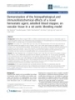
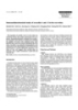
- Cough 2008, 4:9 http://www.coughjournal.com/content/4/1/9 population of neurons that display the functional charac- Background Previous studies have identified a novel vagal sensory teristics of cough receptors [1,2]. In the guinea pig trachea nerve subtype that innervates the large airways (larynx, and larynx, there are very few nodose capsaicin-sensitive trachea and main bronchi) of guinea pigs and is likely nociceptors (tracheal nociceptors are mostly derived from responsible for defensive cough in this species [1]. These the jugular vagal ganglia) and no classically defined rap- sensory neurons (referred to as cough receptors) are idly adapting or slowly adapting stretch receptors [1,2]. derived from the nodose ganglia and are characterized by their insensitivity to capsaicin and their sensitivity to both Anatomical and immunohistochemical studies have also rapid reductions in pH and punctuate (touch-like) provided some information about the nodose cough mechanical stimulation [1-3]. However, unlike other clas- receptor. In the tracheal wall, the peripheral terminals of sically defined low threshold mechanoreceptors which mechanoreceptors (presumably cough receptors) have innervate the airways and lungs, cough receptors display a been differentiated from substance P expressing nocicep- low sensitivity to mechanical stretch (including inflation/ tors using osmium staining techniques [8], the intravital deflation and bronchospasm), conduct action potentials styryl dye FM2-10 [7,9], as well as with immunostaining slower (~5 m/sec for cough receptors compared to > 15 for the alpha3-expressing isozymes of Na+/K+ ATPase and m/sec for intrapulmonary stretch receptors) and are unre- the furosemide sensitive Na+/K+/2Cl- co-transporter sponsive to the purinergic agonist α,β-methylene ATP [1]. NKCC1 [6] (see also Fig 1). Retrograde labeling of affer- Based on these observations, cough receptors are believed ents innervating the guinea pig trachea have shown that to represent a distinct airway afferent nerve in this species the majority of tracheal nodose neurons express neurofil- (reviewed in [4]). ament proteins (associated with myelinated neurons) but are devoid of the neuropeptides substance P, CGRP and Functional and electrophysiological studies have pro- the capsaicin receptor TRPV1 (all associated with capsai- vided key insights into the role of nodose cough receptors cin-sensitive sensory nerves) [2,10,11]. These observa- in the cough reflex. In anesthetized guinea pigs, punctuate tions would also support the suggestion that most nodose mechanical stimulation or rapid acidification of the laryn- neurons innervating the guinea pig trachea and larynx are geal or tracheal mucosa evokes coughing, a response that cough receptors and that these cough receptor neurons can be abolished by selectively disrupting the afferent may be a homogeneous population in the nodose ganglia. pathways from the nodose ganglia [1,5-7]. Extensive elec- However, a detailed neurochemical profile of these neu- trophysiological analyses of the activation profiles of rons has not been performed and as such, the possibility nodose neurons projecting to the guinea pig trachea and of cough receptor heterogeneity cannot be excluded. larynx suggests that the majority (perhaps greater than 95%) of these neurons form a seemingly homogeneous Figure 1 Morphology of cough receptor nerve terminals in the guinea pig trachea Morphology of cough receptor nerve terminals in the guinea pig trachea. Presumed cough receptor nerve terminals labeled (A) with the vital styryl dye FM2-10 and (B) immunohistochemically for α3 Na+/K+ ATPase. Note the terminal struc- tures are arranged parallel to the tracheal muscle fibers (running from top to bottom of the panels). The cough receptor ter- minals (A, B) are clearly differentiated from substance P-containing (SP) tracheal nociceptors (C). The arrow heads and small arrows in panels (A) and (B) illustrate individual cough receptor axons and the nerve bundles from which the axons arise, respectively. The asterisk in panel (C) shows the origin of a primary bronchus at the caudal end of the trachea. The scale bar represents 200 μm in panel (A) and 50 μm in panels (B) and (C). These images were generated, but not used for publication, during previous studies (FM2-10 staining from reference [9] and α3 Na+/K+ ATPase/SP immunohistochemistry from reference [6]). Refer to [6,9] for detailed methods. Page 2 of 16 (page number not for citation purposes)
- Cough 2008, 4:9 http://www.coughjournal.com/content/4/1/9 Immunohistochemical studies of other sensory nerve lular matrix below the epithelium. This procedure would populations have successfully used the expression of pro- be expected to remove some tracheal nociceptors [8] but ton pump isozymes, vesicular glutamate transporters does not disrupt cough receptors [7,9]. (vGluts; a marker for glutamatergic neurons), neuropep- tides and calcium binding proteins (such as calretinin, cal- Immunohistochemistry and microscopy bindin and parvalbumin) as useful markers for Immunohistochemical staining was performed as previ- characterizing sensory nerve subtypes. Therefore, in the ously described [6]. Briefly, nodose ganglia were rapidly frozen in OCT embedding media, and 16 μm cryostat-cut present study we used well characterized antisera raised against these transporters, neurotransmitters and sections were mounted directly onto subbed glass slides. cytosolic proteins to further characterize the guinea pig Slides were incubated for 1 hour in blocking solution cough receptor neurons in the nodose ganglia. (10% horse serum), and then overnight (at room temper- ature) in PBS/0.3% Triton X-100/2% horse serum along with the primary antisera of interest (Table 1). Sections Methods Experiments were approved by the Howard Florey Insti- were washed several times with PBS, and then incubated tute Animal Ethics Committee and conducted on male with the appropriate AlexaFluor-conjugated secondary albino Hartley guinea pigs (200–350 g, n = 36, IVMS, antisera (Table 1). All sections were cover-slipped with South Australia) at the Howard Florey Institute (The Uni- buffer glycerol immediately prior to microscopy. In some versity of Melbourne, Australia). instances, fluorogold was found to be rapidly quenched during microscopy making accurate cell counting and photography difficult. On these occasions, coverslips were Retrograde tracing Guinea pigs (n = 32) were anesthetized with 1.8–2.2 per removed and the sections were incubated with rabbit anti- cent isoflurane in oxygen. The extrathoracic trachea was fluorogold (1:10,000; Fluorochrome LLC, Colorado, exposed via a ventral incision in the animal's neck. Using USA), followed by AlexaFluor 594-congugated donkey a 10 μl Hamilton glass microsyringe fitted with a 32 gauge anti-rabbit antibodies (Table 1). Accordingly, some fluor- needle, 10 μl of 4 per cent fluorogold (Fluorochrome LLC, ogold cells shown in the representative photomicrographs Colorado, USA) was injected into the rostral extrathoracic appear blue (when quenching was not a problem) and tracheal lumen (on to the mucosal surface). Following others appear red (when stabilized with secondary immu- injection, the wound was sutured and the animals were noprocessing processing) (see Fig 2 for example). allowed to recover for 7 days at which time they were anesthetized with sodium pentobarbital (100 mg/kg i.p.) Immunohistochemical processing of tracheal wholem- and transcardially perfused with 10 mM phosphate buff- ounts was performed using a modification of the methods ered saline (PBS) followed by 4% paraformaldehyde in described above for nodose sections. Tissues were first PBS. The nodose ganglia was removed and placed in 4% pinned flat to a sylgard-filled tissue culture dish and incu- paraformaldehyde at 4°C for 2 hours, then cyroprotected bated for 1 hour in blocking solution (10% normal horse in 20% sucrose solution at 4°C overnight prior to immu- serum in 10 mM PBS) and then overnight (at 37°C) in 10 nohistochemical processing (see below). mM PBS/0.3% Triton X-100/2% horse serum containing the primary antisera of interest (refer Table 1). After wash- ing thoroughly with 10 mM PBS (for at least 3 hours), Preparation of tracheal wholemounts Wholemount preparations of guinea pig (n = 4) tracheal wholemounts were then incubated for 1 hour at room segments were prepared using a modification of previ- temperature in the appropriate AlexaFluor-conjugated ously described methods [6,8]. Briefly, animals were secondary antibody (refer Table 1). deeply anesthetized with sodium pentobarbital (80 mg/ kg i.p) and transcardially perfused with 500 mL of 10 mM Labeling of wholemounts and slide mounted sections was phosphate buffered saline (PBS). The entire trachea was visualized using an Olympus BX51 fluorescent micro- removed, cleaned of excess connective tissue, and opened scope equipped with appropriate filters and an Optronics longitudinally via a midline incision along the ventral sur- digital camera. Low and high magnification images were face. The epithelium was gently rubbed off the trachea captured and stored digitally for subsequent off-line anal- with a cotton swab and tracheal segments (8–10 rings in ysis of somal size (see below) and preparation of repre- length) were pinned flat onto a piece of cork board and sentative photomicrographs. Negative control placed in fixative (4% paraformaldehyde) for 2–3 hours experiments, in which the primary antisera were excluded, at 4°C, and then transferred to blocking solution (10 mM were carried out where necessary. PBS and 10% horse serum) for one hour prior to immu- nohistochemical staining (see below). Epithelial removal Data analysis is necessary to visualize cough receptor nerve terminals in Cell counts in a given field of view were performed either the guinea pig trachea which are confined to the extracel- online (during microscopy) or offline (using high resolu- Page 3 of 16 (page number not for citation purposes)
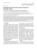
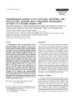
- Cough 2008, 4:9 http://www.coughjournal.com/content/4/1/9 Figure 2 Retrograde labeling of nodose neurons innervating the guinea pig trachea Retrograde labeling of nodose neurons innervating the guinea pig trachea. (A) Low magnification of nodose ganglia showing individual (arrows) and clusters (circle) of fluorogold labeled nodose neurons that have not undergone subsequent secondary immunoprocessing (hence the neurons appear blue). (B) Higher magnification of two fluorogold-labeled nodose neurons that have undergone secondary immunoprocessing and relabeled with a rhodamine fluorophore (hence the neurons appear red). See methods for details. Scale bars represent 150 μm in A and 20 μm in B. (C) Histogram showing the distribution of retrogradely labeled nodose neurons based on somal perimeter. The superimposed line graph shows the moving average calculated from the histogram. See text for details. tion digital images) at 100–200× magnification. 3–10 rep- scale tool. Only cells with a distinct nuclear region were resentative replicate sections were assessed per animal and measured in order to increase the likelihood that perime- a minimum of 4 animals were analyzed per group. For ters were measured close to the middle of the neuron and somal size analysis, stored images were imported into therefore accurately reflected the true somal size. A mini- ImageJ software (NIH, USA http://rsb.info.nih.gov/ij/) mum of 100 labeled cells, taken from at least 3 different and cell edges were traced on screen using a calibrated animals, were used to estimate somal sizes for each Page 4 of 16 (page number not for citation purposes)
- Cough 2008, 4:9 http://www.coughjournal.com/content/4/1/9 Table 1: Details of the primary and secondary antibodies used for immunohistochemical staining. Host Dilution Source Primary Antibodies – Transporters α1 Na+/K+ ATPase (clone 05–369) Mouse 1:100 Millipore, Australia. α3 Na+/K+ ATPase (clone XVIF9-G10) Mouse 1:400 Biomol, PA, USA. NKCC1 Rabbit 1:1000 Gift Dr RJ Turner, National Institute of Dental and Craniofacial Research, USA. vGLUT1 (catalogue# 135 302) Rabbit 1:2000 Synaptic Systems Goettingen, Germany. vGLUT2 (catalogue# 135 402) Rabbit 1:2000 Synaptic Systems Goettingen, Germany. Primary Antibodies – Neurotransmitters CGRP (catalogue# RPN 1842) Rabbit 1:4000 Amersham, UK. Neuronal nitric oxide synthase (nNOS) Sheep 1:4000 Gift Dr Colin Anderson, University of Melbourne, Australia. Somatostatin (catalogue# AB5494) Rabbit 1:100 Millipore, Australia. Substance P (clone NC1) Rat 1:200 Millipore, Australia. Primary Antibodies – Cytosolic Proteins Calbindin D28k (number CB-38A) Rabbit 1:1000 Swant Bellinzona, Switzerland. Calretinin (number 7699/4) Rabbit 1:1000 Swant Bellinzona, Switzerland. Neurofilament 160KD (clone NN18) Mouse 1:400 Millipore, Australia. Parvalbumin (number 235) Mouse 1:400 Swant Bellinzona, Switzerland. Secondary Antibodies (IgG, H+L, 2 mg/ml) AlexaFluor 488 or 594 anti-goat Donkey 1:200 Molecular Probes Eugene, OR, USA AlexaFluor 488 or 594 anti-mouse Donkey 1:200 Molecular Probes Eugene, OR, US AlexaFluor 488 or 594 anti-rabbit Donkey 1:200 Molecular Probes Eugene, OR, USA AlexaFluor 488 anti-rat Goat 1:200 Molecular Probes Eugene, OR, USA Note, AlexaFluor 488 and 594 are green and red fluorphores, respectively. marker. Data are expressed as a mean ± SEM. Differences neurons in the nodose ganglia (data not directly shown but can inferred from Fig 3). Both α1 Na+/K+ ATPase and between group data are compared using a Student's t-test and significance was set at P < 0.05. vGlut2 immunoreactivity was present in cells with a large range of somal sizes (60–190 μm, average 109.3 ± 2.8 μm and 109.6 ± 3.0 μm, respectively), whereas α3 Na+/K+ Results ATPase and vGlut1 immunoreactivity was primarily lim- Fluorogold retrograde labeling ited to medium and larger sized neurons (100–190 μm, Injection of fluorogold into the rostral trachea labeled average 139.1 ± 1.8 μm and 135.7 ± 2.7 μm, respectively) neurons bilaterally in the nodose ganglia (Fig 2A, B). In 4 experiments, fluorogold labeled neurons represented 2.8 (Fig 4B, C). The pattern of labeling observed for the vari- ± 0.4 per cent of the total cell population (assessed using ous transporter markers also varied. NKCC1, vGlut1 and NKCC1 immunoreactivity as a pan-neuronal marker, see vGlut2 immunoreactivity was found throughout the cyto- plasm while α1 and α3 Na+/K+ ATPase immunoreactivity below). As previously reported, retrogradely labeled soma appeared randomly distributed throughout the ganglia was principally confined to the cell membrane (Fig 5). with no obvious topographical organization [2,11]. Most (> 80 percent) traced neurons had somal perimeters rang- NKCC1 immunoreactivity was present in all retrogradely ing between 100–150 μm (average 137.3 ± 6.2 μm), labeled neurons that were assessed for this marker (Fig 3 although neurons as small as 90 μm and up to 200 μm in and Fig 5). The vast majority (84–93 percent) of traced size were less frequently noted (Fig 2C). The percentage of neurons were also immunoreactive for vGlut1 or vGlut2 and many (57–73 per cent) expressed α1 or α3 Na+/K+ fluorogold traced neurons expressing each of the immu- nohistochemical markers tested is summarized in Fig 3 ATPase on their plasma membranes (Fig 3 and Fig 5). and discussed in more detail below. Those populations of neurons that were retrogradely labeled by fluorogold but did not show immunoreactivity for the relevant transporter markers did not significantly Immunohistochemical expression of transporter proteins differ in size from the overall population of traced neu- in nodose ganglia NKCC1 immunoreactivity was present in neurons from a rons (Table 2) and showed no other obvious morpholog- wide range of somal sizes (ranging from 60–200 μm, aver- ical characteristics that would differentiate them from the age 113.9 ± 3.1 μm) (Fig 4A) and likely represents a pan- population of traced cells that expressed the marker. neuronal marker for vagal sensory neurons (Fig 3; [6]). By Exclusion of the primary antisera prevented detectable contrast, α1 and α3 Na+/K+ ATPase, vGlut1 and vGlut2 immunoreactivity in all cases (for example, Fig 5F). immunoreactivity was not universally expressed by all Page 5 of 16 (page number not for citation purposes)
- Cough 2008, 4:9 http://www.coughjournal.com/content/4/1/9 Figure 3 Summary of the neurochemical profile of retrogradely labeled nodose neurons Summary of the neurochemical profile of retrogradely labeled nodose neurons. The data represent the mean ± SEM (minimum 3 nodose sections from n = 4–5 animals) per cent of fluorogold (FG) traced neurons that stained positive for the neurochemical markers. Explanation of neurochemical marker labels: NKCC1, Na+/K+/2Cl- co-transporter 1; vGlut, vesicu- lar glutamate transporter; CGRP, calcitonin gene-related peptide; nNOS all, all cells expressing detectable neuronal nitric oxide synthase; nNOS IFCs, nNOS Intensely fluorescent cells. tion of neuropeptide-negative fluorogold traced neurons Immunohistochemical expression of neurotransmitters in in this ganglia (P < 0.05, Table 2). nodose ganglia Immunoreactivity for the neuropeptides substance P, CGRP and somatostatin was almost exclusively confined Immunoreactivity for nNOS was observed in many neu- to smaller neurons in the nodose ganglia (mean perime- rons in the nodose ganglia, albeit with two quite distinct ters of untraced neurons were 99.1 ± 1.7, 90.0 ± 2.6 and patterns of expression. Most nodose neurons exhibited 80.4 ± 2.2 for substance P, CGRP and somatostatin, nNOS immunoreactivity that was characterized by respectively; P < 0.05 significantly smaller than the mean numerous distinct dense fluorescent clusters throughout perimeter of fluorogold traced neurons) (Fig 6A). Sub- the cytoplasm (Fig 7E). By contrast, less than 10 per cent stance P was present in both soma and nerve fibers of the nNOS positive neurons showed more uniform throughout the nodose ganglia, whereas CGRP was intensely fluorescent cytoplasmic labeling (Fig 7E). The restricted to nerve fibers (substantially fewer cells were cells that exhibited clustered labeling and the intensely immunoreactive for this peptide) (Fig 7). Somatostatin fluorescent cells (IFCs) largely shared overlapping somal immunoreactivity was extremely sparse in both soma and size distributions (Fig 6B), although the nNOS IFCs were fibers and, when seen, was often confined to very small generally slightly smaller (112.6 ± 2.9 versus 98.3 ± 2.6 μm for the cells with clustered labeling and IFCs, respec- neurons (Fig 6A and Fig 7). Of the fluorogold traced neu- rons, 8.7 ± 2.1 per cent (22 out of 260 traced neurons, n = tively; Table 2). Most (> 90 per cent) of the fluorogold- 4 animals) expressed detectable levels of substance P, less traced neurons showed detectable immunoreactivity for than 1 per cent expressed CGRP (1 out of 210 traced neu- nNOS (Fig 3 and 7D). However, only 2.8 ± 0.7 per cent of rons) and none expressed somatostatin (Fig 3 and Fig 7). traced neurons were nNOS IFCs (Fig 3) and these cells The small population of substance P-positive, fluorogold- were significantly (P < 0.05) smaller in size compared to positive neurons identified in the nodose ganglia were sig- the remainder of the fluorogold-traced neurons (Table 2). nificantly smaller in size compared to the larger popula- Immunoreactivity for nNOS was not observed in cough Page 6 of 16 (page number not for citation purposes)
- Cough 2008, 4:9 http://www.coughjournal.com/content/4/1/9 (B) α1 Na+/K+ ATPase size distribution of all nodose neurons or vGlut2 Histograms showing the or α3 Na+/K+ ATPase, and (C) vGlut1 (irrespective of fluorogold tracing) that express (A) NKCC1, Figure 4 Histograms showing the size distribution of all nodose neurons (irrespective of fluorogold tracing) that express (A) NKCC1, (B) α1 Na+/K+ ATPase or α3 Na+/K+ ATPase, and (C) vGlut1 or vGlut2. The superimposed solid lines show the moving averages associated with each histogram and the dashed line references the size distribution of fluoro- gold (FG) traced neurons shown in figure 2. Page 7 of 16 (page number not for citation purposes)
- Cough 2008, 4:9 http://www.coughjournal.com/content/4/1/9 laid with5immunoreactivity for (A) showing nodose neuronsα3 Na+/K+ ATPase, (C) vGlut1, (D) vGlut2,fluorogold (FG) over- Representative photomicrographs α1 Na+/K+ ATPase, (B) retrogradely labeled from the trachea with (E) NKCC1 or (F) Figure negative control (neg) Representative photomicrographs showing nodose neurons retrogradely labeled from the trachea with fluoro- gold (FG) overlaid with immunoreactivity for (A) α1 Na+/K+ ATPase, (B) α3 Na+/K+ ATPase, (C) vGlut1, (D) vGlut2, (E) NKCC1 or (F) negative control (neg). In panels A and B, the arrows point to FG traced neurons that are immunoreactive for α1 or α3 Na+/K+ ATPase, the arrow heads show traced neurons that are not immunoreactive for α1 or α3 Na+/K+ ATPase and the asterisks show FG-negative neurons that are immunolabeled for α1 or α3 Na+/K+ ATPase. Traced neurons appear red in panel A as the tissue underwent secondary immunoprocessing for FG. Scale bar represents 40 μm. Page 8 of 16 (page number not for citation purposes)
- Cough 2008, 4:9 http://www.coughjournal.com/content/4/1/9 Table 2: Mean cell sizes of guinea pig nodose neurons. Average Cell Perimeter (μm) Average Cell Perimeter (μm) Markers Markers Commonly Uncommonly Expressed1 Expressed2 Marker (+) Marker(-)/FG(+) Marker (+) Marker(+)/FG(+) NKCC1 113.9 ± 3.1* None Substance P 99.1 ± 3.7* 108.3 ± 5.9* α1 Na+/K+ ATPase 92.93 * 109.3 ± 4.8* 131.3 ± 8.4 CGRP 90.0 ± 2.6* α3 Na+/K+ ATPase 139.1 ± 1.8 128.5 ± 5.1 Somatostatin 80.4 ± 2.2* None vGlut1 135.7 ± 2.6 126.8 ± 4.7 nNOS (IFCs) 98.3 ± 2.6* 109.5 ± 8.9* vGlut2 109.6 ± 3.0* 138.6 ± 1.8 Calretinin 177.9 ± 2.6* None Neurofilament 142.1 ± 5.7 105.3 ± 2.4* Calbindin 173.3 ± 2.1* None nNOS (all) 112.6 ± 2.9* 126.8 ± 5.9 Parvalbumin None None 1 Defined as a marker that is expressed in more than 50 per cent of the FG traced neurons. 2 Defined as a marker that is expressed in less than 50 per cent of the FG traced neurons. 3 Only one FG traced neuron expressed CGRP. *P < 0.05, significantly different compared to the average size of FG traced neurons, Student's t-test (Note. The average somal perimeter of FG traced neurons = 137.3 ± 6.2). Abbreviations: CGRP; Calcitonin Gene-Related Peptide; FG, Fluorogold; IFCs, Intensely Fluorescent Cells; nNOS, neuronal Nitric Oxide Synthase; vGlut, vesicular Glutamate transporter. receptor nerve terminals in the tracheal submucosa (iden- the majority of nodose neurons projecting to the trachea tified using α3 Na+/K+ ATPase wholemount immunohis- have medium somal sizes and express neurofilament, a tochemistry; [6]) but rather was expressed in a subset of marker for myelinated neurons [2,11,12]. The minor pop- varicose nerve fibers (Fig 7F), resembling those fibers ulation of small sized neurons that were retrogradely immunoreactive for substance P (Fig 1). labeled did not express neurofilament, but rather stained positively for neuropeptides such as substance P or CGRP. All traced neurons in the nodose ganglia expressed the Immunohistochemical expression of cytosolic proteins in Na+/K+/2Cl- co-transporter, NKCC1. By contrast, nodose ganglia As previously reported [2,11], neurofilament immunore- although many medium sized traced neurons (cough receptor neurons) expressed α1 or α3 Na+/K+ ATPase, activity in the nodose ganglia was observed in many medium and large sized neurons (Fig 8 and Fig 9A). Cal- vGlut1 or vGlut2, none of these markers were universally retinin and calbindin immunoreactivity in the nodose expressed by all cough receptor cells. Most neurons in the was confined to nerve fibers and a relatively small number nodose ganglia displayed detectable levels of nNOS of large sized cells (150–200 μm) (Fig 8 and Fig 9B, C). immunoreactivity. However, intense immunolabeling for Parvalbumin immunoreactivity (Fig 9D) was not present nNOS was not characteristic of cough receptor neurons in any nodose structures (although was observed in neu- and nNOS was not observed in cough receptor nerve ter- rons and nerve processes in the guinea pig brainstem, con- minals in the tracheal wall. Furthermore, cough receptors firming that the antisera employed is appropriate for did not express somatostatin, calretinin, calbindin or par- guinea pig tissues, data not shown). valbumin. These data provide a detailed immunohisto- chemical characterization of guinea pig cough receptor Approximately 90 per cent of the neurons retrogradely neurons in the nodose ganglia. Furthermore our data sug- labeled with fluorogold expressed neurofilament (Fig 3 gest that, despite the evidence suggesting homogeneity in and Fig 9A). By contrast there were no fluorogold-positive their peripheral physiology, it is likely that variations exist neurons that exhibited either calretinin or calbindin (or in the neurochemical profile of some cough receptors. parvalbumin) immunoreactivity (Fig 3 and Fig 9B–D). The population (approximately 10 per cent) of fluoro- Characterization of cough receptors in guinea pigs gold-positive neurofilament negative neurons were signif- Previous studies have characterized a novel airway affer- icantly (P < 0.05) smaller in size compared to the traced ent nerve subtype in guinea pigs that appears to be essen- neurons that were neurofilament-positive (Table 2). tial for defensive cough in this species [[1,7], reviewed in [4]]. These cough receptors represent a subset of mechan- ically sensitive afferent nerves innervating the extrapul- Discussion In the present study we investigated the expression of a monary airways. This distribution (at least in guinea pigs) variety of neurochemical markers in cough receptor neu- is in contrast to the terminal location of the classically rons in the nodose ganglia. Retrograde neuronal tracing defined rapidly and slowly adapting receptors (RARs and from the airways confirmed previous studies showing that SARs) which are mainly confined to the intrapulmonary Page 9 of 16 (page number not for citation purposes)
- Cough 2008, 4:9 http://www.coughjournal.com/content/4/1/9 Figure 6 showing the size distribution of all or somatostatin (irrespective of fluorogold tracing) synthase (nNOS) P (SP), calcitonin gene-related peptide (CGRP) nodose neurons (SST), and (B) neuronal nitric oxidethat express (A) substance Histograms Histograms showing the size distribution of all nodose neurons (irrespective of fluorogold tracing) that express (A) substance P (SP), calcitonin gene-related peptide (CGRP) or somatostatin (SST), and (B) neuronal nitric oxide synthase (nNOS). In panel B nNOS all denotes all nNOS immunoreactive cells whereas nNOS IFCs denotes only nNOS intensely fluorescent cells. The superimposed solid lines show the moving averages associated with each histogram and the dashed line references the size distribution of fluorogold (FG) traced neurons shown in figure 2. airways and lungs. Cough receptors also display very dis- The available electrophysiological data suggests that tinct activation profiles and electrophysiological proper- almost all of the nodose neurons projecting to the guinea ties compared to RARs and SARs [1]. Cough receptors are pig trachea display activation profiles that classify them as readily differentiated from bronchopulmonary C-fibers cough receptors [1-3,13,16-18]. Few capsaicin-sensitive by their lack of sensitivity to capsaicin and bradykinin, airway afferents arising from the nodose ganglia innervate faster conduction velocity and lack of expression of sub- the guinea pig trachea (most originate from the jugular stance P and TRPV1 [1,2,13], and from the vagal afferents ganglia) and in guinea pigs the mechanically-sensitive that innervate neuroepithelial bodies (NEBs) by their ter- nodose nerve endings in the trachea don't display the minal locations (sub-epithelial, rather than associated characteristics of RARs or SARs (although other species with specialized epithelial cells, and exclusively extrapul- such as dogs and rabbits possess RARs and/or SARs in the monary) [14]. Guinea pigs also have reportedly very few trachea) [1,2,19,20]. There is also no evidence to suggest NEBs [15]. that individual cough receptors vary significantly in their Page 10 of 16 (page number not for citation purposes)
- Cough 2008, 4:9 http://www.coughjournal.com/content/4/1/9 Figure 7 (see legend on next page) Page 11 of 16 (page number not for citation purposes)
- Cough 2008, 4:9 http://www.coughjournal.com/content/4/1/9 Figure (seenitric oxide synthase substance P (SP), (B) calcitonin gene-related peptide (CGRP), (C) somatostatin (FG) and (D) with7immunoreactivity for (A) showing nodose neurons retrogradely labeled from the trachea with fluorogold(SST),over- laid neuronal previous page) Representative photomicrographs (nNOS) Representative photomicrographs showing nodose neurons retrogradely labeled from the trachea with fluoro- gold (FG) overlaid with immunoreactivity for (A) substance P (SP), (B) calcitonin gene-related peptide (CGRP), (C) somatostatin (SST), and (D) neuronal nitric oxide synthase (nNOS). The arrow heads in panels (B) and (C) point out CGRP-labeled nerve fibers and SST-labeled neurons, respectively. Panel (E) shows low and higher (E') magni- fication of nNOS immunoreactive cells in the nodose ganglia without FG overlaid. Note the clustered labeling associated with most neurons (arrow, E') and smaller population of intensely fluorescent cells (arrow heads, E and E'). In tracheal wholemounts (F), cough receptors identified with α3 Na+/K+ ATPase were not immunoreactive for nNOS, whereas fine varicose fibers (arrows) were nNOS positive (representative of 4 similar experiments). Traced neurons appear red in panel (C) as the tissue underwent secondary immunoprocessing for FG. Scale bar in panel E represents 50 μm in panels (A-E) and 20 μm in panel (E'). Scale bar in panel (F) represents 50 μm. basic physiology. The available anatomical data would The results of the present study, however, provide some also support the assertion that nodose neurons innervat- evidence that not all cough receptor neurons are identical. ing the trachea are relatively homogenous. These observa- Not all neurons in the nodose ganglia that were retro- gradely labeled from the trachea expressed α1 or α3 Na+/ tions make guinea pigs an ideal species for characterizing tracheal cough receptors. Thus, retrograde labeling of tra- K+ ATPase, vGlut1 or vGlut2. Unlike the neurofilament cheal afferent nerves in the guinea pig nodose ganglia negative traced neurons which displayed a significantly reveals a major population (95–99%) of medium to large smaller somal size compared to the neurofilament posi- sized neurons that do not express substance P or TRPV1 tive traced neurons (suggesting that they are small diame- (markers used to define capsaicin-sensitive nociceptors) ter nociceptors), the average somal sizes of the retrogradely labeled neurons that were negative for α1 but do express neurofilament proteins (a marker for mye- and α3 Na+/K+ ATPase, vGlut1 and vGlut2 immunoreac- linated axons) [2,11,12]. These neurons are presumably the cough receptor neurons that have been identified tivity were not significantly different to the size of cough functionally. The minor population of nodose neurons receptor neurons. This would suggest that some cough that project to the trachea (~5%) show the characteristics receptors likely differ in their expression of certain neuro- of small, unmyelinated nociceptors. Our data are consist- nal markers. A similar observation has been made with ent with these observations. respect to the myelinated vagal afferent nerves that inner- Figure 8 showing the ndin or neurofilament size distribution of all nodose neurons (irrespective of fluorogold tracing) that express calretinin, calbi- Histogram Histogram showing the size distribution of all nodose neurons (irrespective of fluorogold tracing) that express calretinin, calbindin or neurofilament. The superimposed solid lines show the moving averages associated with each his- togram and the dashed line references the size distribution of fluorogold (FG) traced neurons shown in figure 2. Parvalbumin is not shown as there were no neurons expressed this marker. Page 12 of 16 (page number not for citation purposes)
- Cough 2008, 4:9 http://www.coughjournal.com/content/4/1/9 Figure showing no immunostaining) laid with9immunoreactivity for (A) showing nodose neurons retrogradelycalretinin, andthe trachea with fluorogold (FG) over- Representative photomicrographs neurofilament (NF), (B) calbindin, (C) labeled from (D) parvalbumin (low magnification Representative photomicrographs showing nodose neurons retrogradely labeled from the trachea with fluoro- gold (FG) overlaid with immunoreactivity for (A) neurofilament (NF), (B) calbindin, (C) calretinin, and (D) parvalbumin (low magnification showing no immunostaining). The arrow heads in panels (B) and (C) point out calbi- ndin and calretinin-labeled nerve fibers, respectively. The inset (B') shows two nodose neurons from an adjacent region of the ganglia that were immunoreactive for calbindin. The scale bar in (B') represents 50 μm. The scale bar shown in panel D repre- sents 50 μm in panels (A-C) and 250 μm in panel (D). vate pulmonary NEBs in rats (two myelinated vagal affer- although NKCC1 is not a specific marker for cough recep- ent types can be differentiated by the expression of α3 tor neurons as seemingly all neurons in the nodose gan- Na+/K+ ATPase, and P2X3 receptors) [21]. Whether glia showed NKCC1 labeling. NKCC1 functions to clearly definable and distinct subsets of cough receptor accumulate intracellular chloride ions above the electro- neurons can be differentiated based on these, or other chemical equilibrium in somatic (and likely vagal) sen- neuronal markers, awaits additional analyses. sory neurons [6,22,23], allowing a depolarizing chloride current to contribute to the regulation of afferent activity. Depolarizing chloride currents in sensory neurons are par- NKCC1 and Na+/K+ ATPase expression by cough ticularly critical to GABA-evoked primary afferent depo- receptors The results from the present study confirm our previous larization and inhibition of neurotransmitter release from data which showed that nodose neurons and cough recep- the central projections of somatic and vagal sensory termi- tor nerve terminals in the airways express NKCC1 [6], nals in the spinal cord and brainstem (reviewed in [24]). Page 13 of 16 (page number not for citation purposes)
- Cough 2008, 4:9 http://www.coughjournal.com/content/4/1/9 More recently, NKCC1-mediated chloride uptake has also investigating the high frequency firing properties of rat been suggested to be an important mechanism regulating calyx of Held nerve terminals also support this assertion the peripheral excitability of sensory neurons [6,23,25] [32]. However, it is not known if the population of nodose mechanoreceptors that lack α3 Na+/K+ ATPase and may represent a useful peripheral target for suppress- ing afferent nerve excitability [6,23,26]. (present study) display different physiological attributes. It is also worth adding that Na+/K+ ATPase activity may be Unlike NKCC1 expression, populations of sensory neu- intrinsically linked to NKCC1 in cough receptors. NKCC1 rons may be differentiated based on the sodium pump would presumably elevate intracellular sodium ion con- isozyme which they express. For example, previous stud- centrations (in addition to chloride) thereby facilitating ies have revealed that in the dorsal root ganglia (DRG) α1 Na+/K+ ATPase activity. Na+/K+ ATPase is commonly expressed across many DRG neurons of varying somal sizes whilst α3 Na+/K+ ATPase Cough receptor neurotransmitters is apparently restricted to medium and large sized neu- Although a rigorous analysis of the neurotransmitters rons that presumably give rise to mechanically sensitive expressed by cough receptors was not conducted, several afferent fibers [27,28]. This is in agreement with our data points are worthy of note. The expression of vGlut1 and/ in the nodose ganglia showing that α3 Na+/K+ ATPase, or vGlut2 by cough receptor neurons would suggest that but not α1 Na+/K+ ATPase, immunolabeling is restricted cough receptors utilize glutamate as a key neurotransmit- to medium and large sized neurons. In the airways, α3 ter. The vGluts are a family of transporters that are respon- Na+/K+ ATPase is not expressed in substance P-containing sible for packaging glutamate into synaptic vesicles, and a C-fibers (Fig 1 of present study; [6]) but is present in the substantial body of evidence shows that these proteins are peripheral terminals of SARs in rabbits and rats [21,29], reliable markers for glutamatergic neurons (e.g., [33]). myelinated afferent fibers associated with NEBs in rats Consistent with this, cough receptor evoked coughing in [21] and cough receptor terminals in guinea pigs (Figs 1 rabbits and guinea pigs is inhibited by selective ionotropic and 7 of present study; [6,30]). Moreover, few neurons in glutamate receptor antagonists injected in to the nucleus the jugular ganglia that project to the trachea (exclusively of the solitary tract [34,35]. C- and Aδ-fiber nociceptors; [1,2]) express α3 Na+/K+ ATPase (S.B. Mazzone and A.E. McGovern, unpublished In line with previous studies, our experiments have also data). There have been no previous reports of α1 Na+/K+ failed to identify any neuropeptides in healthy cough ATPase expression in vagal afferent nerves, but our data receptor neurons. Studies to date have shown convinc- suggest that more than half of the cough receptors in the ingly that cough receptors do not normally express sub- nodose ganglia express detectable levels of α1 Na+/K+ stance P, CGRP, somatostatin or dynorphin [present ATPase immunoreactivity. It is not known what propor- study; [1,11,12,36]]. However, this is not to say that tion of cough receptors express one versus both isozymes cough receptors don't contain a yet to be identified neu- of Na+/K+ ATPase. ropeptide, as many neurons utilize neuropeptides as co- transmitters in the mammalian nervous system. The The apparent selective expression of α1 or α3 containing present data also suggest that cough receptor neurons may isozymes of Na+/K+ ATPase in different sensory neurons not use nitric oxide as a neurotransmitter. However, the raises the question of what specific contribution the vari- results from these experiments are more difficult to inter- ous sodium pump isozymes have on sensory neuron func- pret. In our hands, nNOS immunoreactivity in nodose tion. The more ubiquitous expression of α1 Na+/K+ ganglia neurons showed two distinct patterns: many cells ATPase across neurons of all sizes may suggest that pump possessed dense nNOS immunoreactive clusters, whereas isozymes containing this subunit play more of a house- substantially fewer cells displayed uniform intense somal keeping role in regulating sensory neuron Na+ and K+ gra- labeling. Importantly, very few nodose neurons that pro- dients [31]. By contrast, some evidence suggests that α3 jected to the airways showed intense uniform immunore- Na+/K+ ATPase may be specialized to mechanoreceptors. activity for nNOS (those that did tended to be smaller Indeed, the Na+/K+ ATPase inhibitor ouabain, at doses cells, probably nodose nociceptors). Similar results have that are reportedly selective for inhibiting the α3 subunit, been previously reported in guinea pigs [37]. Moreover, inhibits cough receptor activation and coughing evoked nNOS was not present in cough receptor terminals, but by citric acid, mechanical stimulation or electrical stimu- was expressed by fine varicose fibers, in the guinea pig tra- lation of the tracheal mucosa in anesthetized guinea pigs chea. Nevertheless, given that nNOS expression was not while having no effect on C-fiber dependent reflexes assessed in the central terminals of cough receptors we evoked from the trachea [30]. This effect may be due to cannot conclude definitively that NOS is not a neuro- unique kinetic properties of the α3 Na+/K+ ATPase iso- transmitter of cough receptors. The exact nature of the zyme that facilitate high frequency action potential con- clustered nNOS immunoreactivity is also unclear at duction along the cough receptor axons [30]. Experiments present. It may represent labeling of non-neuronal struc- Page 14 of 16 (page number not for citation purposes)
- Cough 2008, 4:9 http://www.coughjournal.com/content/4/1/9 tures (e.g., glia associated with nodose neurons), labeling script. All authors read and approved the final manu- of nNOS in specific cellular compartments that have no script. relation to neurotransmission, or non-specific labeling produced by the antisera. It is, however, interesting that Acknowledgements nitric oxide has been shown to mediate inter-somal trans- This research was funded by the National Health and Medical Research Council (NH&MRC) of Australia (grant numbers 350333, 454776). mission in the guinea pig nodose ganglia, suggesting the existence of a specific source of NOS for generating releas- References able nitric oxide [38]. 1. Canning BJ, Mazzone SB, Meeker SN, Mori N, Reynolds SM, Undem BJ: Identification of the tracheal and laryngeal afferent neu- Absence of calcium binding proteins in cough receptors rones mediating cough in anaesthetized guinea-pigs. J Physiol 2004, 557(Pt 2):543-58. A final interesting observation to arise from the present 2. Riccio MM, Kummer W, Biglari B, Myers AC, Undem BJ: Intergan- studies was the distinct lack of expression of calretinin, glionic segregation of distinct vagal afferent fibre phenotypes calbindin or parvalbumin in nodose cough receptor neu- in guinea-pig airways. J Physiol 1996, 496(Pt 2):521-30. 3. Kollarik M, Undem BJ: Mechanisms of acid-induced activation of rons. This is in contrast to studies of other vagal mechano- airway afferent nerve fibres in guinea-pig. J Physiol 2002, sensitive and/or myelinated afferent nerve populations. In 543(Pt 2):591-600. 4. Mazzone SB: An overview of the sensory receptors regulating rats, the vagal afferents that innervate NEBs are defined in cough. Cough 2005, 1:2. part by their expression of calbindin [39]. Furthermore, 5. Mazzone SB, Mori N, Canning BJ: Synergistic interactions pulmonary smooth muscle associated receptors (likely between airway afferent nerve subtypes regulating the cough reflex in guinea-pigs. J Physiol 2005, 569(Pt 2):559-73. SARs) and laryngeal mechanosensors express calretinin in 6. Mazzone SB, McGovern AE: Na+-K+-2Cl- cotransporters and Cl- rats [21,40,41], and parvalbumin and calbindin have channels regulate citric acid cough in guinea pigs. J Appl Physiol 2006, 101(2):635-43. been shown to be reliable markers for a variety of other 7. Canning BJ, Farmer DG, Mori N: Mechanistic studies of acid- vagal (or other visceral) mechanosensory neurons [42- evoked coughing in anesthetized guinea pigs. Am J Physiol Regul 44]. In our studies, calretinin and calbindin only labeled Integr Comp Physiol 2006, 291(2):R454-63. 8. Baluk P, Gabella G: Afferent nerve endings in the tracheal mus- a subset of very large neurons in the nodose, whereas par- cle of guinea-pigs and rats. Anat Embryol (Berl) 1991, 183(1):81-7. valbumin failed to label any structures in the nodose gan- 9. Mazzone SB, Mori N, Burman M, Palovich M, Belmonte KE, Canning glia (despite labeling neurons and fibers in the guinea pig BJ: Fluorescent styryl dyes FM1-43 and FM2-10 are mus- carinic receptor antagonists: intravital visualization of recep- brainstem, not shown). Whether the large neurons immu- tor occupancy. J Physiol 2006, 575(Pt 1):23-35. noreactive for calbindin and calretinin consist in part of 10. Lawson SN, Perry MJ, Prabhakar E, McCarthy PW: Primary sensory neurones: neurofilament, neuropeptides, and conduction mechanoreceptive neurons that project to the intrapul- velocity. Brain Res Bull 1993, 30(3–4):239-43. monary airways (i.e., RARs and/or SARs) is unknown, but 11. Mazzone SB, Canning BJ: Synergistic interactions between air- may reveal an alternative method for differentiating way afferent nerve subtypes mediating reflex bronchospasm in guinea pigs. Am J Physiol Regul Integr Comp Physiol 2002, cough receptors from other airway mechanosensors. 283(1):R86-98. 12. Kummer W, Fischer A, Kurkowski R, Heym C: The sensory and sympathetic innervation of guinea-pig lung and trachea as In summary, the present data provide further characteriza- studied by retrograde neuronal tracing and double-labelling tion of the nodose neurons that innervate the guinea pig immunohistochemistry. Neuroscience 1992, 49(3):715-37. trachea and suggest that immunohistochemically distinct 13. McAlexander MA, Myers AC, Undem BJ: Adaptation of guinea-pig vagal airway afferent neurones to mechanical stimulation. J subtypes of presumed cough receptors likely exist. Physiol 1999, 521(Pt 1):239-47. 14. Van Genechten J, Brouns I, Burnstock G, Timmermans JP, Adriaensen List of abbreviations D: Quantification of neuroepithelial bodies and their innerva- tion in fawn-hooded and Wistar rat lungs. Am J Respir Cell Mol CGRP: Calcitonin Gene-Related Peptide; DRG: Dorsal Biol 2004, 30(1):20-30. Root Ganglia; IFCs: Intensely Fluorescent Cells; NEB: 15. Van Lommel A, Lauweryns JM: Postnatal development of the pulmonary neuroepithelial bodies in various animal species. Neuroepithelial Body; NKCC1: Na+/K+/2Cl- Co-trans- J Auton Nerv Syst 1997, 65(1):17-24. porter 1; nNOS: neuronal Nitric Oxide Synthase; RAR: 16. Pedersen KE, Meeker SN, Riccio MM, Undem BJ: Selective stimu- Rapidly Adapting Receptor; SAR: Slowly Adapting Recep- lation of jugular ganglion afferent neurons in guinea pig air- ways by hypertonic saline. J Appl Physiol 1998, 84(2):499-506. tor; TRPV1: Transient Receptor Potential Vanilloid 1; 17. Kajekar R, Proud D, Myers AC, Meeker SN, Undem BJ: Character- vGlut: vesicular Glutamate transporter ization of vagal afferent subtypes stimulated by bradykinin in guinea pig trachea. J Pharmacol Exp Ther 1999, 289(2):682-7. 18. Undem BJ, Oh EJ, Lancaster E, Weinreich D: Effect of extracellular Competing interests calcium on excitability of guinea pig airway vagal afferent The authors declare that they have no competing interests. nerves. J Neurophysiol 2003, 89(3):1196-204. 19. Bergren DR, Sampson SR: Characterization of intrapulmonary, rapidly adapting receptors of guinea pigs. Respir Physiol 1982, Authors' contributions 47(1):83-95. AM carried out the immunohistochemical experiments, 20. Keller E, Kohl J, Koller EA: Location of pulmonary stretch receptors in the guinea-pig. Respir Physiol 1989, 76(2):149-57. performed some data analysis and drafted some sections 21. Brouns I, De Proost I, Pintelon I, Timmermans JP, Adriaensen D: Sen- of the manuscript. SM conceived and designed the study, sory receptors in the airways: neurochemical coding of smooth muscle-associated airway receptors and pulmonary analysed some data and wrote and compiled the manu- Page 15 of 16 (page number not for citation purposes)
- Cough 2008, 4:9 http://www.coughjournal.com/content/4/1/9 neuroepithelial body innervation. Auton Neurosci 2006, 126– 44. Yamamoto Y, Atoji Y, Suzuki Y: Calbindin D28k-immunoreac- 127:307-19. tive afferent nerve endings in the laryngeal mucosa. Anat Rec 22. Sung KW, Kirby M, McDonald MP, Lovinger DM, Delpire E: Abnor- 2000, 259(3):237-47. mal GABAA receptor-mediated currents in dorsal root gan- glion neurons isolated from Na-K-2Cl cotransporter null mice. J Neurosci 2000, 20(20):7531-8. 23. Lee MG, Macglashan DW Jr, Undem BJ: Role of chloride channels in bradykinin-induced guinea pig airway vagal C-fibre activa- tion. J Physiol 2005, 566(Pt 1):205-12. 24. Rudomin P, Schmidt RF: Presynaptic inhibition in the vertebrate spinal cord revisited. Exp Brain Res 1999, 129(1):1-37. 25. Granados-Soto V, Arguelles CF, Alvarez-Leefmans FJ: Peripheral and central antinociceptive action of Na+-K+-2Cl- cotrans- porter blockers on formalin-induced nociception in rats. Pain 2005, 114(1–2):231-8. 26. Mazzone SB, McGovern AE: Sensory neural targets for the treatment of cough. Clin Exp Pharmacol Physiol 2007, 34(10):955-62. 27. Dobretsov M, Hastings SL, Stimers JR: Non-uniform expression of alpha subunit isoforms of the Na+/K+ pump in rat dorsal root ganglia neurons. Brain Res 1999, 821(1):212-7. 28. Dobretsov M, Hastings SL, Sims TJ, Stimers JR, Romanovsky D: Stretch receptor-associated expression of alpha 3 isoform of the Na+, K+-ATPase in rat peripheral nervous system. Neu- roscience 2003, 116(4):1069-80. 29. Yu J, Wang YF, Zhang JW: Structure of slowly adapting pulmo- nary stretch receptors in the lung periphery. J Appl Physiol 2003, 95(1):385-93. 30. Canning BJ, Kollarik M, Undem BJ, Mazzone SB: Demonstration of the essential role of the alpha3-expressing isozyme of the Na+-K+-ATPase in regulating cough receptor activation in guinea pigs. Am J Respir Crit Care Med 2004, 169(7):A799. 31. Dobretsov M, Hastings SL, Stimers JR: Functional Na+/K+ pump in rat dorsal root ganglia neurons. Neuroscience 1999, 93(2):723-9. 32. Kim JH, Sizov I, Dobretsov M, von Gersdorff H: Presynaptic Ca2+ buffers control the strength of a fast post-tetanic hyperpo- larization mediated by the alpha3 Na(+)/K(+)-ATPase. Nat Neurosci 2007, 10(2):196-205. 33. Takamori S, Rhee JS, Rosenmund C, Jahn R: Identification of a vesicular glutamate transporter that defines a glutamatergic phenotype in neurons. Nature 2000, 407(6801):189-94. 34. Mutolo D, Bongianni F, Fontana GA, Pantaleo T: The role of exci- tatory amino acids and substance P in the mediation of the cough reflex within the nucleus tractus solitarii of the rabbit. Brain Res Bull 2007, 74(4):284-93. 35. Canning BJ, Mori N, Mazzone SB: Vagal afferent nerves regulat- ing the cough reflex. Respir Physiol Neurobiol 2006, 152(3):223-42. 36. Myers AC, Kajekar R, Undem BJ: Allergic inflammation-induced neuropeptide production in rapidly adapting afferent nerves in guinea pig airways. Am J Physiol Lung Cell Mol Physiol 2002, 282(4):L775-81. 37. Fischer A, Mayer B, Kummer W: Nitric oxide synthase in vagal sensory and sympathetic neurons innervating the guinea-pig trachea. J Auton Nerv Syst 1996, 56(3):157-60. 38. Moore KA, Taylor GE, Weinreich D: Serotonin unmasks func- tional NK-2 receptors in vagal sensory neurones of the guinea-pig. J Physiol 1999, 514(Pt 1):111-24. 39. Brouns I, Adriaensen D, Burnstock G, Timmermans JP: Intraepithe- lial vagal sensory nerve terminals in rat pulmonary neuroep- ithelial bodies express P2X(3) receptors. Am J Respir Cell Mol Publish with Bio Med Central and every Biol 2000, 23(1):52-61. scientist can read your work free of charge 40. Yamamoto Y, Atoji Y, Kuramoto H, Suzuki Y: Calretinin-immuno- reactive laminar nerve endings in the laryngeal mucosa of "BioMed Central will be the most significant development for the rat. Cell Tissue Res 1998, 292(3):613-7. disseminating the results of biomedical researc h in our lifetime." 41. Yamamoto Y, Atoji Y, Suzuki Y: Calretinin immunoreactive Sir Paul Nurse, Cancer Research UK nerve endings in the trachea and bronchi of the rat. J Vet Med Sci 1999, 61(3):267-9. Your research papers will be: 42. Ichikawa H, Helke CJ: Parvalbumin and calbindin D-28k in vagal available free of charge to the entire biomedical community and glossopharyngeal sensory neurons of the rat. Brain Res 1995, 675(1–2):337-41. peer reviewed and published immediately upon acceptance 43. Ichikawa H, Jin HW, Terayama R, Yamaai T, Jacobowitz DM, Sugim- cited in PubMed and archived on PubMed Central oto T: Calretinin-containing neurons which co-express par- valbumin and calbindin D-28k in the rat spinal and cranial yours — you keep the copyright sensory ganglia; triple immunofluorescence study. Brain Res BioMedcentral 2005, 1061(2):118-23. Submit your manuscript here: http://www.biomedcentral.com/info/publishing_adv.asp Page 16 of 16 (page number not for citation purposes)
CÓ THỂ BẠN MUỐN DOWNLOAD

-

Báo cáo khoa học: "Immunohistochemical study of constitutive neuronal and inducible nitric oxide synthase in the central nervous system of goat with natural listeriosis"
 4 p |
4 p |  41
|
41
|  4
4
-

Báo cáo khoa học: "Immunohistochemical detection of Prion protein (PrP-Sc) and epidemiological study of BSE in Korea"
7 p | 48
| 4
-
Báo cáo y học: "Radiofrequency-induced thermotherapy of nasopharyngeal angiofibroma and immunohistochemical analysis of vessel proliferation: a case report"
5 p | 48
| 4
-
Báo cáo y học: "Demonstration of the histopathological and immunohistochemical effects of a novel hemostatic agent, ankaferd blood stopper, on vascular tissue in a rat aortic bleeding mode"
7 p | 57
| 4
-

Báo cáo khoa học: "Lymphangiosis carcinomatosa in squamous cell carcinomas of larynx and hypopharynx – value of conventional evaluation and additional immunohistochemical staining of D2-40"
8 p | 51
| 4
-
Báo cáo y học: " Immunohistochemical Characteristics of Bone Forming Cells in Pleomorphic Adenoma"
3 p | 53
| 4
-
Báo cáo y học: " Immunohistochemical identification of primary peritoneal serous cystadenocarcinoma mimicking advanced colorectal carcinoma: a case report."
5 p | 58
| 3
-
Báo cáo y học: " Immunohistochemical detection and regulation of a5 nicotinic acetylcholine receptor (nAChR) subunits by FoxA2 during mouse lung organogenesis"
11 p | 40
| 3
-

báo cáo khoa học: "Concomitant pulmonary and thyroid tumors identified by FDG PET/CT and immunohistochemical techniques"
17 p | 39
| 3
-

Báo cáo khoa học: "Immunohistochemical Localization of Bcl-2 in the Spinal Cords of Rats with Experimental Autoimmune Encephalomyelitis"
5 p | 53
| 3
-

Báo cáo khoa học: "Immunohistochemical Study of the Pancreatic Endocrine Cells inthe BALB/c mice: An Unique Distributional Pattern of Glucagon"
7 p | 58
| 3
-

Báo cáo y học: " Morphologically and immunohistochemically undifferentiated gastric neoplasia in a patient with multiple metastatic malignant melanomas: a case report"
5 p | 48
| 3
-
Báo cáo khoa học: "Immunohistochemical study of caveolin-1 and -2 in the rat retina"
4 p | 47
| 2
-

Báo cáo khoa học: " Immunohistochemical Study of the Endocrine Cells in the Pancreas of the Carp, Cyprinus carpio (Cyprinidae)"
12 p | 50
| 2
-

Báo cáo y học: " Immunohistochemical study of the phenotypic change of the mesenchymal cells during portal tract maturation in normal and fibrous (ductal plate malformation) fetal liver"
13 p | 48
| 2
-

Báo cáo y học: "Immunohistochemical localization of mu opioid receptor in the marginal division with comparison to patches in the neostriatum of the rat brain"
9 p | 38
| 2
-
Báo cáo khoa học: "Immunohistochemical Localization of Nerve Growth Factor,Glial Fibrillary Acidic Protein and Ciliary Neurotrophic Factor in Mesencephalon, Rhombencephalon, and Spinal Cord of Developing Mongolian Gerbil"
7 p | 58
| 2


Chịu trách nhiệm nội dung:
Nguyễn Công Hà - Giám đốc Công ty TNHH TÀI LIỆU TRỰC TUYẾN VI NA
LIÊN HỆ
Địa chỉ: P402, 54A Nơ Trang Long, Phường 14, Q.Bình Thạnh, TP.HCM
Hotline: 093 303 0098
Email: support@tailieu.vn
Giấy phép Mạng Xã Hội số: 670/GP-BTTTT cấp ngày 30/11/2015 Copyright © 2022-2032 TaiLieu.VN. All rights reserved.

