


Báo cáo y học: " Immunohistochemical identification of primary peritoneal serous cystadenocarcinoma mimicking advanced colorectal carcinoma: a case report."
lượt xem 3
download
 Download
Vui lòng tải xuống để xem tài liệu đầy đủ
Download
Vui lòng tải xuống để xem tài liệu đầy đủ
Tuyển tập báo cáo các nghiên cứu khoa học quốc tế ngành y học dành cho các bạn tham khảo đề tài: Immunohistochemical identification of primary peritoneal serous cystadenocarcinoma mimicking advanced colorectal carcinoma: a case report...
Bình luận(0) Đăng nhập để gửi bình luận!
Nội dung Text: Báo cáo y học: " Immunohistochemical identification of primary peritoneal serous cystadenocarcinoma mimicking advanced colorectal carcinoma: a case report."
- Journal of Medical Case Reports BioMed Central Open Access Case report Immunohistochemical identification of primary peritoneal serous cystadenocarcinoma mimicking advanced colorectal carcinoma: a case report Wesley B von Riedenauer*1, Sumbul A Janjua1,3, David S Kwon1, Ziying Zhang2 and Vic Velanovich1 Address: 1Department of Surgery, Henry Ford Hospital, Detroit, Michigan, USA, 2Department of Pathology, Henry Ford Hospital, Detroit, Michigan, USA and 3The Aga Khan University Medical College, Karachi, Pakistan Email: Wesley B von Riedenauer* - vonriedenauer@yahoo.com; Sumbul A Janjua - sumbuljanjua@hotmail.com; David S Kwon - dkwon1@hfhs.org; Ziying Zhang - ZZHANG2@hfhs.org; Vic Velanovich - VVELANO1@hfhs.org * Corresponding author Published: 26 November 2007 Received: 24 June 2007 Accepted: 26 November 2007 Journal of Medical Case Reports 2007, 1:150 doi:10.1186/1752-1947-1-150 This article is available from: http://www.jmedicalcasereports.com/content/1/1/150 © 2007 von Riedenauer et al; licensee BioMed Central Ltd. This is an Open Access article distributed under the terms of the Creative Commons Attribution License (http://creativecommons.org/licenses/by/2.0), which permits unrestricted use, distribution, and reproduction in any medium, provided the original work is properly cited. Abstract Primary peritoneal cystadenocarcinoma is a rare tumor of similar histogenic origin as primary ovarian carcinoma. We present a case of primary peritoneal serous cystadenocarcinoma mimicking advanced colorectal cancer in a 68 yr-old African American female. Radiology, endoscopy and cytology yielded only inconclusive findings. Immunohistochemical analysis of percutaneously obtained ascitic fluid provided a correct diagnosis of primary peritoneal cystadenocarcinoma. The discovery of serous ascites at the time of laparotomy confirmed a diagnosis of primary peritoneal serous cystadenocarcinoma. Final surgical pathology reconfirmed the diagnosis of primary peritoneal cystadenocarcinoma. This case demonstrates the utility of immunohistochemistry for accurately diagnosing patients with inconclusive findings in the setting of peritoneal carcinomatosis and primary peritoneal cystadenocarcinoma. ian carcinoma may present as a solitary mass, but is the Introduction Primary peritoneal cystadenocarcinoma is a rare tumor most common cause of carcinomatosis in women. Pri- [1]. Originally described by Swerdlow in 1959, the true mary peritoneal cystadenocarcinoma uniformly presents incidence of primary peritoneal cystadenocarcinoma as disseminated intraperitoneal carcinomatosis. remains unknown although an estimated relative fre- quency to ovarian cancer is 1:10[2]. Better recognition of Both primary ovarian carcinoma and primary peritoneal this entity in recent years has contributed to an increasing cystadenocarcinoma can present with carcinomatosis. diagnostic frequency approaching 18% of laparotomies Clinical presentation results from local tumor effects performed for ovarian carcinoma [2]. Synonyms for pri- involving multiple organs [3]. Common symptoms mary peritoneal cystadenocarcinoma include primary include abdominal distension/ascites, dyspnea, nausea, peritoneal papillary carcinoma, extraovarian peritoneal vomiting and constipation. The most common presenting papillary carcinoma, peritoneal mesothelioma, surface complaints are ascites, abdominal mass and pleural effu- papillary carcinoma, primary peritoneal carcinoma, and sion. Both primary ovarian carcinoma and primary perito- multiple focal extraovarian carcinoma [1]. Primary ovar- neal cystadenocarcinoma have serous and mucinous Page 1 of 5 (page number not for citation purposes)
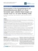
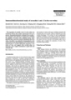
- Journal of Medical Case Reports 2007, 1:150 http://www.jmedicalcasereports.com/content/1/1/150 subtypes. The serous subtypes, primary serous ovarian car- demonstrate any masses. Magnetic resonance imaging of cinoma and primary peritoneal serous cystadenocarci- the abdomen and pelvis revealed pelvic peritoneal masses noma, predominate over the mucinous subtypes in both suspicious for metastatic implants and a 4.1 centimeters primary ovarian carcinoma and primary peritoneal cysta- mass in the appendix. Colonoscopy was performed to the denocarcinoma. Ascites and abdominal distension appear cecum. At 50 centimeters proximal to the anal verge, a 4 more frequently with primary peritoneal cystadenocarci- centimeters subserosal non-circumferential partially noma than with primary ovarian carcinoma. Primary occluding mass was discovered. The mass was biopsied. ovarian carcinoma alternately presents more often with a The remainder of the exam was normal. Consideration palpable pelvic mass. Primary peritoneal cystadenocarci- was given to an endoscopically concealed appendiceal noma and primary ovarian carcinoma are immunohisto- cystadenocarcinoma or primary appendiceal colonic ade- chemically indistinguishable [1,4]. Both malignancies nocarcinoma. Pathology reported the endoscopic tissue stain positive for CK7, ER, Mesothelin and CA125. Pri- biopsy as displaying a moderately well differentiated ade- mary peritoneal cystadenocarcinoma is histologically dis- nocarcinoma with a papillary growth pattern (Fig. 1). tinguished from primary ovarian carcinoma by ovaries of Attendant paracentesis was performed and the ascitic fluid normal size or enlarged secondary to a benign process, obtained was positive for malignant cells consistent with extraovarian involvement greater than ovarian involve- metastatic adenocarcinoma. Immunohistochemical stain- ment, and ovarian surface penetration of less than 5 mm ing of the malignant ascitic cells was positive for CA 125, depth [1,3,4]. WT-1, CK7 and ER but negative for CK20, TTF-1, PR, Cal- retinin and BRST-2. These immunohistochemistry results Management of primary peritoneal cystadenocarcinoma were consistent with primary peritoneal cystadenocarci- has followed treatment of primary ovarian carcinoma noma or primary serous ovarian carcinoma. Ovarian ori- with surgical debulking and adjuvant platinum-contain- gin was believed very unlikely without radiologic ing chemotherapy [4]. Survival in primary peritoneal evidence of a pelvic mass. The gynecologic oncology serv- serous cystadenocarcinoma parallels survival of stage III– ice concurred that the probability of a primary ovarian ori- IV primary serous ovarian carcinoma [1,4,5]. Median sur- gin was remote. Primary peritoneal cystadenocarcinoma vivals for primary peritoneal serous cystadenocarcinoma became the operating diagnosis. Surgical consultation was and primary serous ovarian carcinoma are equivalent and requested. range from 32–40 months [1,5]. Exploratory laparotomy was offered and performed. Pen- Primary peritoneal cystadenocarcinoma and primary etration of the peritoneum immediately presented serous ovarian carcinoma both stain positive for estrogen recep- ascites. The omentum was grossly thickened and adherent tor (ER), cytokeratin 7 (CK7), Wilm's tumor suppressor to the mid transverse colon at 50 centimeters proximal to gene (WT1), and cancer antigen 125 (CA 125). Neither the anus. This section of transverse colon was segmentally entity possesses cytokeratin 20 (CK 20), progesterone receptor (PR), Calretinin, carcinoembryonic antigen (CEA), gross cystic disease fluid protein (BRST-2), and thyroid transcription factor 1 (TTF1). Cytochemical over- lap can occur between different celltypes but the constel- lation of positive and negative antigenicity produces a unique immunohistochemical "fingerprint" that identi- fies cellular origin. Case presentation The patient was a 68 yr-old African American female who presented to the emergency department of Henry Ford Hospital with complaints of shortness of breath and abdominal distension. Her past medical history was sig- nificant for asthma, Type II diabetes mellitus, hyperten- sion, gastroesophageal reflux, adenomatous colonic polyps and breast cancer. Abdominal ascites and a right pleural effusion were present. Ultrasound imaging (US) of Figure 1 growth pattern sigmoid colonic glands with a papillary ing&nests of infiltrating neoplastic mucosal biopsy demonstrat- H E section of the abdomen and pelvis was normal except for the known H & E section of sigmoid colonic mucosal biopsy demonstrat- ascites. Computed tomography (CT) of the abdomen and ing nests of infiltrating neoplastic glands with a papillary pelvis demonstrated moderate ascites, a right pleural effu- growth pattern. sion and omental thickening. US and CT imaging failed to Page 2 of 5 (page number not for citation purposes)
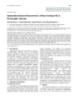
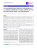
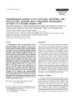
- Journal of Medical Case Reports 2007, 1:150 http://www.jmedicalcasereports.com/content/1/1/150 excised (Fig. 2). Multiple areas of peritoneal studding sig- nificantly involved the small bowel gutter and ascending colon. Foreshortening of the cecal mesentery was noted in conjunction with a soft verrucous appendix. Effective debulking mandated a right hemicolectomy, which was performed. Intestinal continuity was re-established with a standard side-to-side ileocolostomy. Histologic evalua- tion of the submitted omentum and transverse colon specimen revealed a moderately differentiated adenocar- cinoma with an infiltrative papillary growth pattern char- acteristic of primary peritoneal cystadenocarcinoma (Fig. 3). Histologic findings in the surgical and endoscopic biopsy specimens were analogous and confirmed primary peritoneal cystadenocarcinoma. These histologic findings also substantiated the immunohistochemical diagnosis of primary peritoneal cystadenocarcinoma. Postoperative ileus complicated the patient's recovery. She was dis- charged to home tolerating a regular diet on postoperative day 8. Systemic chemotherapy with carboplatin and Taxol was arranged in follow-up. Discussion Figure 3 tumor cells H & E section of omentum showing diffuse infiltration by The patient presented with a diagnosis of metastatic ade- H & E section of omentum showing diffuse infiltration by nocarcinoma of the colon. Her personal history of colonic tumor cells. The tumor cells demonstrate the papillary growth pattern characteristic of primary peritoneal adeno- polyps and her strong family history of colon cancer sup- carcinoma. ported this diagnosis. Endoscopic and pathologic findings were inconsistent with advanced colorectal cancer. Radio- logic imaging demonstrated peritoneal carcinomatosis but could not identify a unique origin. Accurately identi- predicated appropriate therapy as treatment differs fying the origin of the patient's peritoneal carcinomatosis among the varying malignancies causing peritoneal carci- nomatosis. Disease progression and survival also differ among the varying malignancies causing peritoneal carci- nomatosis and correct identification of malignant origin was required for accurate prognosis. Breast and gastrointestinal metastasis may clinically, radi- ologically and biochemically mimic primary ovarian and primary peritoneal adenocarcinoma [6]. Peritoneal mes- othelioma and pulmonary malignancies can also present clinically similar to primary ovarian carcinoma and pri- mary peritoneal cystadenocarcinoma with abdominal pain and ascitic distension. Treatment of primary perito- neal mesothelioma and carcinomatosis from metastatic breast, lung and gastrointestinal cancers differ signifi- cantly from the treatment of primary ovarian and primary peritoneal adenocarcinoma. Surgical debulking with intraoperative hyperthermic chemotherapy provides some survival advantage with peritoneal mesothelioma [7]. Advanced peritoneal mesothelioma is treated with combination systemic chemotherapy [7]. Overall survival with peritoneal mesothelioma is less than twelve months Figure 2 omentum Gross picture of a segment of transverse colon with attached [7]. Peritoneal carcinomatosis resulting from colorectal Gross picture of a segment of transverse colon with attached primaries has also been treated with intraoperative hyper- omentum. The Omentum is firm, nodular, and totally thermic chemotherapy an surgical debulking [8]. Survival replaced by tumor. in patients with peritoneal carcinomatosis from colorectal Page 3 of 5 (page number not for citation purposes)
- Journal of Medical Case Reports 2007, 1:150 http://www.jmedicalcasereports.com/content/1/1/150 primaries varies from 5.3 to 12.6 month [8]. Systemic immunohistochemical findings portrayed primary perito- chemotherapy for peritoneal carcinomatosis resulting neal cystadenocarcinoma. Immunohistochemistry from advanced colorectal cancer is 5-fluorouracil (5-FU) allowed the patient to be diagnosed without surgery. based with the addition of Oxaliplatin, Irinotecan, Bevaci- Without immunohistochemical technology, patients with zumab, and Cetuximab based on the patient's perform- diagnostically uncertain peritoneal carcinomatosis for- ance status and whether prior chemotherapy was merly required open or laparoscopic tissue biopsy with provided [9]. Median survival for peritoneal carcinomato- further operative interventions pending histologic analy- sis secondary to disseminated pulmonary malignancies is sis. Immunohistochemistry facilitated diagnosis without 2 months [10]. Surgical intervention with breast cancer surgery. carcinomatosis shows a variable survival advantage [11]. Chemotherapy and hormonal manipulation prolongs Preoperatively discerning primary peritoneal serous cysta- survival with breast cancer carcinomatosis [11]. Cytore- denocarcinoma from primary peritoneal mucinous aden- ductive surgery combined with systemic cisplatin-based ocarcinoma was not possible as these two entities are multiagent chemotherapy extends survival in both pri- indistinguishable immunohistochemically and histologi- mary ovarian and primary peritoneal cystadenocarcinoma cally. Primary peritoneal serous cystadenocarcinoma was [1]. Survival with carcinomatosis secondary to primary confirmed by the intraoperative finding of clear ascites ovarian carcinoma and primary peritoneal cystadenocar- and the patient was afforded significant tumor debulking. cinoma can extend beyond thirty months [1]. Histo-pathology returned a diagnosis of moderately dif- ferentiated invasive papillary adenocarcinoma, which was Metastatic lung cancer and peritoneal mesothelioma were consistent with primary peritoneal cystadenocarcinoma. excluded based on the absence of pulmonary disease on Our institution does not offer surgical debulking for peri- radiologic imaging and the absence of characteristic his- toneal carcinomatosis secondary to advanced colorectal topathologic features on biopsy, respectively. The patient cancer. Persistent misdiagnosis of advanced colorectal reported a personal history of breast cancer, ductal carci- cancer would have then resulted in suboptimal treatment noma in situ, treated with lumpectomy and radiation ther- of this patient by forgoing surgical debulking and assign- apy five years prior. Surgical margins were negative. ing inappropriate chemotherapy. Tamoxifen was prescribed following completion of her radiation therapy, but was discontinued after one month. Conclusion This history of breast cancer raised our suspicion of a Cognizance of primary peritoneal cystadenocarcinoma breast origin. Metastatic breast cancer was considered and the utility of immunohistochemistry permitted the unlikely given the rarity of peritoneal carcinomatosis from accurate diagnosis of a patient previously misidentified as this disease in the absence of any other evidence of metas- having advanced colorectal adenocarcinoma. Correctly tasis. Primary ovarian carcinoma was considered given the diagnosing primary peritoneal cystadenocarcinoma dra- patient's personal history of breast cancer. Genetic testing matically affected this patient's prognosis as opposed to was not performed. Consultation with the gynecologic metastatic colorectal adenocarcinoma with carcinomato- oncology service disavowed a primary ovarian etiology. sis. The incidence of primary peritoneal cystadenocarci- The possibility of primary peritoneal adenocarcinoma noma has increased dramatically over the past 10 years became prescient. [2]. Perhaps the incident rise in primary peritoneal cysta- denocarcinoma has resulted from a rising awareness of Immunohistochemical analysis lead to the correct identi- this disease and an increased ability to pathologically dis- fication of primary peritoneal cystadenocarcinoma while tinguish this disease. With the known ability of gastroin- excluding alternate diagnoses. Tumor markers ordered testinal malignancies to mimic gynecologic cancer, and included: CK7, CK20, TTF1, ER, PR, Calretinin, WT1, CA with 1.1% of cancers referred to gynecologic oncologists 125, BRST-2, and CEA. Colon adenocarcinoma stains pos- reported to be nongynecologic [12], the ability to distin- itive for CK20 and typically negative for CK7, CA125, guish advanced colorectal cancer from primary peritoneal TTF1 and ER. Breast cancer cells typically stain positive for cystadenocarcinoma and primary ovarian carcinoma has CK7, ER and are negative for CA125, CK20, and TTF1. become more important. Immunohistochemical technol- Ovarian carcinoma binds antigens for CK7, CA125, ER, ogy demonstrated marked utility for distinguishing pri- and is negative for CDX2, CK20, and TTF1. The patient's mary peritoneal cystadenocarcinoma from other cells were immunohistochemically negative for CK20, mimicking diseases in the setting of unknown primary TTF1, Calretinin, PR and BRST-2. Her immunohistochem- peritoneal carcinomatosis. This technology was successful istry was positive for CA125, WT1, CK7, and ER. Primary using only paracentesis and thus obviated the risks and peritoneal cystadenocarcinoma bears antigens CK7, ER, morbidity of surgical biopsy. Immunohistochemistry WT1, and CA125. The constellation of symptoms, endo- facilitated preoperative diagnostic certainty and thereby scopic morphology, imaging results, histo-pathologic and improved surgical planning. Improved patient outcomes Page 4 of 5 (page number not for citation purposes)
- Journal of Medical Case Reports 2007, 1:150 http://www.jmedicalcasereports.com/content/1/1/150 may result from further use of immunohistochemical 3. Piura B, Meirovitz M, Bartfeld M, Yanai-Inbar I, Cohen Y: Peritoneal papillary serous carcinoma: study of 15 cases and compari- analysis to diagnose unknown primary peritoneal carci- son with stage III–IV ovarian papillary serous carcinoma. J nomatosis. Surg Oncol 1998, 68(3):173-178. 4. McCluggage WG, Wilkinson N: Metastatic neoplasms involving the ovary: a review with an emphasis on morphological and Competing interests immunohistochemical features. Histopathology 2005, The author(s) declare that they have no competing inter- 47(3):231-247. 5. Ben-Baruch G, Sivan E, Moran O, Rizel S, Menczer J, Seidman DS: Pri- ests. mary peritoneal serous papillary carcinoma: a study of 25 cases and comparison with stage III–IV ovarian papillary serous carcinoma. Gynecol Oncol 1996, 60(3):393-396. Authors' contributions 6. Hewitt MJ, Anderson K, Hall GD, et al.: Women with peritoneal WV (Chief Resident/HOV) directly assembled patient carcinomatosis of unknown origin: Efficacy of image-guided information, directed further in depth analysis of the biopsy to determine site-specific diagnosis. BJOG 2007, 114(1):46-50. known literature, performed the operative interventions 7. Sugarbaker PH, Yan TD, Stuart OA, Yoo D: Comprehensive man- and managed the recovery of the patient, reviewed and agement of diffuse malignant peritoneal mesothelioma. Eur directed several drafts of the manuscript, and was respon- J Surg Oncol 2006, 32(6):686-691. 8. Koppe MJ, Boerman OC, Oyen WJ, Bleichrodt RP: Peritoneal car- sible for the final construction of the manuscript for sub- cinomatosis of colorectal origin: incidence and current treat- mission to the senior staff/primary investigator. ment strategies. Ann Surg 2006, 243(2):212-222. 9. National Comprehensive Cancer Network (NCCN), Clinical Practice Guide- lines in Oncology; Colon Cancer & Rectal Cancer. Version 2 2007. SJ (MSIV) acquired patient information, researched the 10. Satoh H, Ishikawa H, Yamashita YT, Kurishima K, Ohtsuka M, Seki- known literature, participated in the patient's surgery and zawa K: Peritoneal carcinomatosis in lung cancer patients. Oncol Rep 2001, 8(6):1305-1307. recovery, and constructed the manuscript under guidance. 11. Garg R, Zahurak ML, Trimble EL, Armstrong DK, Bristow RE: In this capacity she directly contributed to the conceptual- Abdominal carcinomatosis in women with a history of breast ization, design and production of this manuscript. Her cancer. Gynecol Oncol 2005, 99(1):65-70. 12. Benoit MF, Hannigan EV, Smith RP, Smith ER, Byers LJ: Primary gas- final manuscript was then submitted to senior members trointestinal cancers presenting as gynecologic malignan- for final approval. cies. Gyne Onc 2004, 95:388-392. DK (HOIII) acquired patient information, instructed the junior investigator (SJ) in conceptualization and construc- tion of the manuscript. ZZ (Pathologist) processed, evaluated and diagnosed the submitted surgical specimens. She also constructed the pathology narrative for the final manuscript. VV (Senior Staff) served as the senior staff reviewer/pri- mary investigator on this case report. He initially con- ceived the possibility of alternative diagnoses, arranged patient testing, scheduled and immediately directed the surgical interventions, supervised the patient's recovery and follow-up. He was responsible for final approval of the submitted manuscript following directed revision. Consent Written informed consent was obtained from the patient for publication of the study. Publish with Bio Med Central and every scientist can read your work free of charge Acknowledgements "BioMed Central will be the most significant development for No sources of funding were involved in the production of this manuscript disseminating the results of biomedical researc h in our lifetime." or the conduct of this study. Sir Paul Nurse, Cancer Research UK References Your research papers will be: 1. Bloss JD, Liao SY, Buller RE, et al.: Extraovarian peritoneal serous available free of charge to the entire biomedical community papillary carcinoma: a case-control retrospective compari- peer reviewed and published immediately upon acceptance son to papillary adenocarcinoma of the ovary. Gynecol Oncol 1993, 50(3):347-351. cited in PubMed and archived on PubMed Central 2. Halperin R, Zehavi S, Langer R, Hadas E, Bukovsky I, Schneider D: Pri- yours — you keep the copyright mary peritoneal serous papillary carcinoma: a new epidemi- ologic trend? A matched-case comparison with ovarian BioMedcentral Submit your manuscript here: serous papillary cancer. Int J Gynecol Cancer 2001, 11(5):403-408. http://www.biomedcentral.com/info/publishing_adv.asp Page 5 of 5 (page number not for citation purposes)
CÓ THỂ BẠN MUỐN DOWNLOAD

-

Báo cáo khoa học: "Immunohistochemical study of constitutive neuronal and inducible nitric oxide synthase in the central nervous system of goat with natural listeriosis"
 4 p |
4 p |  41
|
41
|  4
4
-

Báo cáo khoa học: "Immunohistochemical detection of Prion protein (PrP-Sc) and epidemiological study of BSE in Korea"
7 p | 48
| 4
-
Báo cáo y học: "Radiofrequency-induced thermotherapy of nasopharyngeal angiofibroma and immunohistochemical analysis of vessel proliferation: a case report"
5 p | 48
| 4
-
Báo cáo y học: "Demonstration of the histopathological and immunohistochemical effects of a novel hemostatic agent, ankaferd blood stopper, on vascular tissue in a rat aortic bleeding mode"
7 p | 57
| 4
-

Báo cáo khoa học: "Lymphangiosis carcinomatosa in squamous cell carcinomas of larynx and hypopharynx – value of conventional evaluation and additional immunohistochemical staining of D2-40"
8 p | 51
| 4
-
Báo cáo y học: " Immunohistochemical Characteristics of Bone Forming Cells in Pleomorphic Adenoma"
3 p | 53
| 4
-

Báo cáo y học: " Immunohistochemical characterization of nodose cough receptor neurons projecting to the trachea of guinea pigs"
16 p | 49
| 3
-
Báo cáo y học: " Immunohistochemical detection and regulation of a5 nicotinic acetylcholine receptor (nAChR) subunits by FoxA2 during mouse lung organogenesis"
11 p | 40
| 3
-

báo cáo khoa học: "Concomitant pulmonary and thyroid tumors identified by FDG PET/CT and immunohistochemical techniques"
17 p | 39
| 3
-

Báo cáo khoa học: "Immunohistochemical Localization of Bcl-2 in the Spinal Cords of Rats with Experimental Autoimmune Encephalomyelitis"
5 p | 53
| 3
-

Báo cáo khoa học: "Immunohistochemical Study of the Pancreatic Endocrine Cells inthe BALB/c mice: An Unique Distributional Pattern of Glucagon"
7 p | 58
| 3
-

Báo cáo y học: " Morphologically and immunohistochemically undifferentiated gastric neoplasia in a patient with multiple metastatic malignant melanomas: a case report"
5 p | 48
| 3
-
Báo cáo khoa học: "Immunohistochemical study of caveolin-1 and -2 in the rat retina"
4 p | 47
| 2
-

Báo cáo khoa học: " Immunohistochemical Study of the Endocrine Cells in the Pancreas of the Carp, Cyprinus carpio (Cyprinidae)"
12 p | 50
| 2
-

Báo cáo y học: " Immunohistochemical study of the phenotypic change of the mesenchymal cells during portal tract maturation in normal and fibrous (ductal plate malformation) fetal liver"
13 p | 48
| 2
-

Báo cáo y học: "Immunohistochemical localization of mu opioid receptor in the marginal division with comparison to patches in the neostriatum of the rat brain"
9 p | 38
| 2
-
Báo cáo khoa học: "Immunohistochemical Localization of Nerve Growth Factor,Glial Fibrillary Acidic Protein and Ciliary Neurotrophic Factor in Mesencephalon, Rhombencephalon, and Spinal Cord of Developing Mongolian Gerbil"
7 p | 58
| 2


Chịu trách nhiệm nội dung:
Nguyễn Công Hà - Giám đốc Công ty TNHH TÀI LIỆU TRỰC TUYẾN VI NA
LIÊN HỆ
Địa chỉ: P402, 54A Nơ Trang Long, Phường 14, Q.Bình Thạnh, TP.HCM
Hotline: 093 303 0098
Email: support@tailieu.vn
Giấy phép Mạng Xã Hội số: 670/GP-BTTTT cấp ngày 30/11/2015 Copyright © 2022-2032 TaiLieu.VN. All rights reserved.

